Agonistic Anti-tumor necrosis factor receptor 2 antibodies

a tumor necrosis factor and receptor 2 technology, applied in the field of agonistic antitumor necrosis factor receptor 2 antibodies, can solve the problems that the specific binding of non-human tnfr2 antibodies and antigen-binding fragments thereof may also lack specific binding to a tnfr superfamily, so as to enhance the solubility of the scfv fragment, improve the biophysical stability of the molecule, and increase the resistance of the s

Inactive Publication Date: 2019-05-09
THE GENERAL HOSPITAL CORP
View PDF2 Cites 6 Cited by
  • Summary
  • Abstract
  • Description
  • Claims
  • Application Information

AI Technical Summary

Benefits of technology

[0068]As used herein, the term “regulatory sequence” includes promoters, enhancers and other expression control elements (e.g., polyadenylation signals) that control the transcription or translation of the antibody chain genes. Such regulatory sequences are described, for example, in Goeddel, Gene Expression Technology: Methods in Enzymology 185 (Academic Press, San Diego, Calif., 1990); incorporated herein by reference.
[0069]As used herein, the term “scFv” refers to a single chain Fv antibody in which the variable domains of the heavy chain and the light chain from an antibody have been joined to form one chain. scFv fragments contain a single polypeptide chain that includes the variable region of an antibody light chain (VL) (e.g., CDR-L1, CDR-L2, and/or CDR-L3) and the variable region of an antibody heavy chain (VH) (e.g., CDR-H1, CDR-H2, and/or CDR-H3) separated by a linker. The linker that joins the VL and VH regions of a scFv fragment can be a peptide linker composed of proteinogenic amino acids. Alternative linkers can be used to so as to increase the resistance of the scFv fragment to proteolytic degradation (e.g., linkers containing D-amino acids), in order to enhance the solubility of the scFv fragment (e.g., hydrophilic linkers such as polyethylene glycol-containing linkers or polypeptides containing repeating glycine and serine residues), to improve the biophysical stability of the molecule (e.g., a linker containing cysteine residues that form intramolecular or intermolecular disulfide bonds), or to attenuate the immunogenicity of the scFv fragment (e.g., linkers containing glycosylation sites). scFv molecules are known in the art and are described, e.g., in U.S. Pat. No. 5,892,019; Flo et al., (Gene 77:51, 1989); Bird et al., (Science 242:423, 1988); Pantoliano et al., (Biochemistry 30:10117, 1991); Milenic et al., (Cancer Research 51:6363, 1991); and Takkinen et al., (Protein Engineering 4:837, 1991). The VL and VH domains of an scFv molecule can be derived from one or more antibody molecules. It will also be understood by one of ordinary skill in the art that the variable regions of the scFv molecules of the invention can be modified such that they vary in amino acid sequence from the antibody molecule from which they were derived. For example, in one embodiment, nucleotide or amino acid substitutions leading to conservative substitutions or changes at amino acid residues can be made (e.g., in CDR and/or framework residues). Alternatively or in addition, mutations are made to CDR amino acid residues to optimize antigen-binding using art-recognized techniques. ScFv fragments are described, for example, in WO 2011/084714; incorporated herein by reference.
[0070]As used herein, a small modular immunopharmaceutical (SMIP) protein refers to a protein that contains one or more of the following immunoglobulin domains: an antigen-binding domain, an immunoglobulin hinge region or a domain derived there from, an immunoglobulin heavy chain CH2 constant region or a domain derived there from, and an immunoglobulin heavy chain CH3 constant region or a domain derived there from. Polypeptides containing one or more of these domains can be obtained using methods known in the art or described herein, e.g., by recombinant expression of a polynucleotide encoding one or more of these domains or by chemical synthesis techniques (e.g., solid phase peptide synthesis, see Solid Phase Peptide Synthesis, 2nd ed., 1984 The Pierce Chemical Co., Rockford, 111; the disclosure of which is incorporated herein by reference in its entirety).
[0071]As used herein, the phrase “specifically binds” refers to a binding reaction which is determinative of the presence of an antigen in a heterogeneous population of proteins and other biological molecules that is recognized, e.g., by an antibody or antigen-binding fragment thereof, with particularity. An antibody or antigen-binding fragment thereof that specifically binds to an antigen will bind to the antigen or an epitope(s) thereof with a KD of less than 100 nM (e.g., between 1 pM and 100 nM). An antibody or antigen-binding fragment thereof that does not exhibit specific binding to a particular antigen or epitope thereof will exhibit a KD of greater than 100 nM (e.g., greater than 500 nM, 1 μM, 100 μM, 500 μM, or 1 mM) for that particular antigen or epitope thereof. A variety of immunoassay formats may be used to select antibodies specifically immunoreactive with a particular protein or carboh

Problems solved by technology

In addition, TNFR2 agonist antibodies and antigen-binding fragments thereof that specifically bi

Method used


Image

  • Agonistic Anti-tumor necrosis factor receptor 2 antibodies
  • Agonistic Anti-tumor necrosis factor receptor 2 antibodies
  • Agonistic Anti-tumor necrosis factor receptor 2 antibodies

Examples


example 1

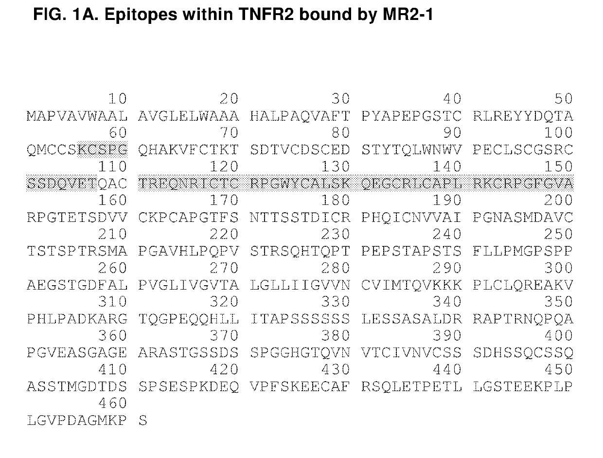
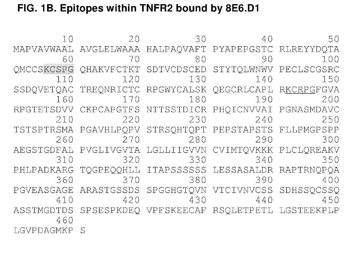
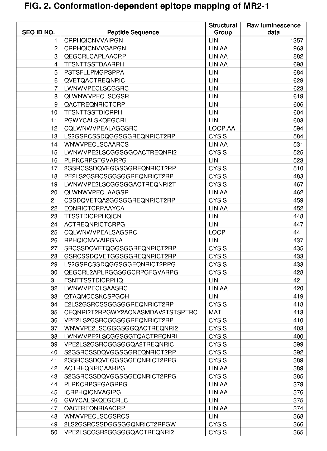
he Discrete Epitopes within TNFR2 that Interact with MR2-1

[0218]Libraries of linear, cyclic, and bicyclic peptides derived from human TNFR2 were screened for distinct sequences within the protein that exhibit high affinity for TNFR2 antibody MR2-1. In order to screen conformational epitopes within TNFR2, peptides from distinct regions of the primary protein sequence were conjugated to one another to form chimeric peptides. These peptides contained cysteine residues at strategic positions within their primary sequences (see, e.g., FIG. 2A, SEQ ID NOs: 53, 69, 75, 118, and 233). This facilitated an intramolecular cross-linking strategy that was used to constrain individual peptides to a one of a wide array of three dimensional conformations. Unprotected thiols of cysteine residues were cross-linked via nucleophilic substitution reactions with divalent and trivalent electrophiles, such as 2,6-bis(bromomethyl)pyridine and 1,3,5-tris(bromomethyl)benzene, so as to form conformationally re...

example 2

TNFR2 Antibodies Induce T-Reg Cell Proliferation

Materials and Methods

[0223]HUMAN T-REG FLOW™ Kit (BioLegend, Cat. No. 320401)[0224]Cocktail Anti-human CD4 PE-Cy5 / CD25 PE (BioLegend, Part No. 78930)[0225]ALEXA FLUOR® 488 Anti-human FOXP3, Clone 259D (BioLegend, Part No. 79467)[0226]ALEXA FLUOR® 488 Mouse IgG1, k Isotype Ctrl (ICFC), Clone MOPC-21 (BioLegend, Part No. 79486)[0227]FOXP3 Fix / Perm Buffer (4×) (BioLegend, Cat. No. 421401)[0228]FOXP3 Perm Buffer (10×) (BioLegend, Cat. No. 421402)[0229]PE anti-human CD25, Clone: BC96 (BioLegend, Cat. No. 302606)[0230]ALEXA FLUOR® 488 Anti-human FOXP3, Clone 259D (BioLegend, Cat. No. 320212)[0231]PBS pH 7.4 (1×) (Gibco Cat. No. 10010-023)[0232]HBSS (1×) (Gibco Cat. No. 14175-095)[0233]FBS (heat inactivated)[0234]15 ml tubes[0235]Bench top centrifuge with swing bucket rotor for 15 ml tubes (set speed 1100 rpm or 200 g)

[0236]Agonistic TNFR2 antibodies (MR2-1 and 8E6.D1) were tested for the ability to induce the proliferation of T-reg cells. Cu...

example 3

g Agonistic TNFR2 Antibodies by Phage Display

[0239]An exemplary method for in vitro protein evolution of agonistic TNFR2 antibodies of the invention is phage display, a technique which is well known in the art. Phage display libraries can be created by making a designed series of mutations or variations within a coding sequence for the CDRs of an antibody or the analogous regions of an antibody-like scaffold (e.g., the BC, CD, and DE loops of 10Fn3 domains). The template antibody-encoding sequence into which these mutations are introduced may be, e.g., a naive human germline sequence as described herein. These mutations can be performed using standard mutagenesis techniques described herein or known in the art. Each mutant sequence thus encodes an antibody corresponding in overall structure to the template except having one or more amino acid variations in the sequence of the template. Retroviral and phage display vectors can be engineered using standard vector construction techniqu...


PUM

PropertyMeasurementUnit
Molar densityaaaaaaaaaa
Digital informationaaaaaaaaaa
Therapeuticaaaaaaaaaa

Abstract

The invention provides agonistic TNFR2 antibodies and antigen-binding fragments thereof and encompasses the use of these antibodies as therapeutics to promote the proliferation of regulatory T cells (T-reg) for the treatment of immunological diseases. Antibodies of the invention can be used to potentiate the T-reg-mediated deactivation of self- and allergen-reactive T- and B-eases. Antibodies and can thus be used to treat a wide variety of indications, including autoimmune diseases, allergic reactions, asthma, graft-versus-host disease, and allograft rejection, among others.

Description

FIELD OF THE INVENTION[0001]The invention relates to antibodies capable of potentiating tumor necrosis factor receptor 2 signalling and their use for modulating the activity of T-reg cells, and provides therapies for immunological disorders or conditions, such as multiple sclerosis, asthma, allergic reactions, graft-versus-host disease, and transplantation graft rejection.BACKGROUND OF THE INVENTION[0002]Maintaining control of the cell-mediated and humoral immune responses is an important facet of healthy immune system activity. The aberrant regulation of T-cell and B-cell driven immune reactions has been associated with a wide array of human diseases, as the inappropriate mounting of an immune response against various self and foreign antigens plays a causal role in such pathologies as autoimmune disorders, asthma, allergic reactions, graft-versus-host disease, transplantation graft rejection, and a variety of other immunological disorders. These diseases are mediated by T- and B-l...

Claims


Application Information

Patent Timeline
IPC IPC(8): C07K16/28G01N33/68A61K38/19A61K39/395A61P37/06
CPCC07K16/2878G01N33/6854A61K38/191A61K39/3955A61P37/06C07K2317/34C07K2317/92C07K2317/75C07K2317/622G01N2333/7151
Inventor FAUSTMAN, DENISE L.
Owner THE GENERAL HOSPITAL CORP
Why Eureka
Try Eureka