Unlock instant, AI-driven research and patent intelligence for your innovation.

Method for scanning along a 3-dimensional line and method for scanning a region of interest by scanning a plurality of 3-dimensional lines

a technology of 3-dimensional lines and scanning methods, applied in the field of scanning along a 3-dimensional line and scanning a region of interest by scanning a plurality of 3-dimensional lines, can solve the problems of method faces, limited sampling rate, and loss of fluorescence data, and achieve the effect of reducing the amplitude of motion artifacts and fast functional measuremen

Active Publication Date: 2019-09-19
FEMTONICS
View PDF0 Cites 1 Cited by
  • Summary
  • Abstract
  • Description
  • Claims
  • Application Information

AI Technical Summary

Benefits of technology

The present invention provides a new method for 3D microscopy called 3D drift AO microscopy. This method allows the excitation spot to drift in any direction with any desired speed in 3D space while continuously recording fluorescence data. This is achieved by using non-linear chirps in the AO deflectors with parabolic frequency profiles. The partial drift compensation realized with these profiles allows the directed and continuous movement of the focal spot in arbitrary directions and with arbitrary velocities determined by the temporal shape of the chirped acoustic signals. During these fast 3D drifts of the focal spot, the fluorescence collection is uninterrupted, lifting the pixel dwell time limitation of the previously used point scanning. This allows pre-selected individual scanning points to extend to small 3D lines, surfaces, or volume elements to cover not only the pre-selected ROIs but also the neighboring background areas or volume elements. The scanning methods also allow for motion artifact correction on a fine spatial scale and, hence, the in vivo measurement of fine structures in behaving animals. This enables the fast functional measurement of neuronal somata and fine neuronal processes, such as dendritic spines and dendrites, even in moving, behaving animals in a z-scanning range of more than 650 μm in 3D.

Problems solved by technology

Despite the evident benefits of 3D random-access AO microscopy, the method faces two major technical limitations: i) fluorescence data are lost or contaminated with large amplitude movement artifacts during in vivo recordings; and ii) sampling rate is limited by the large optical aperture size of AO deflectors, which must be filled by an acoustic wave to address a given scanning point.
The first technical limitation occurs because the actual location of the recorded ROIs is continuously changing during in vivo measurements due to tissue movement caused by heartbeats, blood flow in nearby vessels, respiration, and physical motion.
This results in fluorescence artifacts because of the spatial inhomogeneity in the baseline fluorescence signal of all kinds of fluorescent labelling.
Moreover, there is also a spatial inhomogeneity in relative fluorescence changes within recorded compartments; therefore, measurement locations within a somata or dendritic compartment are not equivalent.
Therefore it is really difficult to separate post-hoc the genuine fluorescence changes associated with neural activity from the artifacts caused by brain movement.
The second technical problem with 3D point-by-point scanning is the relatively long switching time, which limits either the measurement speed or the number of ROIs.
However, to fill these large apertures with an acoustic signal takes considerable time.
Therefore, the resulting long-duration AO switching time does not allow volume or surface elements to be generated from single points in an appropriate time period.
In this way, the point scanning method yields high pointing stability but requires relatively long switching times, because it is necessary to fill the large AO deflector apertures each time when addressing a new point in 3D.
Nature methods 9:201-208) allows shorter pixel dwell times, but in this case, the fast lateral scans are restricted to two dimensions; 3D trajectory scans, however, still need to be interrupted by time-consuming jumps when moving along the z axis.
In other words, scanning along the z axis still suffers from the same limitation as during point-by-point scanning.

Method used


Image

  • Method for scanning along a 3-dimensional line and method for scanning a region of interest by scanning a plurality of 3-dimensional lines
  • Method for scanning along a 3-dimensional line and method for scanning a region of interest by scanning a plurality of 3-dimensional lines
  • Method for scanning along a 3-dimensional line and method for scanning a region of interest by scanning a plurality of 3-dimensional lines

Examples


example 1

Scanning to Compensate In Vivo Motion Artifacts

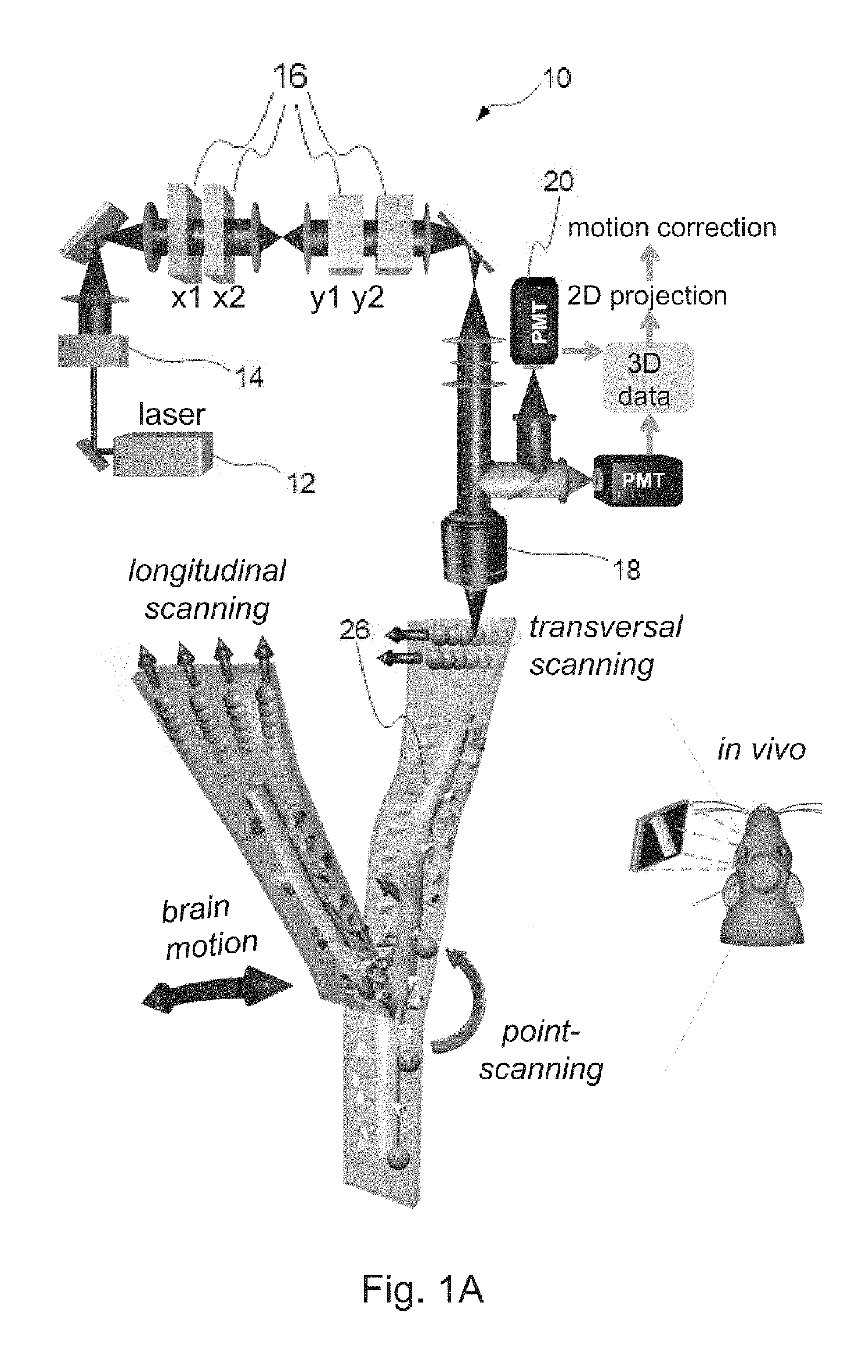
[0096]To demonstrate 3D ribbon scanning we labelled a small portion of pyramidal neurons in the V1 region of the visual cortex with a Ca2+ sensor, GCaMP6f, using an AAV vector for delivery. Then, according to the z-stack taken in advance, we selected guiding points and fitted the 3D trajectory which covered a spiny dendritic segment of a labelled pyramidal cell (FIG. 1C). FIG. 1C shows a 3D image of a dendritic segment of a selected GCaMP6f-labelled neuron. Cre-dependent GCaMP6f-expressing AAV vectors were used to induce sparse labelling. A 3D ribbon (indicated with dashed lines) was selected for fast 3D drift AO scanning within the cuboid

[0097]We used transversal drifts to scan along the selected 3D ribbons to measure the selected 140 μm dendritic segment and spines with 70.1 Hz (FIG. 1D). Raw fluorescence data (raw) were measured along the selected 3D ribbon and were projected into 2D along the longitudinal and traversal axes of the r...

example 2

of Spiny Dendritic Segments with Multiple 3D Ribbon Scanning

[0109]Recently it has been reported that for many cortical neurons, synaptic integration occurs not only at the axon initial segment but also within the apical and basal dendritic tree. Here, dendritic segments form non-linear computational subunits which also interact with each other, for example through local regenerative activities generated by non-linear voltage-gated ion channels. However, in many cases, the direct result of local dendritic computational events remains hidden in somatic recordings. Therefore, to understand computation in neuronal networks we also need novel methods for the simultaneous measurement of multiple spiny dendritic segments. Although previous studies have demonstrated the simultaneous recording of multiple dendritic segments under in vitro conditions, in vivo recording over large z-scanning ranges has remained an unsolved problem because the brain movement generated by heartbeat, breathing, o...

example 3

er, Multi-Frame Imaging of Neuronal Networks: Chessboard Scanning

[0113]To understand neuronal computation, it is also important to record not only assemblies of spines and dendrites, but also populations of somata. Random-access point scanning is a fast method which provides good signal-to-noise ratio for population imaging in in vitro measurements and in anesthetized mouse models; however, point scanning generates large motion artifacts during recording in awake, behaving animals for two reasons. First, the amplitude of motion artifacts is at the level of the diameter of the soma. Second, baseline and relative fluorescence is not homogeneous in space, especially when GECIs are used for labelling (FIG. 2C). Therefore, we need to detect fluorescence information not only from a single point from each soma, but also from surrounding neighbouring ROIs, in order to preserve somatic fluorescence information during movement. To achieve this, we extended each scanning point to small squares...


PUM


Abstract

The invention relates to a method for scanning along a substantially straight line (3D line) lying at an arbitrary direction in a 3D space with a given speed using a 3D laser scanning microscope having a first pair of acousto-optic deflectors deflecting a laser beam in the x-z plane (x axis deflectors) and a second pair of acousto-optic deflectors deflecting the laser beam in the y-z plane (y axis deflectors) for focusing the laser beam in 3D.The invention further relates to a method for scanning a region of interest with a 3D laser scanning microscope having acousto-optic deflectors for focusing a laser beam within a 3D space defined by an optical axis (Z) of the microscope and X, Y axes that are perpendicular to the optical axis and to each other

Description

CROSS-REFERENCE TO RELATED APPLICATIONS[0001]This application is a continuation-in-part of PCT / HU2017 / 050035, filed on Aug. 31, 2017, which claims priority of Hungarian Patent Application No. P1600519, filed on Sep. 2, 2016, each of which is incorporated herein by reference.TECHNICAL FIELD[0002]The present invention relates to a method for scanning along a substantially straight line (3D line) lying at an arbitrary direction in a 3D space with a given speed using a 3D laser scanning microscope.[0003]The invention further relates to a method for scanning a region of interest with a 3D laser scanning microscope having acousto-optic deflectors for focusing a laser beam within a 3D spaceBACKGROUND OF INVENTION[0004]Neuronal diversity, layer specificity of information processing, area wise specialization of neural mechanisms, internally generated patterns, and dynamic network properties all show that understanding neural computation requires fast read out of information flow and processi...

Claims


Application Information

Patent Timeline
IPC IPC(8): G02B21/00G06K9/32
CPCG02B21/008G06K9/3233G02B21/0076G02B21/0048G02B21/0044G02B21/0036G02F1/33
Inventor ROZSA, BALAZSKATONA, GERGELYMAAK, PALVERESS, MATEFEHER, ANDRASSZALAY, GERGELYMATYAS, PETER
Owner FEMTONICS