Antigen-presenting cell-mimetic scaffolds and methods for making and using the same

Active Publication Date: 2019-09-26
PRESIDENT & FELLOWS OF HARVARD COLLEGE
View PDF0 Cites 13 Cited by
  • Summary
  • Abstract
  • Description
  • Claims
  • Application Information

AI Technical Summary

Benefits of technology

The present invention provides methods for manipulating T-cells to improve their activation, stimulation, and expansion for use in treating various diseases. The methods involve using a scaffold containing T-cell activating molecules and co-stimulatory molecules to activate and maintain T-cells. The scaffold was found to improve the metabolic activity of T-cells and increase their expansion compared to other methods. These manipulated T-cells may be used for therapy of many diseases where T-cells are necessary.

Problems solved by technology

However, since adoptive T cell immunotherapy clinical trials often require billions of cells (Riddell et al., 1995), existing in vitro T-cell expansion protocols are often inadequate to meet the demands of such trials.
This is a very labor-intensive process and results in cell loss, cell damage, increased risk of contamination and increased cost of processing.
Because of the tight association of the immobilized monoclonal antibodies on the beads with the corresponding ligands on the surface of the target T-cells, the removal of the beads from the T-cells is difficult.
This technique can cause damage to the T-cells and can also cause the ligated antigens on the T-cells to be removed from the cell surface (Rubbi et al., Journal of Immunology Methods, 166, 233-241, 1993).
In addition, since activated T-cells are often most-desired for use in cell therapy protocols and the desirable properties of the cells are lost during the 24-72 hour waiting time, paramagnetic separation has a limited use in the adoptive cell-therapy setting.
Techniques for separation and purification of cells attached to paramagnetic beads are also unusable in the clinical context.
This process, while greatly reducing the number of beads remaining with the T-cells, does not completely eliminate the beads.
Implantation of compositions containing beads into patients can cause toxic effects.

Method used


Image

  • Antigen-presenting cell-mimetic scaffolds and methods for making and using the same
  • Antigen-presenting cell-mimetic scaffolds and methods for making and using the same
  • Antigen-presenting cell-mimetic scaffolds and methods for making and using the same

Examples


example 1

ion of Scaffolds and Microscopic Analysis of the Assembled Structures

[0391]Antigen-presenting cells-mimetic scaffolds (APC-MS) were assembled using the methodology described below. Briefly, a base layer containing high surface area mesoporous silica micro-rods (MSR) was first provided, onto which various T-cell homeostatic agents, e.g., interleukins such as IL2 and / or cytokines such as TGF-beta, are optionally loaded. In certain embodiments, it may be preferable to load the homeostatic agents on to the MSR layer. Then, a continuous, fluid-supported lipid bilayer (SLB) was layered on the base layer, thereby generating an MSR-SLB scaffold. If the homeostatic agents are not directly loaded on the MSR layer, then they can be loaded after SLB payload has been applied on top of the MSR layer. Then, a blocking agent such as BSA may be applied to block non-specific integration sites in the MSR-SLB scaffold, after which, one or more T-cell activating molecule(s) and T-cell co-stimulatory mol...

example 2

of the Functional Properties of the APC-MS Release of Homeostatic Factors such as IL-2

[0396]APC-MS containing mesoporous silica rods (MSR) and supported lipid bilayer (SLB), which further contain IL-2 were manufactured using the methods described in Example 1. The release of IL-2 from these MSR-SLB compositions was analyzed using staining techniques and / or binding assays. The results are presented in FIGS. 6A and 6B. As illustrated in the electron micrograph of FIG. 6A, high surface area pores of MSRs are available for potential adsorption of IL2 or other soluble payloads (scale bar=100 nm). The plot showing cumulative IL-2 release over a 15-day period (FIG. 6B) shows that the APC-MS of the invention are capable of releasing homeostatic agents such as IL-2 in a controlled and sustained manner during the entire course of the two-week study period.

Association with T-Cells

[0397]Antigen presenting cell-mimetic scaffolds containing MSR-SLB scaffolds (APC-MS) were incubated with media con...

example 3

Loading

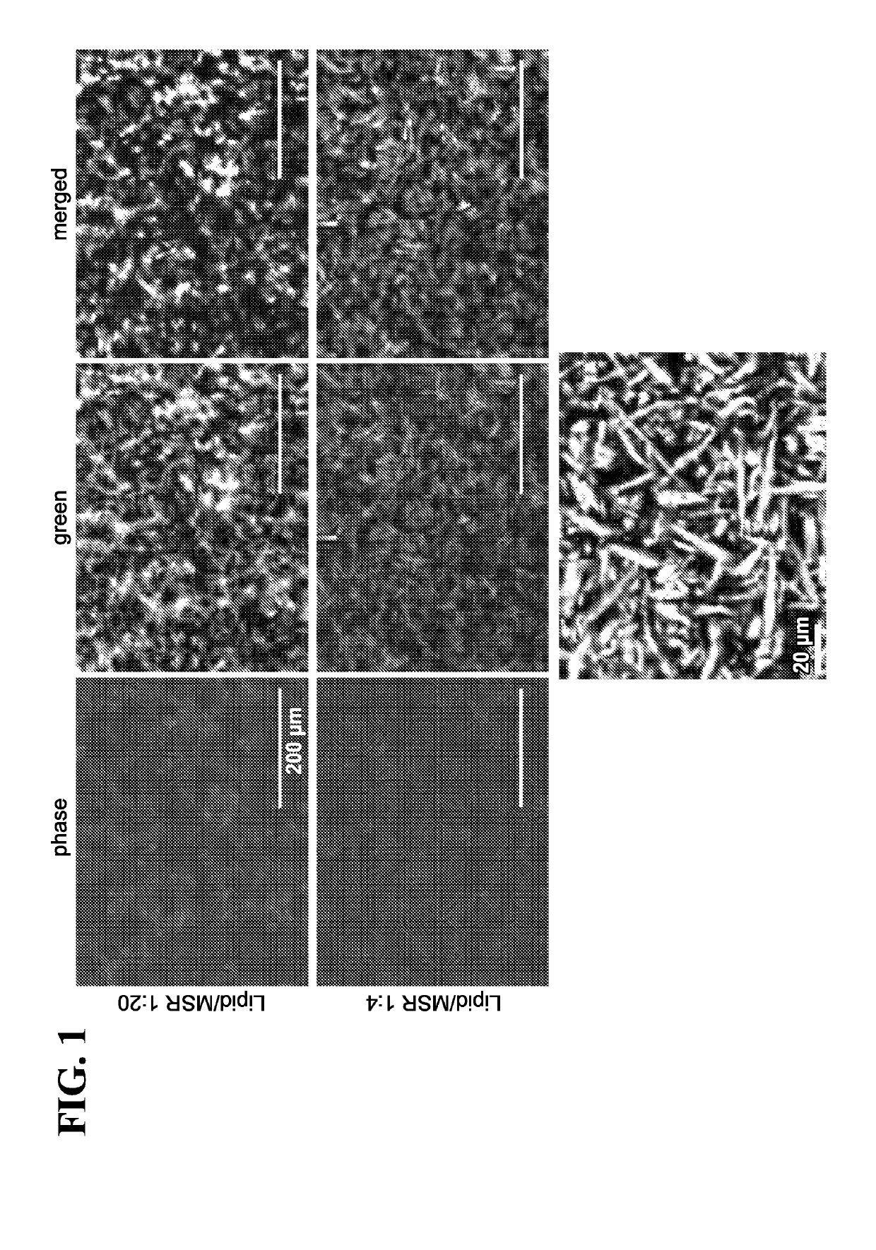
[0398]The APC-MS containing MSR-SLB were then loaded with various stimulatory molecules, co-stimulatory molecules and / or T-cell homeostatic agents and the resulting structures were analyzed with fluorescence microscopy. Four different types of MSR-SLB scaffolds were analyzed—(1) nude MSR-SLB scaffold (control); (2) MSR-SLB containing conjugated antibodies; (3) MSR-SLB containing IL-2; and (4) MSR-SLB containing conjugated antibodies and IL-2. The photomicrographs are shown in FIG. 8 (low resolution images are on the left and high resolution images are on the right). The top panel (greyscale images) contains phase-contrast microscope images of each of the aforementioned MSR-SLB scaffolds. The bottom panel merges images capturing lipid fluorescence with the greyscale images of mesoporous silica microrods (MSR). The images on the right show MSR-SLB scaffolds at high magnification (scale bar=20 μm).


PUM

PropertyMeasurementUnit
Timeaaaaaaaaaa
Timeaaaaaaaaaa
Timeaaaaaaaaaa

Abstract

Embodiments herein described provide antigen-presenting cell-mimetic scaffolds (APC-MS) and use of such scaffolds to manipulating T-cells. More specifically, the scaffolds are useful for promoting growth, division, differentiation, expansion, proliferation, activity, viability, exhaustion, anergy, quiescence, apoptosis, or death of T-cells in various settings, e.g., in vitro, ex vivo, or in vivo. Embodiments described herein further relate to pharmaceutical compositions, kits, and packages containing such scaffolds. Additional embodiments relate to methods for making the scaffolds, compositions, and kits / packages. Also described herein are methods for using the scaffolds, compositions, and / or kits in the diagnosis or therapy of diseases such as cancers, immunodeficiency disorders, and / or autoimmune disorders.

Description

RELATED APPLICATIONS[0001]This application claims priority to U.S. Provisional Patent Application No. 62 / 361,891, filed on Jul. 13, 2016, the entire contents of which are expressly incorporated herein by reference.STATEMENT OF GOVERNMENT SUPPORT[0002]This invention was made with Government support under Grant Nos. EB015498, EB014703, and DE013033, awarded by the U.S. National Institutes of Health. The Government has certain rights in the invention.BACKGROUND OF THE INVENTION[0003]Immunotherapy involving the priming and expansion of T lymphocytes (T cells) holds promise for the treatment of cancer and infectious diseases, particularly in humans (Melief et al., Immunol. Rev. 145: 167-177 (1995); Riddell et al., Annu. Rev. Immunol. 13:545-586 (1995)). Current studies of adoptive transfer in patients with viral infections and / or cancer involve the infusion of T cells that have been stimulated, cloned and expanded for many weeks in vitro on autologous dendritic cells (DC), virally infect...

Claims


Application Information

Patent Timeline
IPC IPC(8): C12N5/00C12N5/0783A61K38/20A61K39/395A61K38/19A61K39/00A61K9/00A61K35/17A61P35/00A61L27/38A61L27/54A61L27/56A61L27/02A61L27/28A61K38/18
CPCC12N2533/20A61K38/1841C12N2501/22A61L2300/45A61K39/395A61K9/0019A61L27/3804C12N2501/15A61K38/2013A61K39/0011A61K35/17C12N5/0075A61L27/54A61K38/193A61P35/00A61L27/28A61L27/025C12N2533/00C12N2501/998C12N2501/2302A61L2300/426A61L27/56C12N5/0638C12N2501/515C12N2533/10A61L2300/416A61L2400/06A61K39/4644A61K39/4611A61K39/464838A61K2300/00
Inventor CHEUNG, ALEXANDER SINGMOONEY, DAVID J.
Owner PRESIDENT & FELLOWS OF HARVARD COLLEGE
Why Eureka
Try Eureka