Analysis and sorting of motile cells
a technology of motile cells and analysis methods, applied in the field of systems and methods for the analysis and sorting of motile cells, to achieve the effects of high motility, high yield of motile cells, and convenient identification and selection
- Summary
- Abstract
- Description
- Claims
- Application Information
AI Technical Summary
Benefits of technology
Problems solved by technology
Method used
Image
Examples
example 1
ple Preparation
[0094]Semen samples were retrieved from B6D2F1 mice aged 7 to 12 weeks from Jackson Laboratories (Bar Harbor, Me.). Mice were exposed to CO2 until movement ceased and then euthanized by cervical dislocation. A small incision was made over the midsection, the skin was reflected back, and the peritoneum was entered with sharp dissection to expose the viscera. Both fat pads were pulled down to expose the testes and epididymides. The section of cauda epididymis and vas deferens was excised and placed into a center-well dish containing 300 μL of Human Tubal Fluid (HTF) (Irvine Scientific, Santa Ana, Calif.) supplemented with 10 mg mL−1 bovine serum albumin (BSA) (Sigma, St Louis, Mo.). Under a dissection microscope, while holding the epididymis in place with a pair of forceps, incisions were made in the distal parts of the epididymis to allow the sperm to flow out. Spermatozoa were pushed out of the vas deferens by stabilizing the organ with an insulin needle and slowly wa...
example 2
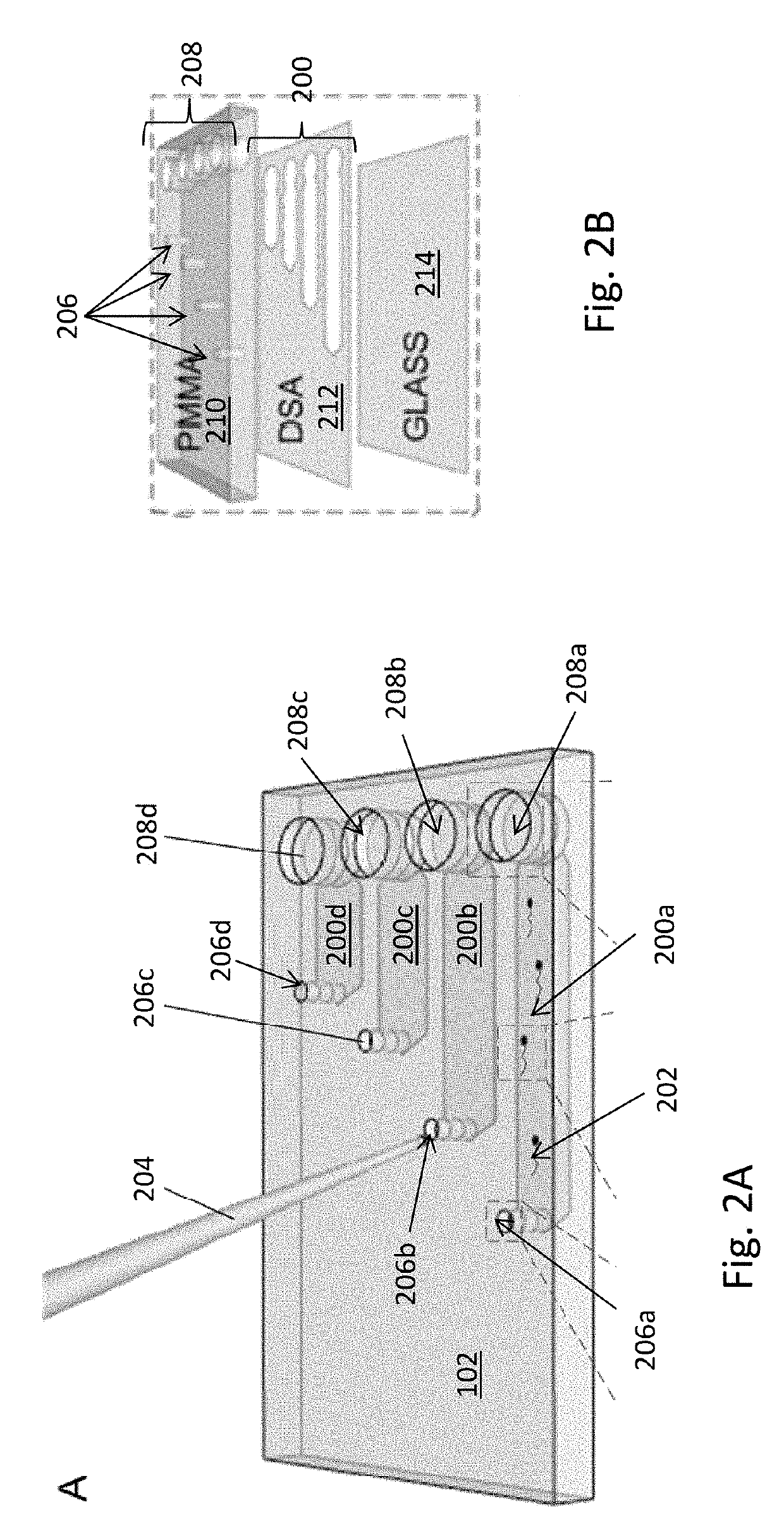
[0098]To demonstrate the capability of the microfluidic chip to sort sperm based on motility, sperm motilities at the input port and output port were compared to sperm motilities of non-sorted sperm based on sequenced images obtained using an optical microscope. This sorting test used a microfluidic chip having a channel length of 7 mm, a channel width of 4 mm, a channel height of 50 μm, an inlet port diameter of 0.65 mm, and an outlet port diameter of 2 mm. The large outlet port diameter was designed for easy extraction of sperm from the channel.
[0099]The channel was filled with fresh HTF medium containing 10 mg mL−1 BSA. The outlet port was filled with 2 μL of HTF / BSA medium and a thin layer of mineral oil was placed on top to avoid evaporation. 1 μL of capacitated sperm was removed after capacitation and added to the inlet port. The microfluidic chip was placed into an incubator at 37° C. for 30 minutes. At the end of the incubation period, 20 sequenced image...
example 3
Channel Orientation on Sperm Sorting
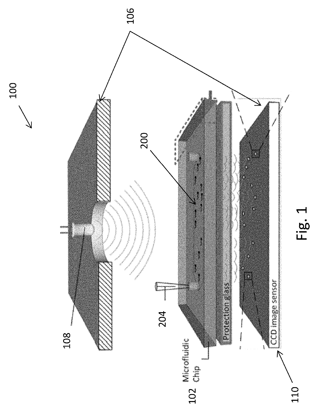
[0103]To determine the effect of channel orientation on sperm motility, sperm motion in both horizontal and vertical channel orientations was recorded. The channel had a length of 7 mm, a width of 4 mm, and a height of 50 am. The channel was filled with fresh HTF medium supplemented with 10 mg mL−1 of BSA. 1 μL of sperm sample was taken from the very top of a swim-up column as described above and pipetted into the input port of the channel. Fifteen sequenced shadow images were recorded using a lensless CCD sensor at a rate of one frame per second. The CCD sensor covered the entire channel such that all of the sperm in the channel were recorded. Motile sperm were identified and tracked using Photoshop (Adobe, San Jose, Calif.).
[0104]To image sperm in the vertical configuration, the microfluidic chip was clamped to the CCD sensor and the entire system was rotated by 90 degrees. Once the microfluidic chip was situated vertically, the above preparatio...
PUM
| Property | Measurement | Unit |
|---|---|---|
| height | aaaaa | aaaaa |
| height | aaaaa | aaaaa |
| height | aaaaa | aaaaa |
Abstract
Description
Claims
Application Information
 - R&D
- Intellectual Property
- Life Sciences
- Materials
- Tech Scout
- Unparalleled Data Quality
- Higher Quality Content
- 60% Fewer Hallucinations
Browse by: Latest US Patents, China's latest patents, Technical Efficacy Thesaurus, Application Domain, Technology Topic, Popular Technical Reports.
© 2025 PatSnap. All rights reserved.Legal|Privacy policy|Modern Slavery Act Transparency Statement|Sitemap|About US| Contact US: help@patsnap.com


