Implantable nerve guidance conduits having polymer fiber guidance channel

a technology of nerve guidance and polymer fibers, can solve the problems of inability to perform end-to-end suturing, each strategy suffers from a number of limitations, and cannot be applied in the field of tissue engineering, so as to facilitate nerve tissue regeneration, increase the surface area available, and restore the function to the site of the injury

Active Publication Date: 2019-10-31
STEVENS INSTITUTE OF TECHNOLOGY
View PDF2 Cites 10 Cited by
  • Summary
  • Abstract
  • Description
  • Claims
  • Application Information

AI Technical Summary

Benefits of technology

[0008]An embodiment of the present invention provides a fabricated implantable NGC. In some embodiments, the NGC comprises an inner spiral structured porous sheet. In some embodiments, the NGC comprises multiple inner spiral-structured porous sheets and / or multiple substantially cylindrical porous sheets. Such conduits have the potential to serve as medical devices to treat PNI and restore function to the site of the injury. This may be achieved by the spiral structure's ability to facilitate regeneration of nerve tissues.
[0009]In another embodiment of the present invention, the NGC has an integrated spiral structured porous sheet decorated with surface channels, multiple spiral structured porous sheets decorated with surface channels and / or multiple substantially cylindrical porous sheets decorated with surface channels. Such structures increase the surface area available for cell migration and attachment, and may reduce the length of time needed for recovery. Additionally, such structures can reduce the wear and tear that is often observed with single lumen tubular NGCs. A highly-aligned set of electrospun fibers are present within the surface channels, and may also be present on the backs thereof. The presence of aligned fibers in such areas ensures that the regenerating nerve will come into contact with aligned fibers.
[0010]In order to place and suture the nerve tissue without tension, there are two reserved chambers at the proximal and distal ends of the conduit. The chambers allow for nerve stumps to be sutured without tension because the chambers provide space to house the nerve in place with an optimal grip. An outer tube comprising a dense layer of randomly-oriented fibers can greatly improve the mechanical properties of the NGC and provides integrated structural support for nerve regeneration. The NGC and its component parts are tunable such that their length and diameter can be varied controllably depending on how the NGC is to be used. The method of fabricating the NGC does not limit its length, thus enabling the application for longer gap repair / regeneration for PNI.

Problems solved by technology

The most frequent causes include motor vehicle accidents, gunshot wounds, stabbings, and birth trauma.
However, each strategy suffers from a number of limitations.
For example, end-to-end suturing cannot be performed when the nerve gap is larger than 1 cm.
The use of autograft results in potential donor site morbidity for the patient and can potentially exacerbate the condition.
The use of allograft tissue has an associated risk of immunogenicity.
Although FDA-approved tissue engineered nerve devices have been available in the market for several years, these implant devices do not possess the proper physical topography or chemical cues for nerve repair and regeneration.
Also, most of them are currently limited to a critical nerve gap of approximately 4 cm.
To design an optimal NGC for enhancing PNR still remains a challenge.
Current laboratorial NGCs developed using haptotactic strategies alone are not yet comparable to autograft.
For example, multichannel NGCs may have an insufficient cross sectional area and or inhibit cell-cell interaction between each of the individual channels.
This may lead to functional mismatches and an insufficient level of regeneration.
Alternatively, microfilaments can mislead cell migration which can result in uneven distribution of cells within the NGC.
These failures in NGCs may be attributed to the inadequate design of intra-luminal guidance channels / filament, forming incomplete fibrin cables during the initial stages of regeneration.
Without the formation of this aligned bridge of extracellular material (ECM), further mechanisms for nerve repair are limited.
Therefore, it still remains a challenge to design an optimal NGC for enhancing PNR, when compared to the use of autografts.

Method used


Image

  • Implantable nerve guidance conduits having polymer fiber guidance channel
  • Implantable nerve guidance conduits having polymer fiber guidance channel
  • Implantable nerve guidance conduits having polymer fiber guidance channel

Examples


first embodiment

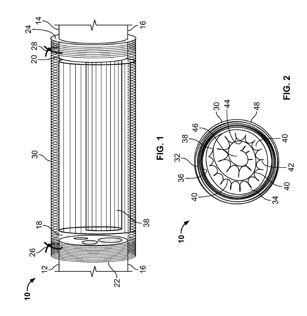
[0045]In the present invention, the NGC has a spiral guidance channel, the spiral structure including a highly porous polycaprolactone (PCL) sheet, which may be formed as a spiral-wound sheet using known methods and decorated with surface channels on a surface of the spiral wound sheet, coated with a thin layer of aligned electrospun fibers on the surface channels, and a dense randomly-oriented fibrous tube on the outside of the NGC. Other bioresorbable materials known for use in the biomedical arts may be used in place of PCL for the sheet and fibers (e.g., collagen / PCL blends for the fibers).

second embodiment

[0046]In the present invention, the NGC has multiple spiral or cylindrically wound structures within a dense randomly-oriented fibrous tube. The spiral or cylindrically wound structures which may be formed as a spiral-wound sheet using known methods. The spiral or cylindrically wound structures may include multiple grooves along the spiral wall and, in some embodiments, in the inner wall of the fibrous tube. The wound structures and / or the inner wall of the fibrous tube, may also be decorated with nanofibers and / or other micro-architectural features that improve cell adhesion and control the direction of nerve regeneration, and make a more favorable environment for nerve regeneration. Methods of making micro-architectural features in polymer sheets and other polymer structures are known in the art. Other bioresorbable materials known for use in the biomedical arts may be used in place of PCL for the sheet and fibers (e.g., collagen / PCL blends for the fibers).

[0047]Other embodiments ...


PUM

PropertyMeasurementUnit
lengthaaaaaaaaaa
diameteraaaaaaaaaa
diameteraaaaaaaaaa

Abstract

A nerve guidance conduit includes one or more guidance channels formed as porous polymeric structures. The guidance channels are within an outer tubular structure that includes randomly-oriented nanofibers. The guidance channels may have electrospun nanofibers on their inner and outer surfaces in a parallel alignment with the guidance channels. Such aligned nanofibers may also be present on the inner surface of the outer tubular structure. The outer surfaces of the guidance channels and the inner surface of the tubular structure define additional guidance channels. Such a nerve guidance conduit provides augmented surface areas for providing directional guidance and enhancing peripheral nerve regeneration. The structure also has the mechanical and nutrient transport requirements required over long regeneration periods.

Description

CROSS-REFERENCE TO RELATED APPLICATIONS[0001]The present application claims the benefit of U.S. Provisional Application No. 62 / 315,755, filed on Mar. 31, 2016, and is a continuation-in-part of U.S. patent Ser. No. 14 / 313,384, filed on Jun. 24, 2014, which claims the benefit of U.S. Provisional Patent Application No. 61 / 838,553, filed on Jun. 24, 2013, all of which applications are incorporated by reference herein.STATEMENT REGARDING FEDERALLY SPONSORED RESEARCH[0002]This invention was made with government support under NIH Grant No. RO3NS058595, NIH Grant No. R15 NS074404, and the Office of the Assistant Secretary of Defense for Health Affairs through the Peer Reviewed Orthopaedic Research Program under Award No. W81XWH-13-02301 and Award No. W81XWH-13-1-0320. The U.S. government has certain rights in the invention.FIELD OF THE INVENTION OR TECHNICAL FIELD[0003]The present invention relates to biomedical engineering, and, more particularly, to tissue engineering.BACKGROUND OF THE IN...

Claims


Application Information

Patent Timeline
Patent Type & Authority Applications(United States)
IPC IPC(8): A61B17/11A61L27/18A61L27/50
CPCA61L27/18A61B17/1128A61L2400/12A61L27/50A61B2017/1132A61B2017/00526A61L2430/32A61L27/56C08L67/04
Inventor YU, XIAOJUNSHAH, MUNISH B.CHANG, WEI
Owner STEVENS INSTITUTE OF TECHNOLOGY
Why Eureka
Try Eureka