
Modular wireless large bore vacuum universal endoscope and vacuumscope

a technology of endoscope and vacuum, which is applied in the field of medical science, can solve the problems of high rate of recurrent stone disease, persistent renal obstruction and permanent renal damage, and increased patient debility and expens

Inactive Publication Date: 2019-11-28
RGT UNIV OF CALIFORNIA
View PDF0 Cites 1 Cited by
  • Summary
  • Abstract
  • Description
  • Claims
  • Application Information

AI Technical Summary

Benefits of technology

The patent describes a device that can be used to remove biopsied tissue or stones from an individual. The device has an optical instrument connected to a narrow vacuum channel, which can be used to capture and remove the biopsied tissue or stone particles. The device can be used in various medical procedures and is designed for visualization and manipulation through a control mechanism. The device also has a battery pack and can be connected to Bluetooth or Wi-Fi. The technical effect of the patent is a more efficient and safe tool for the removal of biopsied tissue or stones from individuals.

Problems solved by technology

Nephrolithiasis may lead to persistent renal obstruction and permanent renal damage if left unresolved.
The failure to completely render the stone bearing kidney free of all stone material results in a high rate of recurrent stone disease and further patient debility and expense.
Of note, the major drawback to both shock wave lithotripsy as well as ureteroscopy is that the stone clearance rate is no better than 50% on computed tomography (CT) scans; leaving even small stone fragments behind, puts the patient at high risk for recurrent stone disease within 2-4 years.

Method used


Image

  • Modular wireless large bore vacuum universal endoscope and vacuumscope
  • Modular wireless large bore vacuum universal endoscope and vacuumscope
  • Modular wireless large bore vacuum universal endoscope and vacuumscope

Examples


example 1

Generally

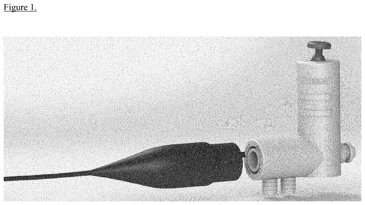
[0033]In accordance with various embodiments herein, the inventors developed a sterile, disposable or re-useable ureteroscope that integrates at minimum 6 French to 26 French vacuum / working (Vacuumoscopy) channel to ensure removal of all stone particle debris at the end of a laser ablation of a renal stone after ureteroscope (i.e. 6F channel) or percutaneous nephrolithotomy (up to 21F). Further, the device may expand indications for ureteroscopic laser stone fragmentation to stones larger than 1 cm. In another embodiment, the device is a wireless and modular universal endoscope that could be used for bronchoscopy, gastroscopy, colonoscopy, arthroscopy, and laryngoscopy to cut down on costs of usability and make endoscopy more portable.

example 2

Overview

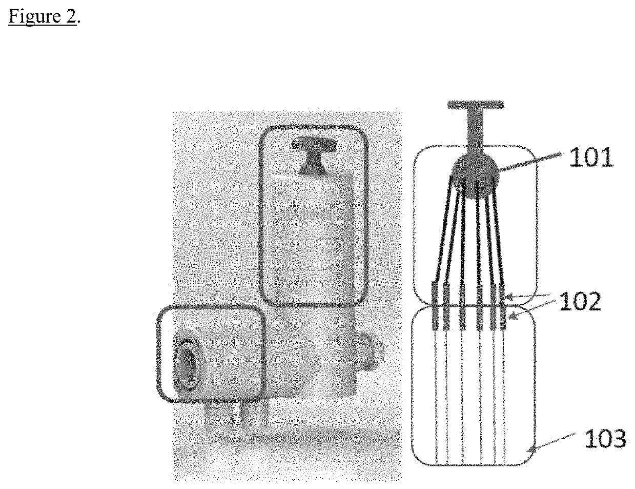
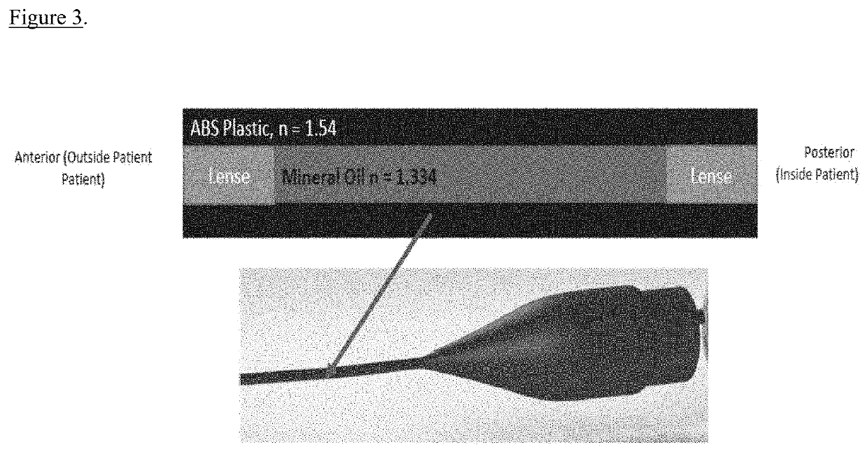
[0034]In one embodiment, the invention will be used in all ureteroscopic or percutaneous nephrolithotomy laser lithotripsy procedures in which a kidney stone is broken into smaller fragments. Typical ureteroscopes have an approximate 1.2 mm channel that does not provide for an adequate channel to suction and remove debris following stone laser fragmentation; indeed this same channel is used for irrigation for visualization within the kidney. The invention will overcome such shortcoming by utilizing a larger bore central vacuum channel (2.5 mm to 7 mm) with a drainage connection that does not compromise the endoscope's central vaccum channel; with ongoing irrigation coming from smaller channels arrayed around the central vacuum channel. The vacuumscope will vary in length and diameter (French) dependent upon the intended use. During ureteroscopy procedures, visualization and manipulation of the endoscope will be achieved through an array of materials around the central vacuum...

example 3

Some Advantages

[0037]At minimum one large bore (i.e. 7 French to 21 French) vacuum port to evacuate all stone fragments [0038]Disposable thereby eliminating risks of infection or endoscope malfunction due to processing.[0039]Wireless video capabilities via Bluetooth and / or Wi-Fi thereby eliminating entangling cords and expensive high power light sources and fixed camera equipment.[0040]Superior universal 360 degree deflection based on joystick steering—eliminating the need for the surgeon to employ excessive wrist movement to guide the tip of the endoscope.[0041]Modular design capable of adapting various endoscope shafts to attach to the same universal handle.[0042]Unique ergonomic handle design allowing for single-handed control of irrigation, suction, tip deflection, and instrument proj ection / retraction.


PUM


Abstract

The present technology relates to ureteroscopy, laser ablation of ureteral and renal stone, capture and removal of stone fragments. In one embodiment, the device includes an optical instrument operably connected to a large vacuum channel between 1.5 mm and 8.0 mm in width. In another embodiment, the device includes two single-time use disposable or potentially reusable units, such as a large vacuum endoscope removal tip and wireless and modular battery powered hand piece.

Description

CROSS-REFERENCE TO RELATED APPLICATIONS[0001]The present application claims the benefit of priority under 35 U.S.C. § 119(e) of provisional application Ser. No. 62 / 675,929, filed May 24, 2018, the contents of which are hereby incorporated by reference.FIELD OF THE INVENTION[0002]The present disclosure relates to the medical field, and more specifically, ureteroscopy and surgical procedures.BACKGROUND OF THE INVENTION[0003]All publications herein are incorporated by reference to the same extent as if each individual publication or patent application was specifically and individually indicated to be incorporated by reference. The following description includes information that may be useful in understanding the present invention. It is not an admission that any of the information provided herein is prior art or relevant to the presently claimed invention, or that any publication specifically or implicitly referenced is prior art.[0004]Nephrolithiasis, or the presence of renal calculi ...

Claims


Application Information

Patent Timeline
Patent Type & Authority Applications(United States)
IPC IPC(8): A61B1/307A61B1/00A61B1/06A61M1/00
CPCA61B1/307A61B2017/22079A61M1/0023A61B1/0676A61B1/00119A61B1/00094A61B18/26A61B1/015A61B1/00103A61B1/00096A61B1/0017A61B2018/00511A61B2018/00577A61B2018/00517A61B2018/00982A61M1/84A61B1/00042A61M1/774
Inventor CLAYMAN, RALPH V.KALER, KAMALJOT S.HEIDARI, ANDREW E.
Owner RGT UNIV OF CALIFORNIA
Learn More