Additive Manufacturing of Functional Myocardial Tissue

a functional and myocardial tissue technology, applied in the field of additive manufacturing systems and methods, can solve the problems of affecting the development of biologically relevant cardiac tissues, unable to achieve the directionality characterized by the physiological myocardium of the construct, and the limited self-renewal potential of the mature cardiomyocy

Active Publication Date: 2020-01-23
THE UNITED STATES OF AMERICA AS REPRESENTED BY THE SECRETARY OF THE NAVY
View PDF0 Cites 4 Cited by
  • Summary
  • Abstract
  • Description
  • Claims
  • Application Information

AI Technical Summary

Benefits of technology

[0009]A bioreactor and methods for using said bioreactor for drug screening, can include a microfibrous hydrogel scaffold, that can be made of a composite alginate-gelatin methacryloyl (GeIMA) bioink, and that can have endothelial cells directly embedded within the scaffold. The scaffold can further be seeded with cardiomyocytes so that said bioreactor has a controlled anisotropy, and the scaffold can be placed in a chamber defined by a PDMS half pieces, that compress the scaffold slightly when the PDMS half pieces are fastened to each other. The chamber could certainly be made of other materials as chosen by the end user, including but not limited to, thermoplastics, glass, etc.

Problems solved by technology

One of the first challenges in engineering cardiac organoids and their on-chip forms in the prior art can be the fact that mature cardiomyocytes exhibit limited self-renewing potential.
Second, the alignment of cardiomyocytes and their organization into bundles characterized by spontaneous and synchronous contraction further complicate the development of biologically relevant cardiac tissues.
While the cardiac spheroids have served an important role in drug testing and have been widely used due to the ease of preparation, these constructs can lack the directionality characterized by the physiological myocardium, which can be critical to maintain the long-term functionality of the engineered cardiac tissues.
However, generating volumetric cardiac tissues containing embedded endothelial networks remains challenging.
Particularly, bioprinting overcomes major drawbacks of conventional scaffold-based approaches including limited control over the 3D structures of engineered tissues and thus reduced reproducibility.
Additionally, the bioprinting process is usually biocompatible, allowing for direct encapsulation of bioactive molecules and cells.

Method used


Image

  • Additive Manufacturing of Functional Myocardial Tissue
  • Additive Manufacturing of Functional Myocardial Tissue
  • Additive Manufacturing of Functional Myocardial Tissue

Examples


Embodiment Construction

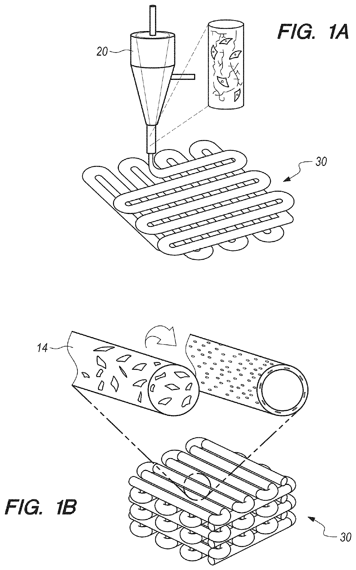
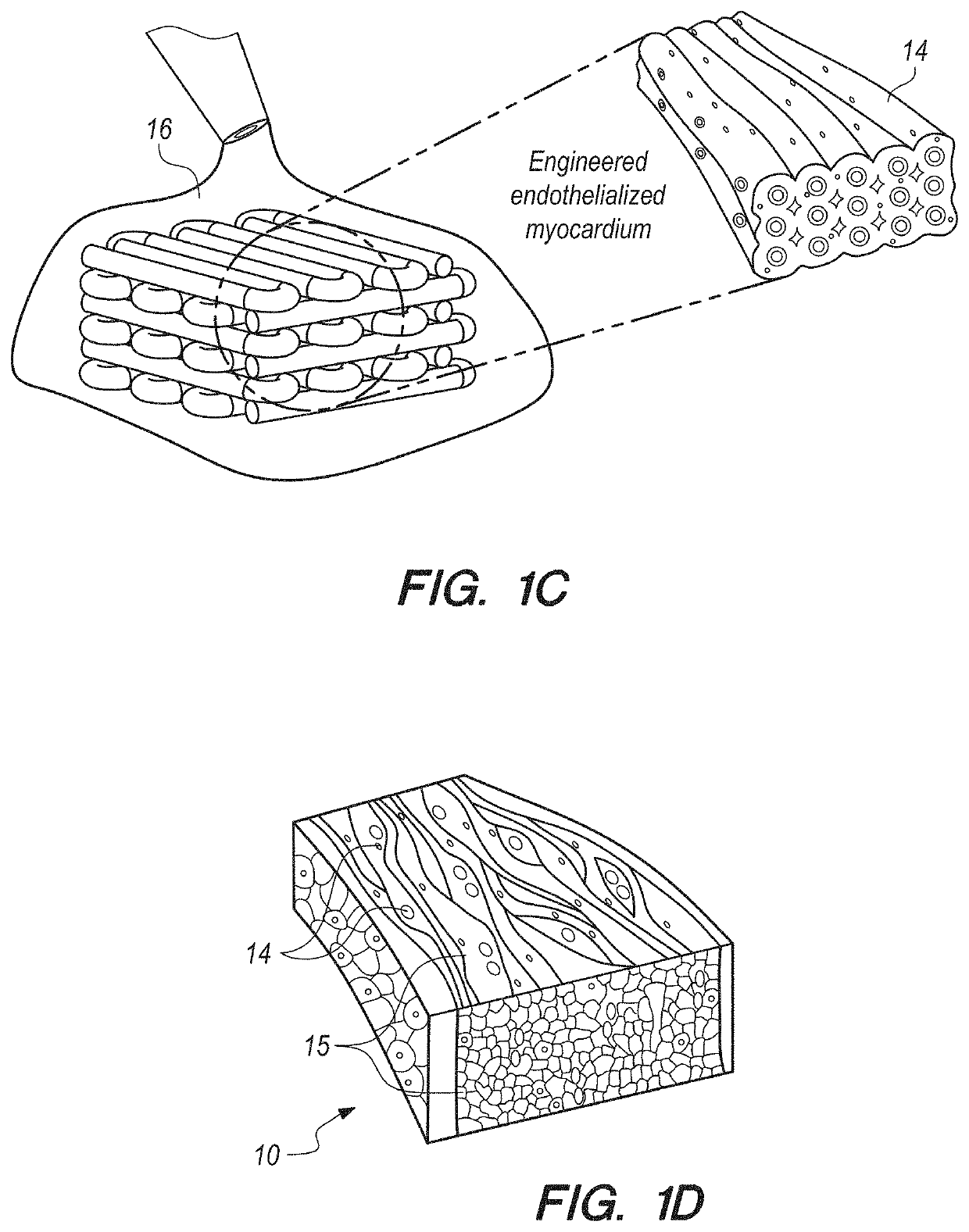
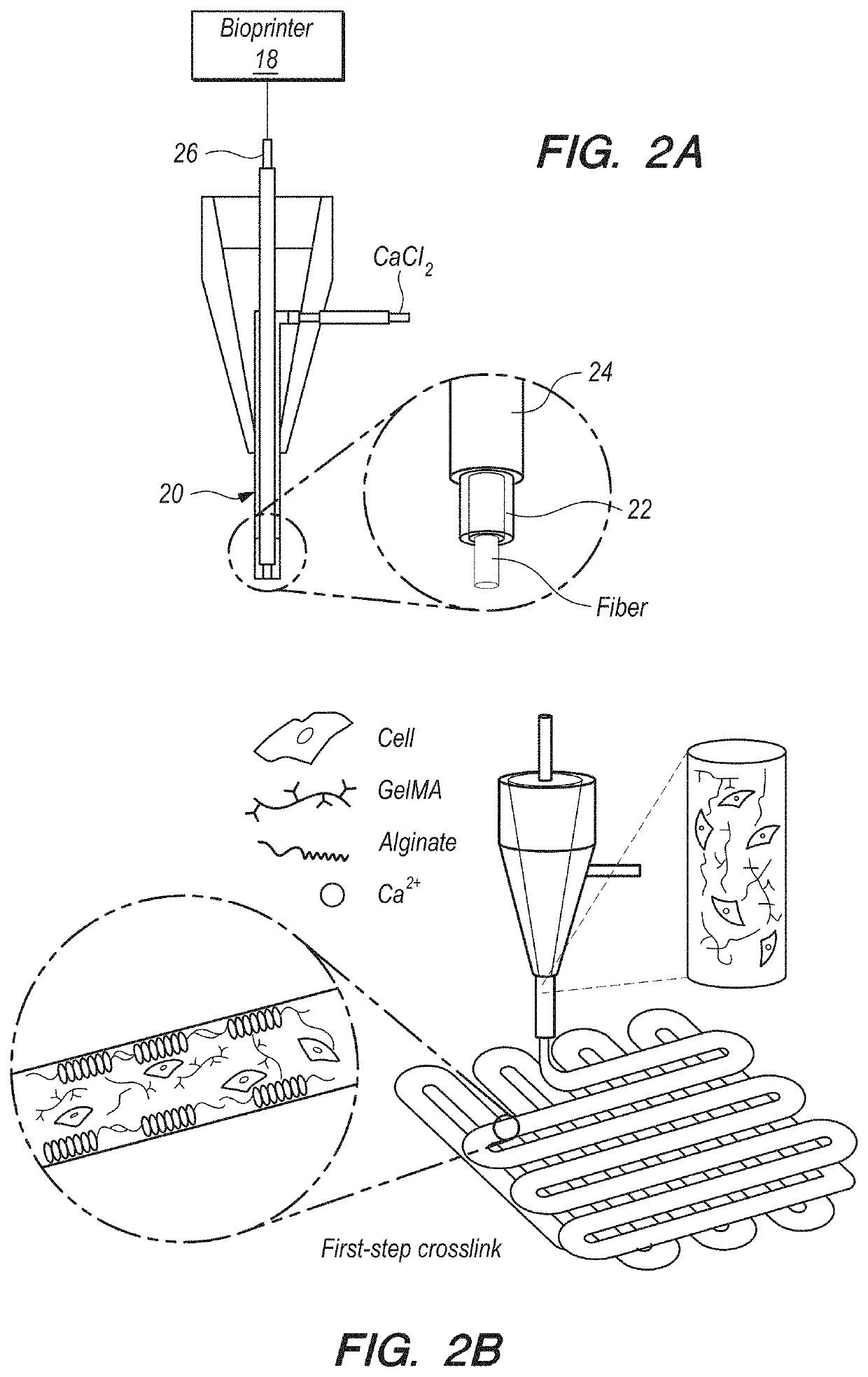
[0086]In brief overview, and referring initially to FIGS. 1A-1C, in this work a novel hybrid strategy based on 3D bioprinting can be presented (as used in this Specification, “bioprinting”, “3D bioprinting” and “additive manufacturing” can be taken to mean the same thing), to engineer endothelialized myocardial tissue 10 (FIG. 1D). Based on the microfluidic technology that has been developed in the prior art, it can be demonstrated that endothelial cells 11 can be directly encapsulated within the bioprinted microfibrous scaffold 30, as shown in FIG. 1A. The cells can gradually migrate towards the peripheries of the microfibers to form a layer of confluent endothelium. Such assembly of the endothelial cells 11 can resemble a blood vessel structure 14 (see FIGS. 1B-1C). The assembly can be enabled by the use of composite alginate-GeIMA bioink with a dual-step crosslinking process, and can be potentially facilitated by the intrinsic polarization tendency of these cells as well as prese...


PUM

PropertyMeasurementUnit
flow rateaaaaaaaaaa
pHaaaaaaaaaa
speedaaaaaaaaaa

Abstract

A bioreactor and methods for use can include a microfibrous scaffold, that can be made of a composite bioink, and that can have endothelial cells directly embedded within the scaffold using an additive manufacturing process. The scaffold can further be seeded with cardiomyocytes. The hydrogel scaffold can be composed of a plurality of serpentine layers, with each serpentine layers, which can be placed on each other in a cross-hatch configuration, so that the primary axes of successive layers are perpendicular. This configuration can establish an aspect ratio for the scaffold, which can be selectively varied. For greater strength, the successive layers that have a primary axis in the same direction can be placed in the scaffold so that they are slight offset from each other. The scaffold can be placed in the bioreactor with perfusion, for use in cardiovascular drug screening and other nanomedicine endeavors.

Description

CROSS REFERENCE TO RELATED APPLICATIONS[0001]This application claims the benefit of U.S. Provisional Application 62 / 552,878, filed Aug. 31, 2017, by Yu Shrike Zhang et al., entitled “Additive Manufacturing of Functional Myocardial Tissue”. The contents of the '878 application are hereby incorporated by reference into this specification.FEDERALLY SPONSORED RESEARCH AND DEVELOPMENT[0002]The United States Government has ownership rights in this invention, pursuant to passing of title to a Subject Invention under Government Contract N66001-13-C-2027 (Wake Forest University). Licensing inquiries may be directed to Office of Research and Technical Applications, Space and Naval Warfare Systems Center, Pacific, Code 72120, San Diego, Calif., 92152; telephone (619) 553-5118; email: ssc pac t2@navy.mil, referencing 105804.FIELD OF THE INVENTION[0003]The present invention pertains generally to additive manufacturing systems and methods. More specifically, the present invention can pertain to s...

Claims


Application Information

Patent Timeline
Patent Type & Authority Applications(United States)
IPC IPC(8): C12M3/00G01N33/50C12N5/074C12N5/077C12N5/071C12M3/06
CPCC12M23/16G01N33/5061G01N33/5064B33Y10/00C12N5/0607C12N5/0657B33Y80/00G01N33/5088C12M21/08B33Y70/00C12N5/069C12M25/14C12M29/10B29C64/106C12N2502/1329C12N2502/28C12N2503/04C12N2503/02C12N2513/00C12N2506/45C12N5/0697C12N2535/00C12N2533/74C12N2533/54C12N2533/30B33Y70/10C12M35/04
Inventor ZHANG, YU SHRIKEKHADEMHOSSEINI, ALI
Owner THE UNITED STATES OF AMERICA AS REPRESENTED BY THE SECRETARY OF THE NAVY
Why Eureka
Try Eureka