

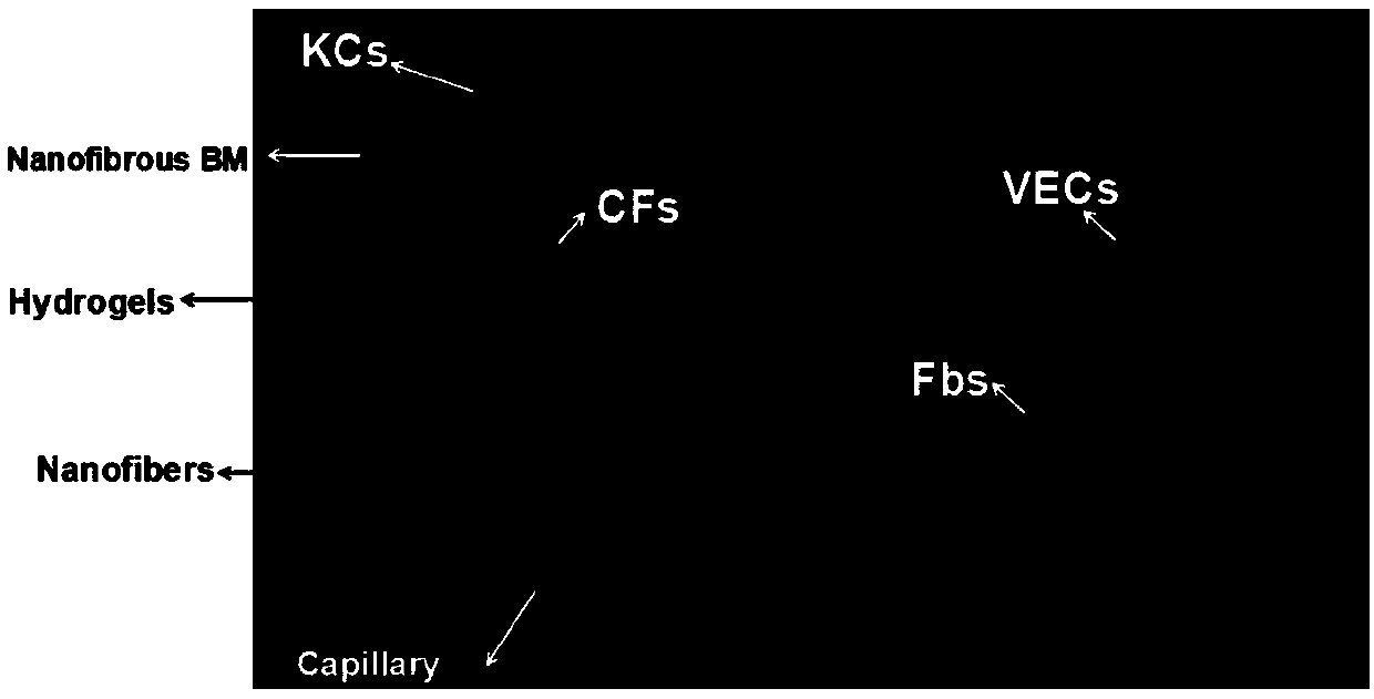
Vascularized full-thickness tissue-engineered skin layer-by-layer assembled by hydrogel, nanofiber scaffolds and skin cells and preparation method thereof
A tissue engineering skin and nanofiber technology, applied in the field of polymer materials and biomedical materials, can solve the problems of unfavorable cell migration, proliferation, no significant improvement of cell distribution and wound healing promotion effect, and no wound healing effect, etc., to achieve Improve tissue distribution, promote wound healing, and reduce scar formation
- Summary
- Abstract
- Description
- Claims
- Application Information
AI Technical Summary
Problems solved by technology
Method used
Image

Examples
Embodiment 1
[0053] Embodiment 1, keratinocyte separation and purification
[0054] The foreskin was taken out and washed several times with sterile PBS to clarify it. After removing fascia and other tissues, cut into strips; add Dispase, and place in a refrigerator at 4°C for overnight digestion. The foreskin digested overnight was rewarmed at 37°C, and the epidermis and dermis were separated after removal, and the separated epidermis was washed in PBS. The isolated epidermis was digested with trypsin (containing EDTA) at 37°C. The digestive juice was filtered through a strainer, and the filtrate was collected and centrifuged. After washing with PBS, the cells were resuspended in K-SFM medium at 1×10 5 / cm 2 Cell density Cells were inoculated and cultured in a 37°C incubator. Change the medium for the first time after 2 days, and change the medium every 3 days thereafter.
Embodiment 2
[0055] Embodiment 2, circulating fibrocyte separation and purification
[0056] Add human lymphocyte separation medium to the L tube, centrifuge to the lower layer, dilute the blood with PBS, add the diluted blood to the L tube, and centrifuge to separate layers. Discard the upper layer of plasma, suck out the buffy coat layer and add it to a centrifuge tube, add PBS to dilute and centrifuge, repeat the above steps 3 times. Then resuspended in PBS, counted cells, at 1×10 6 / mL density inoculated in Petri dishes.
Embodiment 3
[0057] Embodiment 3, separation and purification of vascular endothelial cells
[0058] The HUVEC cell line purchased from Sciencell was resuscitated at 37°C, seeded in a 100mm culture dish, and after the cells were congested, trypsinized and passaged.
PUM
| Property | Measurement | Unit |
|---|---|---|
| diameter | aaaaa | aaaaa |
| thickness | aaaaa | aaaaa |
| diameter | aaaaa | aaaaa |
Abstract
Description
Claims
Application Information