Systems and methods for detecting tissue ischemia and evaluating organ function using serum luminescence

a tissue ischemia and luminescence technology, applied in the field of noninvasive detection and/or evaluation of organ function, can solve the problems of ischemia-reperfusion damage-induced organ failure, major risks, and inability to detect other ischemia, such as bowel ischemia or acute mesenteric ischemia, and achieve the effect of improving the diagnostic accuracy and reducing the risk of organ failur

Pending Publication Date: 2020-03-26
UNIVERSITY OF MACAU
View PDF1 Cites 2 Cited by
  • Summary
  • Abstract
  • Description
  • Claims
  • Application Information

AI Technical Summary

Benefits of technology

[0007]Embodiments of the present disclosure solve one or more of the foregoing or other problems in the art with detecting tissue ischemia and/or evaluating organ dysfunction. In particular, one or more embodiments can include methods for detecting tissue ischemia within a mammal by at least (i) iso

Problems solved by technology

The foregoing illustrates that ischemia-reperfusion damage induced organ failures are major risks associated with organ transplantation.
Thrombosis or embolism caused by obstruction of blood flow from plaques, blood clots, fat emboli, foreign objects, or other bodily substance within the circulation system may also induce organ failure.
Other ischemia, such as bowel ischemia or acute mesenteric ischemia, are more difficult to diagnose due to non-specific symptoms like abdominal pain.
This can be problematic as late diagnosis is associated with a 60-80% mortality rate.
Similarly, deep vein thrombosis can lead to a lethal pulmonary embolism, but there is a lack of distinctive physiological symptoms to enable rapid and accurate diagnoses of deep vein thrombosis.
None of these systems in

Method used


Image

  • Systems and methods for detecting tissue ischemia and evaluating organ function using serum luminescence
  • Systems and methods for detecting tissue ischemia and evaluating organ function using serum luminescence
  • Systems and methods for detecting tissue ischemia and evaluating organ function using serum luminescence

Examples


example 1

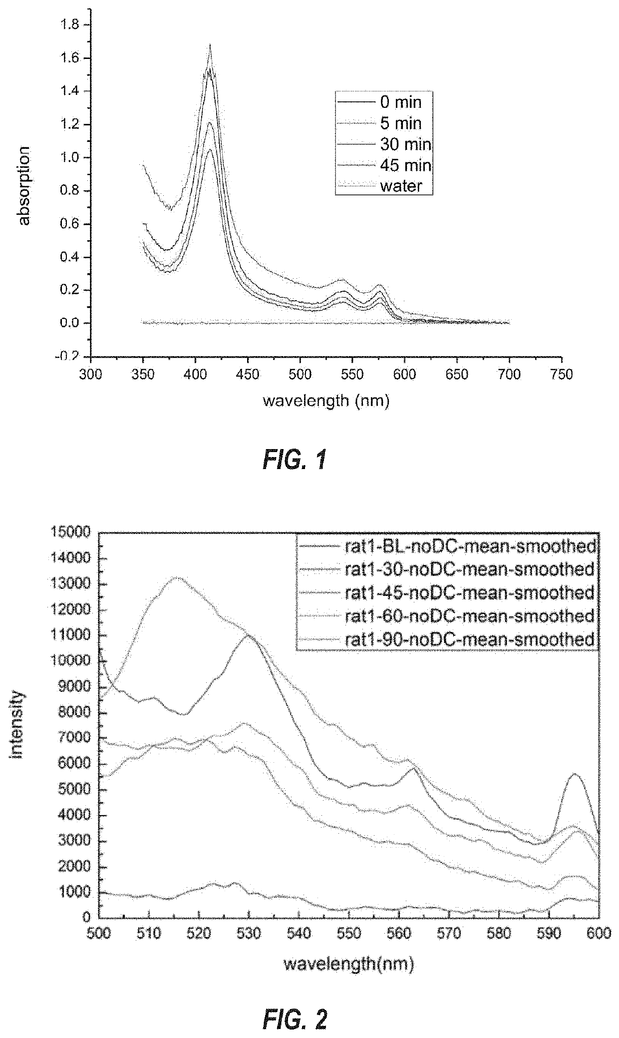
[0112]Methods for detecting tissue ischemia, as provided herein, were validated using a rat model of mesenteric ischemia. Referring now to FIG. 1, the absorption spectra of water and rat serum at 0-, 5-, 20-, and 45-mintues poste mesenteric ischemia are shown. Briefly, rat serum was obtained from male Wistar rats aged between 7 and 9 weeks with a mass of 250-300 g that were subjected to a model of mesenteric ischemia. Rats were initially anesthetized with gas and the the fistul at the rats' carotid artery was grafted. The rats' abdominal cavity was opened, and prior to using two clips to clip the superior mesenteric artery (SMA), a “baseline” blood sample was collected from the fistula. The SMA was then clipped to stop the blood flow to the intestines, thereby simulating human mesenteric ischemia in the rat model. Blood samples were collected at different ischemic time points, and blood was reperfused in the SMA at the end of the ischemia stage by removing the clips. After reperfusi...

example 2

[0124]Embodiments of the present disclosure enable systems and methods for detecting organ dysfunction and were validated using two optical systems to perform ICG fluorescence dosimetry on rats developing hepatocellular carcinoma (HCC).

[0125]One optical system used was a multiphoton microscope, which can non-invasively identify the location of vessels in vivo from sectioning two-photon fluorescence imaging. Time-course dynamics of concentration decay were also analyzed by the signal intensity within vessels. The other tested optical system was a point excitation and time course measurement using a 785 nm laser. The results demonstrate that ICG fluorescence dosimetry can reflect the disorder of at least liver function with the development of HCC and likely extends to the detection of other organ dysfunctions.

[0126]As shown in the diagram of FIG. 11, the multiphoton microscopy system used the scanning inverted microscope system of Leica DMI 3000M. The laser source was the femtosecond ...


PUM


Abstract

Methods for the early sensing of tissue ischemia and/or infarctions within organs using the spectroscopic features of serum luminescence include (i) isolating serum from a blood sample, (ii) directing an excitation light at the serum, (iii) receiving an endogenous serum chromophore emission light from the excited serum, and (iv) determining a presence of an ischemic condition based on the intensity of the endogenous serum chromophore emission light. Methods of detecting organ dysfunction can include transmitting excitation light to luminal contents of a mammalian blood vessel comprising trace amounts of exogenous chromophores administered to the subject mammal via the circulatory system. Transdermal luminescence measurements from the trace amounts of exogenous chromophores can then be used to determine organ dysfunction. Such methods enable routine monitoring of blood luminescence to bridge the diagnosis gap between sensitive symptom diagnosis and specific marker diagnosis, resulting in an effective early screening modality.

Description

CROSS-REFERENCE TO RELATED APPLICATIONS[0001]This application claims priority to and the benefit of U.S. Provisional Patent Application Ser. No. 62 / 734,961, filed Sep. 21, 2018 and titled “A SERUM LUMINESCENCE APPROACH TO DETECT THE TISSUE ISCHEMIA AND EVALUATE THE ORGAN FUNCTION,” which is incorporated herein by this reference in its entirety.BACKGROUNDTechnical Field[0002]This disclosure generally relates to the non-invasive detection and / or evaluation of organ function. More specifically, this disclosure relates to the non-invasive detection of tissue ischemia from serum luminescence to detect and / or evaluate organ dysfunction.Related Technology[0003]The rate of organ failure after liver transplantation has historically been around 32%. Patients who experience organ failure following liver transplantation have an average mortality rate of about 42% compared to the 2% mortality rate of patients without organ failure. Among the organs that fail following liver transplantation, kidn...

Claims


Application Information

Patent Timeline
IPC IPC(8): A61B5/00G01N21/64
CPCA61B5/4244A61B5/489G01N2021/6417G01N21/6428A61B5/0071A61B5/7203G01N21/6486A61B5/201
Inventor LIU, TZU-MING
Owner UNIVERSITY OF MACAU
Why Eureka
Try Eureka