Method for detection and diagnosis of lung and pancreatic cancers from imaging scans

a technology of imaging scans and lung cancer, applied in the field of methods of detecting and diagnosing cancer, can solve the problems of computationally expensive multi-stage frameworks, and achieve the effects of improving the risk stratification of cancer tumors in radiology images, improving performance, and improving performan

Active Publication Date: 2020-05-21
UNIV OF CENT FLORIDA RES FOUND INC
View PDF6 Cites 27 Cited by
  • Summary
  • Abstract
  • Description
  • Claims
  • Application Information

AI Technical Summary

Benefits of technology

[0025]For detection of nodules from an imaging scan, current lung nodule detection studies rely on computationally expensive multi-stage frameworks to detect nodules from CT scans. To address this computational challenge and provide better performance, the inventors propose S4ND, a new deep learning-based method for lung nodule detection. The approach uses a single feed forward pass of a single network for detection and provides better performance when compared to the current literature. The whole detection pipeline is designed as a single 3D Convolutional Neural Network (CNN) with dense connections, trained in an end-to-end manner. S4ND does not require any further post-processing or user guidance to refine detection results. Experimentally, the inventors compared the network with the current state-of-the-art object detection network (SSD) in computer vision as well as the state-of-the-art published method for lung nodule detection (3D DCNN). Publicly available 888 CT scans from LUNA challenge dataset were used and showed that the proposed method outperforms the current literature both in terms of efficiency and accuracy by achieving an average FROC-score of 0:897. The inventors also provide an in-depth analysis of the proposed network to shed light on the unclear paradigms of tiny object detection.
[0026]With regard to diagnosing malignancy in a detected nodule, risk stratification of cancer tumors in radiology images can be improved with computer-aided diagnosis (CAD) tools which can be made faster and more accurate. Tumor characterization through CADs can enable non-invasive cancer staging and prognosis and foster personalized treatment planning as a part of precision medicine. The inventors propose both supervised and unsupervised machine learning strategies to improve tumor characterization. In the supervised learning method, the inventors demonstrate significant gains in deep learning algorithms, particularly by utilizing a 3D Convolutional Neural Network along with transfer learning. For lung nodule characterization, the inventors present a 3D CNN based supervised learning approach to fully appreciate the anatomical information in 3D, which would be otherwise lost in the conventional 2D approaches. The inventors use a fine-tuning strategy to avoid the requirement for a large number of volumetric training examples for 3D CNN. In order to fine-tune, the inventors use a pre-trained network (trained on 1 million videos) and fine-tune it on the CT data. Motivated by the radiologists' interpretations of the scans, the inventors introduce a graph regularized sparse Multi-Task Learning (MTL) platform to integrate the complementary features from lung nodule attributes to improve malignancy prediction. The inventors were able to achieve high-level lung nodule attributes having varying levels of prominence.
[0027]In the unsupervised learning method, the inventors developed an unsupervised scheme to address the limited availability of labeled training data, a common problem in medical imaging applications. Inspired by learning from label proportion (LLP) approaches, the inventors developed a new algorithm, proportion-SVM (aSVM), to characterize tumor types. In the proposed unsupervised learning algorithm, instead of hard assigning labels, the inventors estimate the label proportions in a data-driven manner. To alleviate the effect of noisy labels (i.e. mislabeling) obtained during clustering, aSVM is employed, which is trained on label proportions only.

Problems solved by technology

For detection of nodules from an imaging scan, current lung nodule detection studies rely on computationally expensive multi-stage frameworks to detect nodules from CT scans.

Method used


Image

  • Method for detection and diagnosis of lung and pancreatic cancers from imaging scans
  • Method for detection and diagnosis of lung and pancreatic cancers from imaging scans
  • Method for detection and diagnosis of lung and pancreatic cancers from imaging scans

Examples


example 1

Nodule Detection in a Single Shot

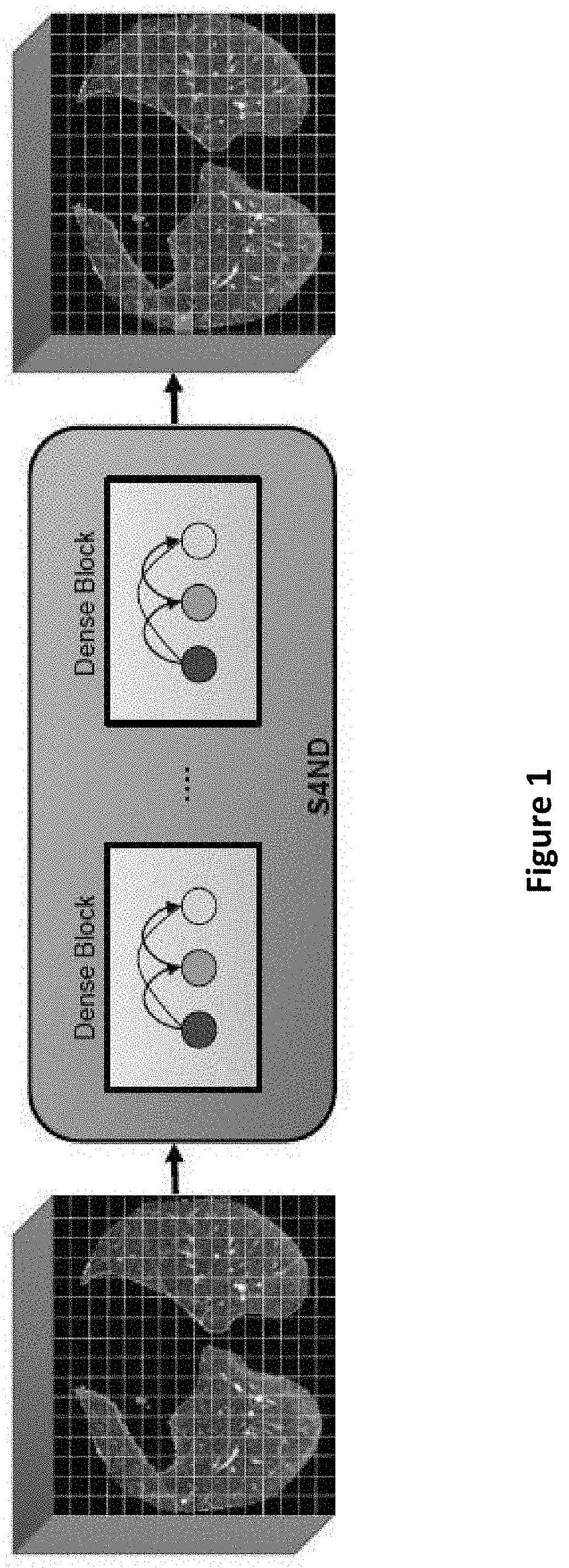
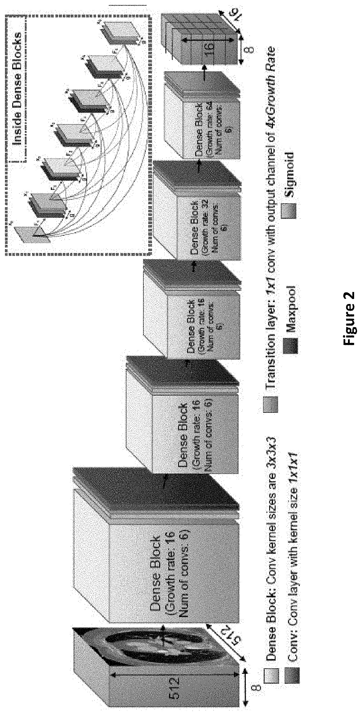
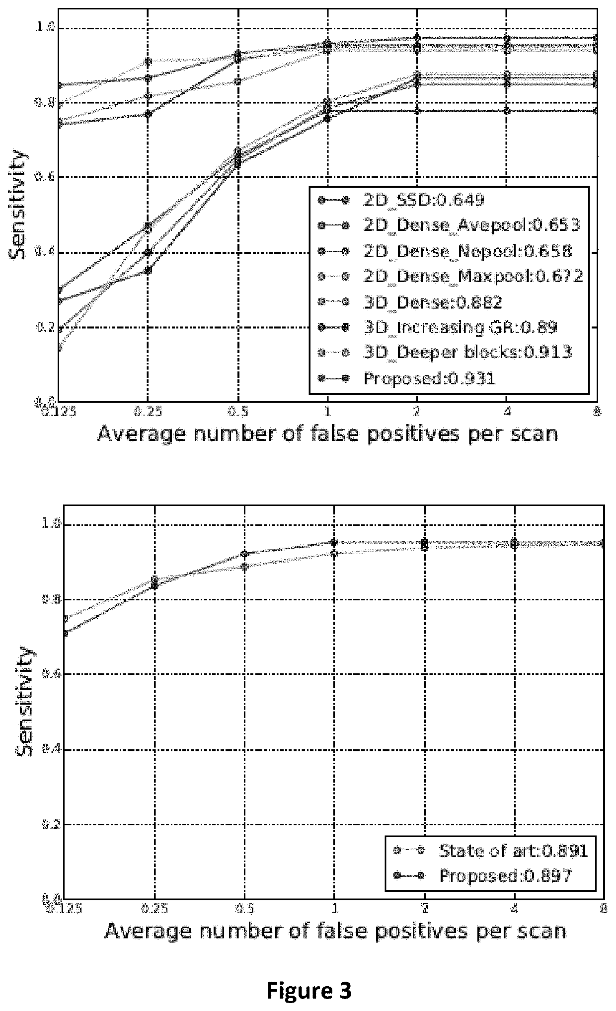
[0093]Current lung nodule detection studies rely on computationally expensive multi-stage frameworks to detect nodules from CT scans. To address this computational challenge and provide better performance, the inventors have developed a new deep learning-based method for lung nodule detection termed S4ND. The approach uses a single feed forward pass of a single network for detection and provides better performance when compared to the current literature. The whole detection pipeline is designed as a single 3D Convolutional Neural Network (CNN) with dense connections, trained in an end-to-end manner. S4ND does not require any further post-processing or user guidance to refine detection results. The inventors compared the network with the current state of the art object detection network (SSD) in computer vision as well as the state-of-the-art published method for lung nodule detection (3D DCNN). Using publicly available 888 CT scans from the LUNA chal...

example 2

Tumor Characterization Using Supervised Learning

[0118]Risk stratification (characterization) of tumors from radiology images can be more accurate and faster with computer-aided diagnosis (CAD) tools. Tumor characterization through such tools can also enable non-invasive cancer staging, prognosis, and foster personalized treatment planning as a part of precision medicine. Tumor characterization based on supervised learning demonstrates significant gains with deep learning algorithms, particularly by utilizing a 3D Convolutional Neural Network and Transfer Learning. Motivated by the radiologists' interpretations of the scans, the inventors illustrate how to incorporate task dependent feature representations into a CAD system via a graph-regularized sparse Multi-Task Learning (MTL) framework.

[0119]The inventors have developed a novel supervised learning strategy to perform risk-stratification of lung nodules from low-dose CT scans. For this strategy, a 3D CNN based discriminative featu...

example 3

Tumor Characterization Using Unsupervised Learning

[0151]Since annotating medical images is laborious, expensive and time-consuming, the inventors also developed an unsupervised learning method to classify lung nodules and IPMN by using a novel algorithm to address the limited availability of labeled training data, a common problem in medical imaging applications. Inspired by learning from label proportion (LLP) approaches in computer vision, the inventors used proportion-SVM for characterizing tumors. First, the inventors extracted discriminative information from a large amount of unlabeled imaging data. The inventors analyzed both hand-crafted and deep learning features and assessed how good those features were when applied to tumor characterization. In order to obtain an initial set of labels in an unsupervised fashion, the samples are clustered into different groups in the feature domain. The inventors then trained Proportion-Support Vector Machine (aSVM) algorithm using label pr...


PUM


Abstract

A method of detecting and diagnosing cancers characterized by the presence of at least one nodule/neoplasm from an imaging scan is presented. To detect nodules in an imaging scan, a 3D CNN using a single feed forward pass of a single network is used. After detection, risk stratification is performed using a supervised or an unsupervised deep learning method to assist in characterizing the detected nodule/neoplasm as benign or malignant. The supervised learning method relies on a 3D CNN used with transfer learning and a graph regularized sparse MTL to determine malignancy. The unsupervised learning method uses clustering to generate labels after which label proportions are used with a novel algorithm to classify malignancy. The method assists radiologists in improving detection rates of lung nodules to facilitate early detection and minimizing errors in diagnosis.

Description

CROSS REFERENCE TO RELATED APPLICATIONS[0001]This application claims priority to U.S. Provisional Application No. 62 / 755,018 entitled “Fast and Reliable Detection and Diagnosis of Lung Cancers from CT Scans”, filed Nov. 2, 2018, the contents of which are hereby incorporated by reference into this disclosure.FIELD OF INVENTION[0002]This invention relates to methods of detecting and diagnosing cancer. Specifically, the invention describes a method of screening and diagnosis of lung cancer from CT scans.BACKGROUND OF THE INVENTION[0003]According to the American Cancer Society, approximately 40% of people will be diagnosed with cancer at some point during their lifetime with an overall mortality of 171.2 per 100,000 people per year (based on deaths between 2008-2012). Lung and pancreatic cancers are two of the most common cancers. While lung cancer is the largest cause of cancer-related deaths in the world, pancreatic cancer has the poorest prognosis with a 5-year survival rate of only ...

Claims


Application Information

Patent Timeline
Patent Type & Authority Applications(United States)
IPC IPC(8): G16H50/20G06T7/00G06K9/62G06N3/04G06N20/10G06N5/04G06N3/08G16H30/40G16H70/60A61B5/055A61B6/03A61B6/00A61B8/08G06V10/764
CPCA61B5/055G06T2207/30096G06N5/04G16H30/40G16H50/20G06N3/08G06K9/6269G06T7/0012G06T2207/20084A61B6/037A61B6/4417A61B8/481A61B8/5223G06K9/6218G16H70/60A61B6/5217G06T2207/30064G06T2207/20081G06K9/6257G06T2207/20076G06N3/04A61B6/032G06K2209/053G06T2207/10081G06N20/10A61B6/12A61B8/085A61B5/7267G06V2201/03G06V10/82G06V10/764G06N3/045G06V2201/032G06F18/23G06F18/2148G06F18/2411
Inventor BAGCI, ULASKHOSRAVAN, NAJIHUSSEIN, SARFARAZ
Owner UNIV OF CENT FLORIDA RES FOUND INC
Why Eureka
Try Eureka