Method for separating and culturing mesenchymal stem cells from wharton's jelly tissue of umbilical cord

Active Publication Date: 2020-05-28
GUO LEI +1
View PDF0 Cites 3 Cited by
  • Summary
  • Abstract
  • Description
  • Claims
  • Application Information

AI Technical Summary

Benefits of technology

[0073]The present invention employs red blood cell lysis buffer to extract the umbilical cord mesenchymal stem cells, which can significantly mitigate the interference from red blood cells in the primary culture of umbilical cord mesenchymal stem cells. Specifically, the treatment with red blood cell lysis buffer used in the primary culture avoids the interference from red blood cells on the migration, adherence and proliferation of mesenchymal stem cells, ensures the successful separation of the mesenchymal stem cells, and improves cell purity, thereby meeting the requirements for the quantity of mesenchymal stem cells of clinical level, and further guaranteeing the viability of the stem cells as well. As a result, sufficient and viable mesenchymal stem cells are available for subsequent passage culture, cryopreservation and resuscitation, even clinical application, which is beneficial to the clinical conversion and application of the stem cells in the future.
[0074]In addition, the present invention combines the use of red blood cell lys

Problems solved by technology

However, during separation and extraction, the interference from red blood cells is a

Method used


Image

  • Method for separating and culturing mesenchymal stem cells from wharton's jelly tissue of umbilical cord
  • Method for separating and culturing mesenchymal stem cells from wharton's jelly tissue of umbilical cord
  • Method for separating and culturing mesenchymal stem cells from wharton's jelly tissue of umbilical cord

Examples


example 1

Screening of the Components of Serum-Free Medium for Mesenchymal Stem Cells

(1) Screening of the Content of Serum Substitute

[0087]Medium to be tested: 0.1 parts by volume of β-mercapto, 10 ng / ml recombinant human basic fibroblast growth factor (b-FGF, Peprotech), 1 part by volume of aqueous solution of non-essential amino acids (Catalog No. 11140, Gibco), 1, 2, 5, 8, 10, 12, 15, or 20 parts by volume of Knockout FBS serum substitute (Catalog No. 10828-028, Gibco), and 89 parts by volume of a-MEM.

[0088]In a biosafety cabinet, the third-passage hUC-MSCs separated from Wharton's jelly tissue of umbilical cord from a healthy neoborn by natural delivery were inoculated into a T75 cell culture flask at a density of 2×10⁴ cells / cm2, and 12-15 ml of medium commercially available were added as well to culture the cells. The cells were cultured till cells were observed to have completely adhered to walls of the flask, and the medium was replaced with 15 ml of medium to be tested. The growth of...

example 2

Red Blood Cell Lysis Buffer Assisted Method of Extracting Umbilical Cord Mesenchymal Stem Cells

[0096]Collecting and Transporting the Sample:

[0097]A umbilical cord tissue sample from a newborn by cesarean section delivery was collected aseptically, put into the preservation and transportion solution for umbilical cord comprising penicillin sodium, streptomycin sulfate, gentamnicin and amphotericin B (the final concentration of each of penicillin sodium, streptomycin sulfate and gentamicin in D-Hank's was 150 U / ml respectively, while that of amphotericin B was 300 U / ml.), then transported in ice to a clean GMP cell laboratory within 2 hours.

[0098]Washing and Sterilizing the Sample:

[0099]In a biosafety cabinet, the fresh umbilical cord sample was placed in a 50 ml aseptic centrifuge tube, washed 2 times with 75% aqueous ethanol, then washed 3 times with sterile saline.

[0100]Pre-Processing the Umbilical Cord Sample:

[0101]The umbilical cord was cut into segments each of about 2-3 cm in l...

example 3

Red Blood Cell Lysis Buffer Assisted Method of Extracting Umbilical Cord Mesenchymal Stem Cells

[0108]The red blood cell lysis buffer used in this Example comprised 10 g / L NH4Cl and 0.1 mM Na2-EDTA, pH 7.2-7.4.

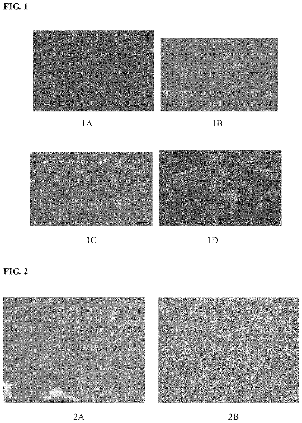
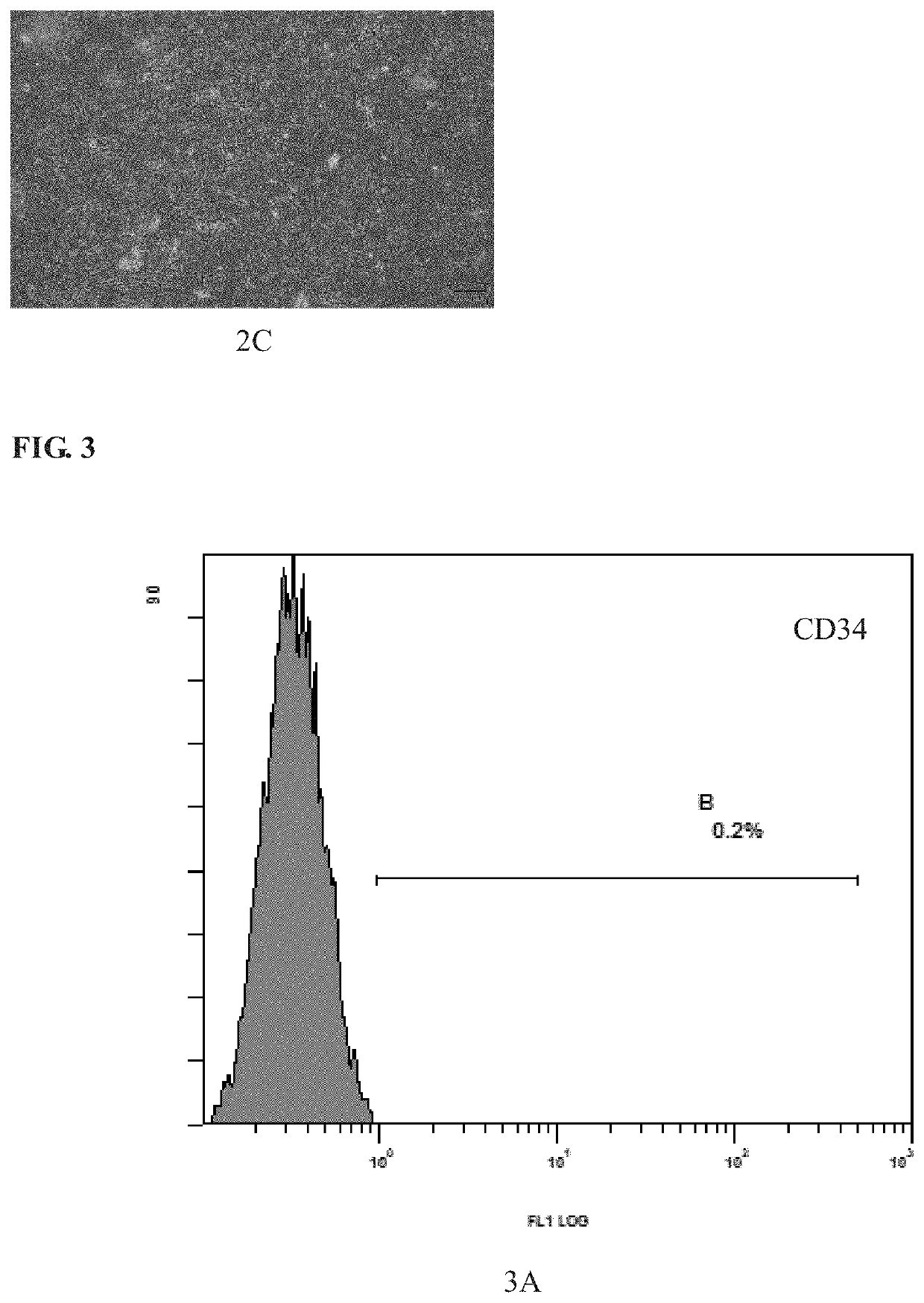
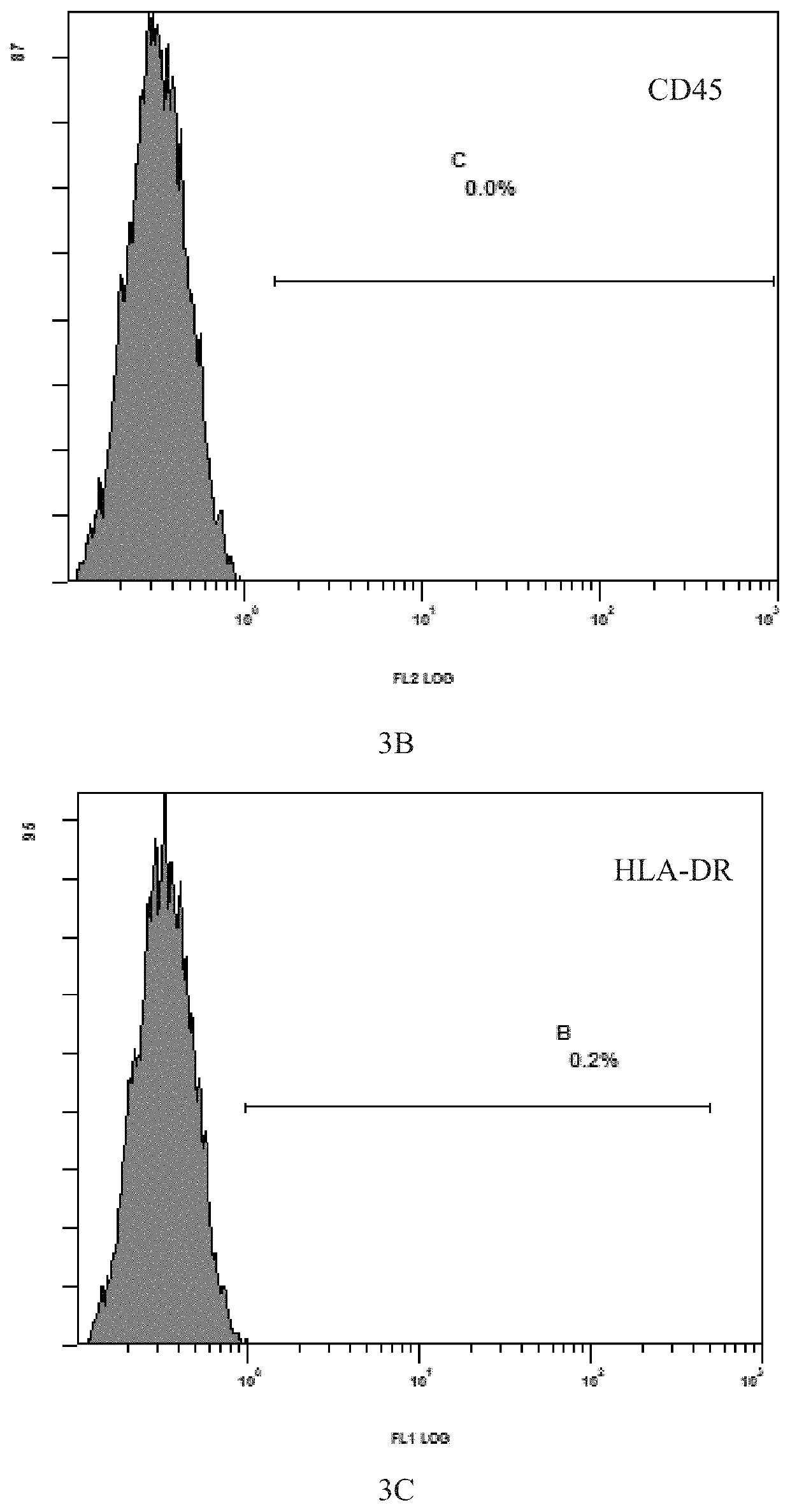
[0109]The process was carried out with reference to the method described in Example 2 above. The mesenchymal stem cells grew out of the bottom of the tissue blocks on the 5th day of culture, and formed cell-clusters in whorls around the 7th day of culture. After the removal of the tissue blocks and replacement with fresh medium, 60% confluence was reached around 12th day, and the cells were passaged after trypsin digestion. The purity of the cells after three passages was greater than 99% while the viability was more than 95%.


PUM


Abstract

Provided is a method for separating and extracting mesenchymal stem cells from the human umbilical cord. The method uses healthy neonatal umbilical cord tissue; after cleaning and disinfection, mechanically pulverising same, separating the Wharton's jelly, and after treating with erythrocyte lysate, carrying out suspension culture in a serum-free culture medium. Replacing the liquid every 3-5 days; after the plate adherence rate reaches 30-70%, carrying out trypsin digestion, and then collecting the cells by centrifugation for passage amplification, until the rate of confluence of the cells reaches 80-90% confluence, thereby obtaining high purity umbilical cord mesenchymal stem cells.

Description

TECHNICAL FIELD[0001]The invention relates to a stable method for separating and extracting mesenchymal stem cells from umbilical cord, especially a method for separating and culturing mesenchymal stem cells from Wharton's jelly tissue of umbilical cord.BACKGROUND OF THE INVENTION[0002]Mesenchymal stem cell (MSC) is an important member of stem cell family, which derives from the mesoderm and ectoderm of early development stage and exists in the connective tissues and mesenchymes in organs of the whole body. MSC was found in bone marrow for the first time in the late 1970s; however, until the 90s of last century, only when the multiple differentiation potential of MSC was recognized, such a stem cell began to draw more and more attention.[0003]Mesenchymal stem cell belongs to pluripotent stem cells. At present, previous study has shown that MSCs can be induced to differentiate into many types of tissue cells and the like cells, such as fat cells, bone cells, bone ligament cells, nerv...

Claims


Application Information

Patent Timeline
IPC IPC(8): C12N5/0775C12N5/00
CPCC12N2500/24C12N5/0031C12N5/0665C12N2500/90C12N2500/32C12N2500/60C12N2501/115C12N2500/44C12N5/0668
Inventor GUO, LEI
Owner GUO LEI
Why Eureka
Try Eureka