Materials and methods for protecting against neuromas

a neuroma and material technology, applied in the field of materials and methods for protecting against neuroma, can solve the problems of affecting the treatment effect, so as to facilitate the insertion of the terminal nerve end, facilitate the insertion, and facilitate the treatment

Active Publication Date: 2020-06-04
AXOGEN CORP
View PDF1 Cites 2 Cited by
  • Summary
  • Abstract
  • Description
  • Claims
  • Application Information

AI Technical Summary

Benefits of technology

[0013]In one embodiment, the partitioning of the internal chamber of the cap is in the form of a spiral-shaped wall that forms a longitudinal spiraled channel with one edge facing the open end. As nerve tissue grows, it can fill in the space between the spirals, so that the outer nerve tissue is substantially separated, subdivided, or partially subdivided from inner nerve tissue. In an alternative embodiment, the internal chamber is partitioned with at least one panel, directed longitudinally with the internal chamber, such that a portion of the internal chamber, opposite to the single open end, is divided into two or more distinct and separate channels. The division of the neuroma tissue into separate or subdivided channels, to form smaller, disconnected, neuroma masses, can mitigate the cascade effect caused when one area of the neuroma is stimulated and the effect is felt through the entire tissue mass. The dimensions of the cylindrical cap can be about 1 mm to about 25 mm in diameter and about 1 mm to about 100 mm in length.
[0014]In addition, the material of the cylindrical cap can remodel into a tissue cushion after implantation, thereby enveloping and further isolating the new neuronal growth, which protects the neuroma from being stimulated and inhibits pain. Remodeling can include, but is not limited to, expansion or enlargement of the material, reshaping of the material, incorporation of the material around nerve tissue, and other changes that can affect the size and shape of the material. Preferably, after the device is implanted, it will remodel into the surrounding tissue to protect the forming neuroma and the sections of smaller, disconnected neuromas mass. In one embodiment, the material of the cap is a biomaterial that can remodel to generate a volume of protective connective tissue around a neuroma (rather than the current use of a biomaterial to contain a neuroma volumetrically). In a preferred embodiment, the material of the cap is a membrane biomaterial, such as, for example, small intestine submucosa (SIS), amnion, dermis, or decellularized fascia.
[0015]In one embodiment of a method of use, a device is provided that contains an internal chamber with a retaining cavity at or near the open end to allow insertion of the nerve stump through the open end, so that it is near or abuts against the divided, or separate, channels facing the open end. A dense layer of biomaterial on the exterior surface can operate to mechanically isolate the neuroma and prevent axons from escaping the internal chamber of the device. The internal chamber contain partitions or dividers of biomaterial to subdivide the neuroma mass 13 into smaller, disconnected neuroma masses 17 as it grows that will form from the nerve stump.
[0017]In one embodiment, to facilitate insertion of a terminal nerve end, a cap can have one or more tabs arranged on and around the external surface 16 of the cap. In an alternative embodiment, a tab can be arranged at or near the open end. The tabs can be used to grasp the cap and manipulate it around the terminal nerve end. Grasping the tabs rather than the exterior surface can help to maintain the shape of the cap, in particular the open end.
[0018]The open end leading into a retaining cavity in which the terminal nerve end can be secured can also be made temporarily larger or more accessible, so that the terminal nerve end can be more easily and gently placed therein. In one embodiment, there is a slit in the cap that is integral with and perpendicular to the opening, such that the opening can be temporarily widened. In another embodiment, the retaining cavity has one or more wings or flaps that allow it to be partially spread open. This can widen the open end sufficiently that a terminal nerve end can be placed or set down within the retaining cavity, making it easier to see how it is placed relative to the dividers. The wings or flaps can then be wrapped over and around the nerve end and secured to hold the nerve in place and inhibit escape of neuronal tissue growth.
[0019]The combination of limiting the growth and size of a neuroma mass through physical partitioning of the neuromas mass into smaller, disconnected sections along with the creation of a connective tissue cushion is unique. This novel combination of features and design effectively uses the natural response of reconstructing implanted biomaterials to minimize the size, isolate, and protect the neuroma.

Problems solved by technology

The up-regulation of certain channels and receptors during neuroma development can also cause abnormal sensitivity and spontaneous activity of injured axons (Curtin C and Carroll I.
Neuromas of the nerve stump are unavoidable consequences of nerve injury when the nerve is not, or cannot be, repaired and can result in debilitating pain.
It has been estimated that approximately 30% of neuromas become painful and problematic.
This is particularly likely if the neuroma is present at or near the skin surface as physical stimulation induces signaling in the nerve resulting in a sensation of pain.
Due to a variety of factors, current methods of neuroma mitigation / prevention have an unacceptable level of efficacy.
However this procedure can greatly complicate surgery, as significant additional dissection of otherwise healthy tissue is required to emplace the nerve stump.
Clinical and pre-clinical evidence has shown, however, that this procedure can cause a painful neuroma to form behind the ligation.
Furthermore, the ligated nerve is generally not positioned to minimize mechanical stimulation of the neuroma, since it is anticipated that the scar tissue will provide sufficient protection to the nerve end.
These devices and methods necessitate insertion of the nerve into the opening of the device, which can be difficult and can further damage the nerve end.
Unfortunately, current methods for addressing the formation of and pain caused by neuromas have not been generally successful and therefore are rarely utilized.
The formation of neuromas at a severed nerve end can be difficult to prevent.

Method used


Image

  • Materials and methods for protecting against neuromas
  • Materials and methods for protecting against neuromas
  • Materials and methods for protecting against neuromas

Examples


Embodiment Construction

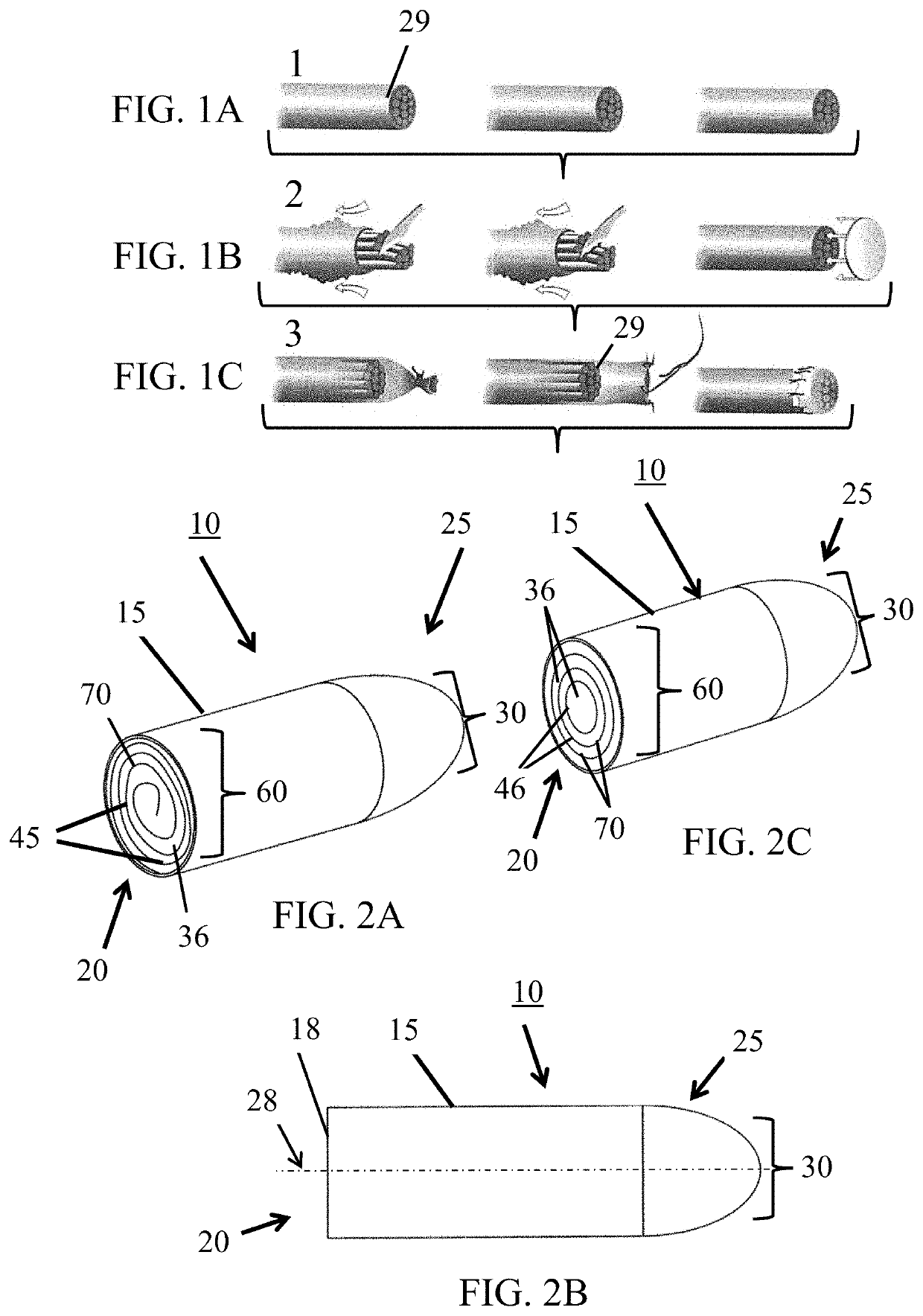
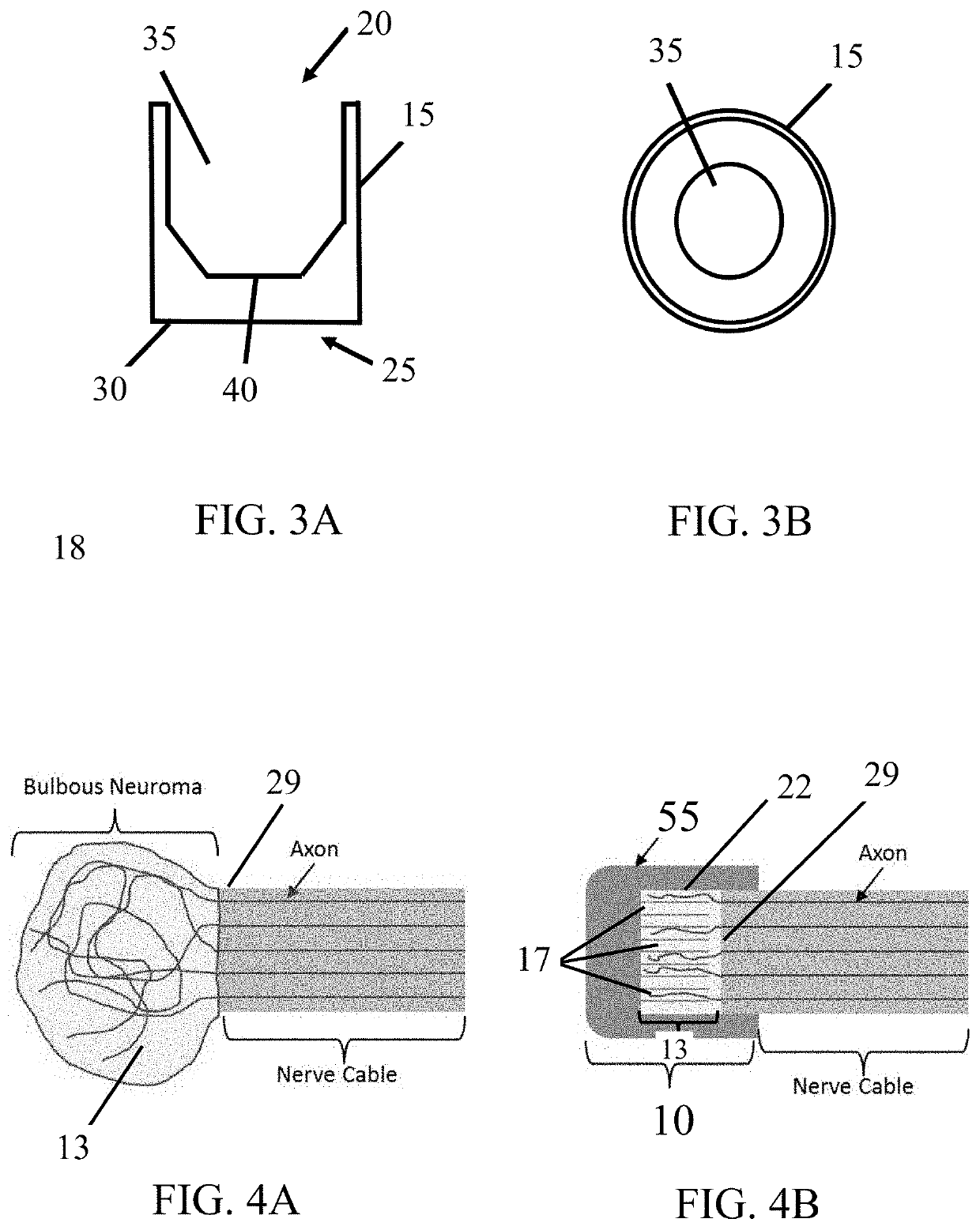
[0030]The subject invention provides devices and methods for alleviating discomfort associated with neuromas. More specifically, the subject invention pertains to devices and methods for limiting neuroma size and physically surrounding the neuroma to inhibit stimulation that elicits neuropathic pain. In certain embodiments, the subject invention is directed to a tissue-engineered scaffold that provides: a barrier that limits the size of a neuroma, dividers that subdivide the neuroma volume or mass into smaller, disconnected neuromas masses to reduce axonal cross-talk or the “cascade effect,” and mechanical isolation of the neuroma to inhibit stimulation.

[0031]A device of the subject invention is designed to form a protective, connective tissue covering or cap surrounding the terminal nerve end 29, thereby surrounding any resulting neuroma formation. Within the volume created by the barrier, sub-dividing the neuroma volume as it forms can limit the amount of interaction between axons...


PUM

PropertyMeasurementUnit
lengthaaaaaaaaaa
lengthaaaaaaaaaa
lengthaaaaaaaaaa

Abstract

The subject invention provides devices and methods for alleviating discomfort associated with neuroma formation. The devices and methods of the invention effectively use the body's natural response of reconstructing implanted biomaterials to minimize the size of isolate, and protect a neuroma. In preferred embodiments, the subject device is a cylindrical cap, wherein the internal chamber of the cylindrical cap physically partitions the nerve to enable an arrangement of nerve fibers (as opposed to haphazardly arranged nerve fibers often produced in neuromas). Tabs arranged on the outside of the cap can be used to manipulate the cap into place on a nerve. The open end can also be configured with flaps that can be used to widen the open end for easier insertion of the nerve into the cap. In addition, the cap's material remodels into a tissue cushion after implantation, which protects the neuroma from being stimulated and inducing pain.

Description

CROSS-REFERENCE TO RELATED APPLICATIONS[0001]This application is a continuation-in-part of U.S. application Ser. No. 14 / 036,405, filed Sep. 25, 2013, now U.S. Pat. No. 9,629,997, which claims the benefit of U.S. provisional application Ser. No. 61 / 705,251, filed Sep. 25, 2012, which are incorporated herein by reference in their entirety.BACKGROUND OF INVENTION[0002]Neuromas develop as a part of a normal reparative process following peripheral nerve injury. They are formed when nerve recovery towards the distal nerve end or target organ fails and nerve fibers improperly and irregularly regenerate into the surrounding scar tissue. Neuromas consist of a deranged architecture of tangled axons, Schwann cells, endoneurial cells, and perineurial cells in a dense collagenous matrix with surrounding fibroblasts (Mackinnon S E et al. 1985. Alteration of neuroma formation by manipulation of its microenvironment. Plast Reconstr Surg. 76:345-53). The up-regulation of certain channels and recepto...

Claims


Application Information

Patent Timeline
Patent Type & Authority Applications(United States)
IPC IPC(8): A61B90/00A61L31/00A61L31/04A61N1/05A61L31/06A61L31/14
CPCA61N1/05A61L31/04A61L31/048A61B2090/0815A61L31/005A61L31/06A61L2430/32A61L31/14A61B90/08A61N1/0558C08L23/06C08L71/02
Inventor DEISTER, CURTSIMON, CRYSTALFALERIS, JENNIFER
Owner AXOGEN CORP
Why Eureka
Try Eureka