
Purification of RNA fractions using a hydrophilic polymeric material

a polymer material and rna fraction technology, applied in the field of rna fraction purification using a hydrophilic polymeric material, can solve the problems of high cost involved in the freezing transportation and processing of whole blood, time-consuming and expensive techniques,

Active Publication Date: 2020-06-25
HUMMINGBIRD DIAGNOSTICS GMBH
View PDF0 Cites 0 Cited by
  • Summary
  • Abstract
  • Description
  • Claims
  • Application Information

AI Technical Summary

Benefits of technology

The present invention is about a method for reducing the complexity of a whole blood sample using the bloodspot technique. This technique can remove certain RNA molecules with a certain length and purify them. The purified RNA molecules have good quality expression profiles. The blood sample can be dried and stored at different temperatures and humidity levels. The invention also provides a way to diagnose or differentially diagnose a disease by analyzing the expression profiles of RNA molecules. The absorbent probe used in the invention may contain a reference standard, an anti-coagulant, and a stabilizer. The use of these substances helps maintain the homogeneity of the blood sample and prevents degradation. The invention also allows for the determination of the level of RNA molecules, such as miRNAs, which simplifies sample processing and reduces costs.

Problems solved by technology

High costs are involved with the freezing transportation and processing of whole blood.
In addition, this techniques are very time consuming and expensive.

Method used


Image

  • Purification of RNA fractions using a hydrophilic polymeric material
  • Purification of RNA fractions using a hydrophilic polymeric material
  • Purification of RNA fractions using a hydrophilic polymeric material

Examples


example 1

ple Collection Using Hydrophilic Polymeric Absorbent Device (Mitra Microsampling Device)

[0312]Blood was collected from different individuals (n=3) by puncture of the middle finger of the left hand using sterile safety lancets (Safety-Lancet Extra 18G, Sarstedt, Nümbrecht, Germany) and four Mitra Microsampling Devices (neoteryx, Torrance, Calif., USA, Ordering No. 10005) per individual. Blood on Mitra Microsampling Device (FIG. 6) was dried for 2 hours at ambient temperature. Four blood filled Sampler Tips per individual were removed from Sampler Body of the Mitra Microsampling Device and transferred to a 2 ml Tube (Eppendorf, Hamburg, Germany)

example 2

n of Small RNA from Mitra Microsampling Device

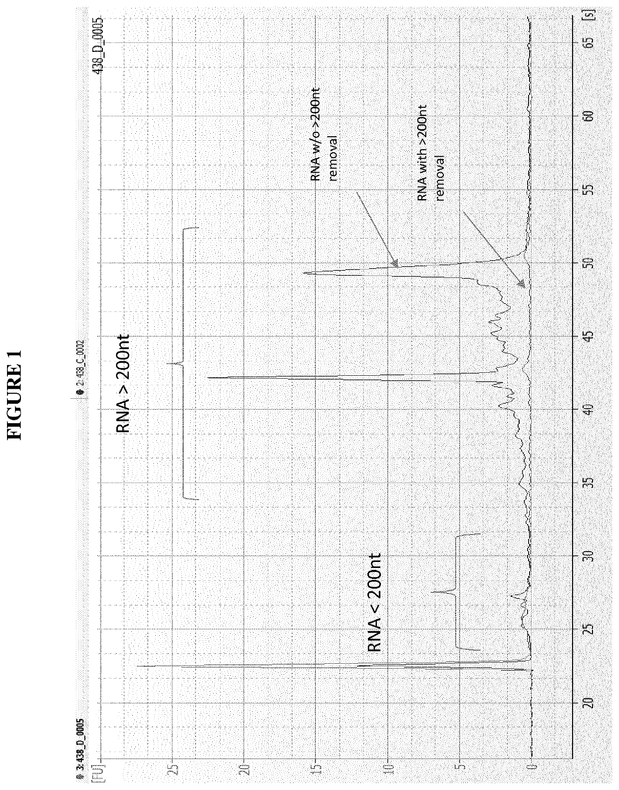
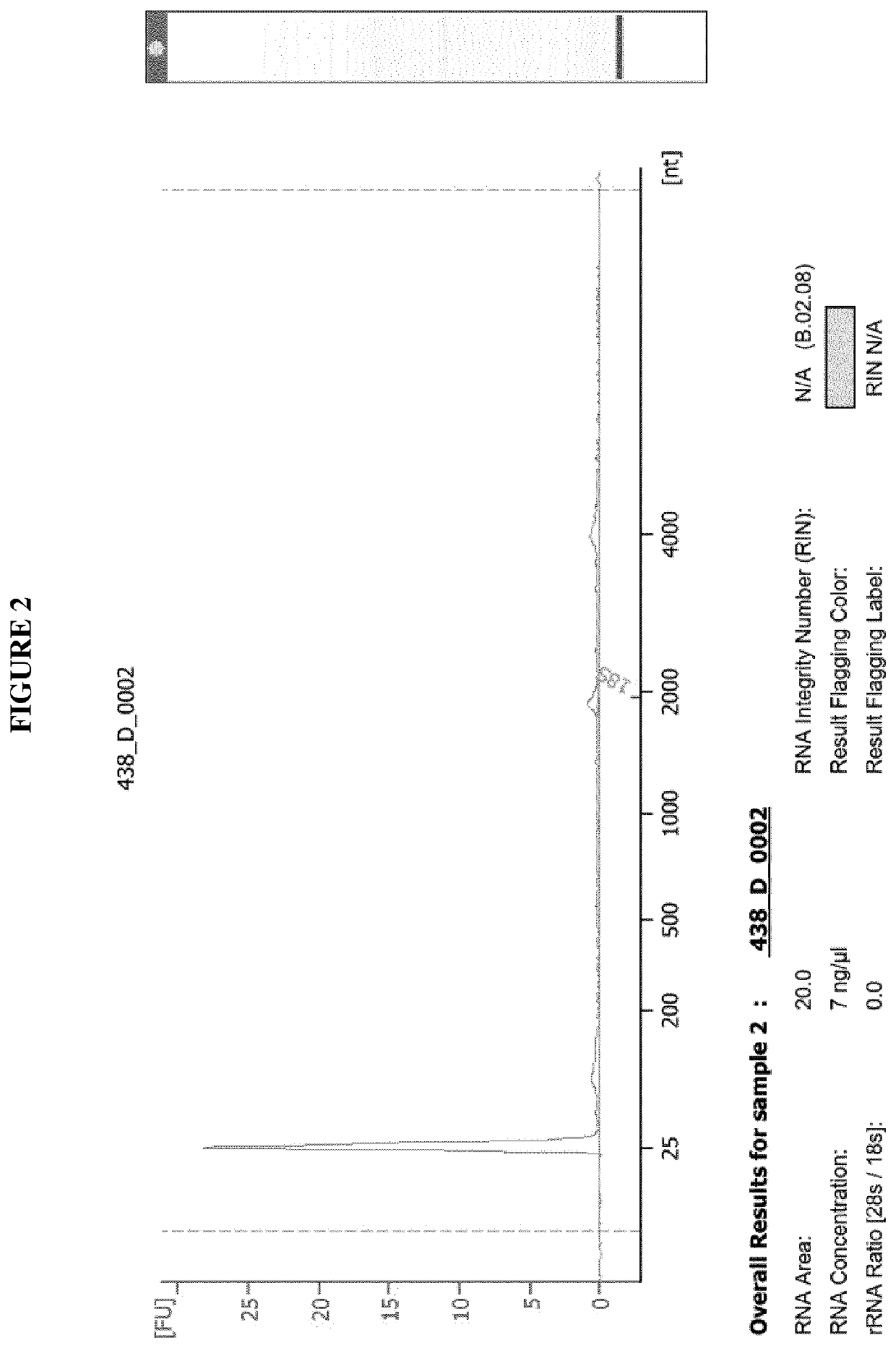
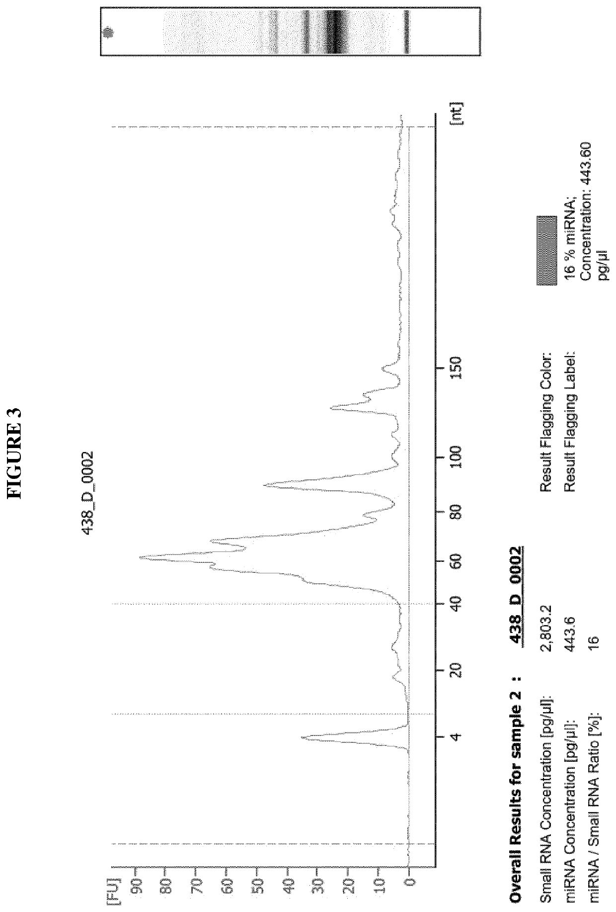
[0313]Small RNA extraction with length <200 nt (including the microRNA-fraction) was carried out using Phenol-Chloroform extraction technique. Purification of the small RNA was performed by use of the miRNeasy Serum Plasma Kit (Qiagen GmbH, Hilden, Germany). Herein, 1 mL Qiazol reagent (Qiagen GmbH, Hilden, Germany, comprising guanidinium thiocyanate as a chaotropic reagent and phenol) was pipetted to the 2 mL tube containing four Mitra Sampler Tips (see Example 1, blood sample collection). The tube was then incubated at 4° C. on a shaker at 1,000 rpm for 16 hours. Afterwards, complete supernatant was transferred to a fresh 2 mL Eppendorf tube. After addition of 200 μl Chloroform, mixture was thoroughly vortexed for 15 sec and incubated for 2 min at room temperature, followed by centrifugation at 12,000×g for 15 min at 4° C. Afterwards, the upper, aqueous phase was transferred to a new 1 mL tube without touching the other two phases. 1.5...

example 3

ple Collection Using a Hydrophilic Cellulose Comprising Absorbent Device

[0314]Blood was collected from different individuals (n=3) by puncture of the ring finger of the left hand using sterile safety lancets (Safety-Lancet Extra 18G, Sarstedt, Nümbrecht, Germany). Two blood drops per individual were added to the hydrophilic cellulose comprising absorbent device (cellulose comprising filter paper from various manufactures, including Non-Indicating FTA Classic Card (GE Healthcare Life Science, Buckinghamshire, Great Britain), Non-Indicating FTA Elute Micro Card (GE Healthcare Life Science, Buckinghamshire, Great Britain), HemaSpot HF (Spot On Sciences, Austin, Tex., USA), HemaSpot SE (Spot On Sciences, Austin, Tex., USA), TNF (Munktell, Bärenstein, Germany), TNF-Di (Munktell, Bärenstein, Germany)) covering an area of approximate 0.5-4.0 square cm. The various hydrophilic cellulose comprising absorbent probes were dried for 2 hours and then stored for 1 week at ambient temperature. Blo...


PUM

PropertyMeasurementUnit
densityaaaaaaaaaa
densityaaaaaaaaaa
volumesaaaaaaaaaa

Abstract

The present invention relates to a method of removing an RNA fraction with ≥200 nucleotides in length from a whole blood sample. The present invention also relates to a method of purifying an RNA fraction with <200 nucleotides in length from a whole blood sample. The present invention further relates to a method of determining the level of RNA molecules with <200 nucleotides in length. In addition, the present invention relates to a method for diagnosing a disease in an individual. Moreover, the present invention relates to a kit which is useful for carrying out the methods of the present invention.

Description

CROSS REFERENCE TO RELATED APPLICATIONS[0001]This application is a U.S. National Stage of PCT / EP2017 / 054582, filed Feb. 28, 2017 which claims priority to European Patent Application No. 16161077.9, filed Mar. 18, 2016, the contents of which are incorporated by reference in the entirety for all purposes.REFERENCE TO A “SEQUENCE LISTING” SUBMITTED AS ASCII TEXT FILES VIA EFS-WEB[0002]The Sequence Listing written in file 095697-1099969-002600US_SequenceListing.txt created on Aug. 21, 2018, 71,381 bytes, machine format IBM-PC, MS-Windows operating system, in accordance with 37 C.F.R. §§ 1.821- to 1.825, is hereby incorporated by reference in its entirety for all purposes.[0003]The present invention relates to a method of removing an RNA fraction with ≥200 nucleotides in length from a whole blood sample. The present invention also relates to a method of purifying an RNA fraction with <200 nucleotides in length from a whole blood sample. The present invention further relates to a metho...

Claims


Application Information

Patent Timeline
Patent Type & Authority Applications(United States)
IPC IPC(8): C12Q1/6883C12N15/10G01N1/34G01N1/40
CPCC12Q1/6883G01N1/34G01N1/4055G01N1/405C12N15/1006
Inventor LAUFER, THOMASBEIER, MARKUSKAHRAMAN, MUSTAFAKELLER, ANDREAS
Owner HUMMINGBIRD DIAGNOSTICS GMBH
Learn More