Method, apparatus, device and storage medium for extracting a cardiovascular vessel from a CTA image

a cardiovascular vessel and image technology, applied in the field of medical image processing, can solve the problems of coronary enhancement method, low signal-to-noise ratio in each structural region, and inability to obtain good results, etc., and achieve the effect of accurate visualization of aortic structure and morphology

Pending Publication Date: 2020-07-30
SHENZHEN INST OF ADVANCED TECH
View PDF0 Cites 0 Cited by
  • Summary
  • Abstract
  • Description
  • Claims
  • Application Information

AI Technical Summary

Benefits of technology

[0014]The present invention performs erosion operation and dilation operation on the image data to erode the irrelevant structures of the lungs and retain the heart and aortic structures, acquires the first ascending aortic structure via the slice-by-slice transformation, and synthesizes a second ascending aortic structure by combining with a binarized sphere structure and performs erosion via a morphological opening operation...

Problems solved by technology

The axial projection after multi-scale filtering processing produces a higher response in the lung region with high contrast between blood vessel and the background, but has a small or no response in the heart region with low contrast between blood vessel and the background, because the heart a...

Method used


Image

  • Method, apparatus, device and storage medium for extracting a cardiovascular vessel from a CTA image
  • Method, apparatus, device and storage medium for extracting a cardiovascular vessel from a CTA image
  • Method, apparatus, device and storage medium for extracting a cardiovascular vessel from a CTA image

Examples


embodiment 1

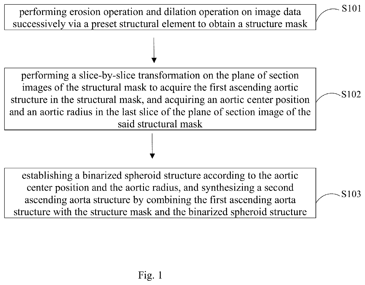
[0022]FIG. 1 shows an implementation process of a method for extracting a cardiovascular vessel from a CTA image provided in Embodiment 1 of the present invention, for the convenience of illustration, only the parts related to the embodiment of the present invention are shown, and the details are as follows:

[0023]In step S101, erosion operation and dilation operation are performed on image data successively via a preset structural element to obtain a structure mask.

[0024]In an embodiment of the present invention, the above image data is a coronary angiography image after down-sampling processing targeted at large-size original CTA data, in order to quickly extract a large-size ascending aortic structure without affecting the precision of the structure extraction. The image size may be down-sampled to half of the original size; some noise in the above image data and some structures that are not related to the aorta are suppressed or attenuated. Erosion operation is performed on image...

embodiment 2

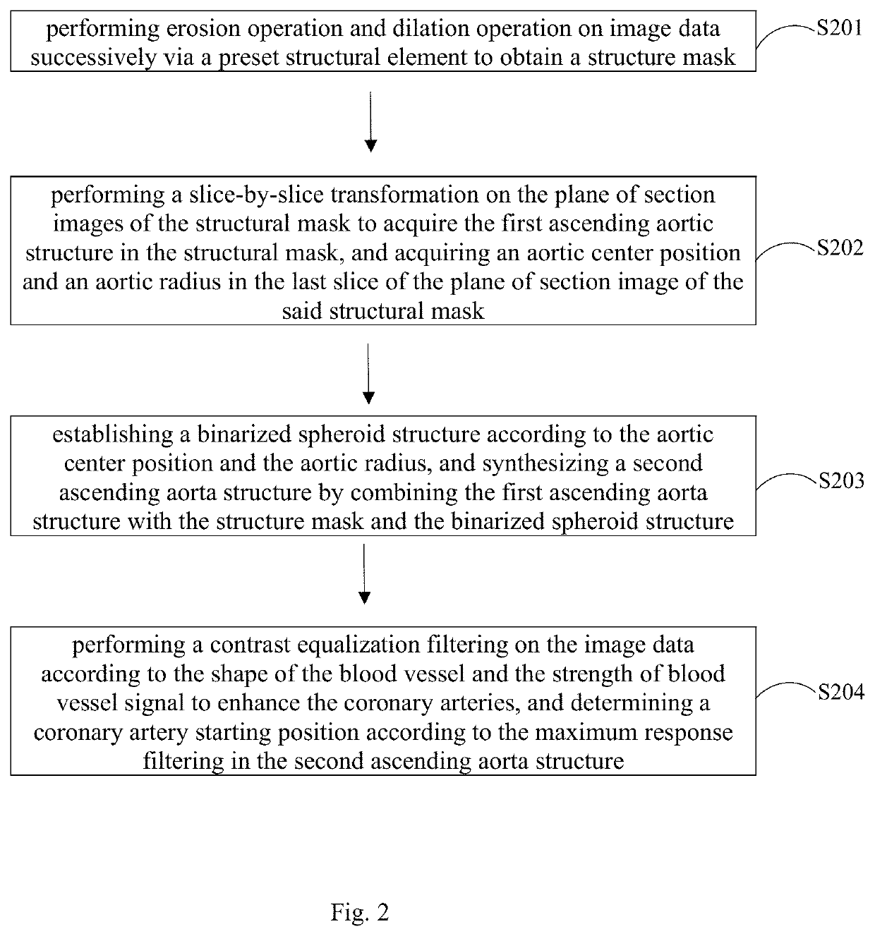
[0035]FIG. 2 shows an implementation process of a method for extracting a cardiovascular vessel from a CTA image provided in Embodiment 2 of the present invention, for the convenience of illustration, only the parts related to the embodiment of the present invention are shown, and the details are as follows:

[0036]In step S201, erosion operation and dilation operation are performed on image data successively via a preset structural element to obtain a structure mask.

[0037]In step S202, a slice-by-slice transformation is performed on plane of section images of the structural mask to acquire the first ascending aortic structure in the structural mask, and an aortic center position and an aortic radius are acquired in the last slice of the plane of section image of the structural mask.

[0038]In step S203, a binarized sphere structure is established according to the aortic center position and the aortic radius, and a second ascending aortic structure is synthesized by combining the first ...

embodiment 3

[0043]FIG. 4 shows a structural representation of an apparatus for extracting a cardiovascular vessel from a CTA image provided in Embodiment 3 of the present invention, for the convenience of illustration, only the parts related to the embodiment of the present invention are shown, and the apparatus for extracting a cardiovascular vessel from a CTA image comprises:

[0044]A structure mask acquiring unit 41, configured for performing erosion operation and dilation operation on image data successively via a preset structural element to obtain a structure mask. The image data is a coronary angiography image after down-sampling processing, and the structural mask is a structure excluding the lung region.

[0045]In an embodiment of the present invention, the above image data is a coronary angiography image after down-sampling processing targeted at large-size original CTA data, in order to quickly extract a large-size ascending aortic structure without affecting the precision of the structu...


PUM


Abstract

The present invention is applicable to the technical field of medical image processing, and provides a method, an apparatus, a device, and a storage medium for extracting a cardiovascular vessel from a CTA image. The method comprises: performing erosion operation and dilation operation on image data successively via a preset structural element to obtain a structure mask; performing a slice-by-slice transformation on the plane of section images of the structural mask to acquire the first ascending aortic structure in the structural mask, and acquiring an aortic center position and an aortic radius in the last slice of the plane of section image of the said structural mask; establishing a binarized sphere structure according to the aortic center position and the aortic radius, and synthesizing a second ascending aorta structure by combining the first ascending aorta structure with the structure mask and the binarized sphere structure. The present invention realizes extracting shapes of ascending aorta and root aortic sinus, so that the aortic structure and morphology can be accurately visualized, thereby greatly improving the level and ability of medical imaging researches to solve clinical issues.

Description

REFERENCE TO RELATED APPLICATIONS[0001]This application claims the benefits of the filing dates of Chinese patent application Serial No. 201710618659.7 filed on Jul. 26, 2017 and PCT Patent Application Serial No. PCT / CN2017 / 094476 filed on Jul. 26, 2017, entitled “Method for extracting blood vessels in CTA image, device, equipment and storage medium”. The teachings of the entire referenced applications are incorporated herein by reference.FIELD OF THE INVENTION[0002]The invention relates to the technical field of medical image processing, particularly a method, an apparatus, a device, and a storage medium for extracting a cardiovascular vessel from a CTA image.BACKGROUND OF THE INVENTION[0003]As an image preprocessing method, cardiovascular vessel extraction is of great significance for the visualization of clinical diagnosis, interventional surgery planning and interventional therapy processes. It extracts an ascending section of heart aorta from a 3D medical angiography image and ...

Claims


Application Information

Patent Timeline
IPC IPC(8): G06T7/00G06K9/46G06T5/30G06T7/262G06V10/34
CPCG06T5/30G06T7/0012G06T2207/20024G06K9/46G06T2211/404G06T7/262G06T2207/30104G06T7/11G06T7/155G06T7/174G06T2207/10081G06T2207/20016G06T2207/30048G06T2207/30101G06T2207/30172G06V10/34G06V2201/03
Inventor ZHOU, SHOUJUNZHANG, BAOCHANGLI, BAOLINWANG, CHENGLU, PEI
Owner SHENZHEN INST OF ADVANCED TECH
Why Eureka
Try Eureka