
Fluorine-18 labeled compositions and their use in imaging of biological tissue

a composition and fluorine technology, applied in the field of fluorine-18 labeled compositions, can solve the problems of limited use of advanced bioimaging and bioengineering equipment, availability of contrast agents, etc., and achieve the effect of superior resolution and superior spectral results

Pending Publication Date: 2020-10-22
CORNELL UNIVERSITY
View PDF1 Cites 0 Cited by
  • Summary
  • Abstract
  • Description
  • Claims
  • Application Information

AI Technical Summary

Benefits of technology

The patent describes a new imaging method using a positron-emitting fluorescing compound that can be used to diagnose and monitor various conditions in the body, such as transplant rejection, cancer, and hemorrhage. This method provides higher resolution and quicker diagnostics than other imaging methods like computed tomography (CT), magnetic resonance imaging (MRI), and magnetic resonance angiography (MRA). The method uses blood cells that have been labeled with a positron-emitting fluorescing imaging compound containing at least one fluorine-18 radionuclide bound directly or indirectly to a fluorophore. The fluorophore can be used to image internal biological tissue by PET or SPECT. The imaging agents described in this patent provide higher resolution, lower quantity, and lower activities than current contrast agents, making them useful for imaging small hemorrhages. This non-invasive imaging method can also be used in image-guided surgery and allows for pre-surgical planning, intra-operative guidance, and post-surgical fluorescence activated cell sorting of cells with desired characteristics.

Problems solved by technology

Yet, few fluorescent and 18F-PET tracers are approved for human use.
This lack of availability of contrast agents currently limits the use of advanced bioimaging and bioengineering equipment, including the PET / MR scanner and robotic platforms for image-guided surgery.

Method used


Image

  • Fluorine-18 labeled compositions and their use in imaging of biological tissue
  • Fluorine-18 labeled compositions and their use in imaging of biological tissue
  • Fluorine-18 labeled compositions and their use in imaging of biological tissue

Examples


examples

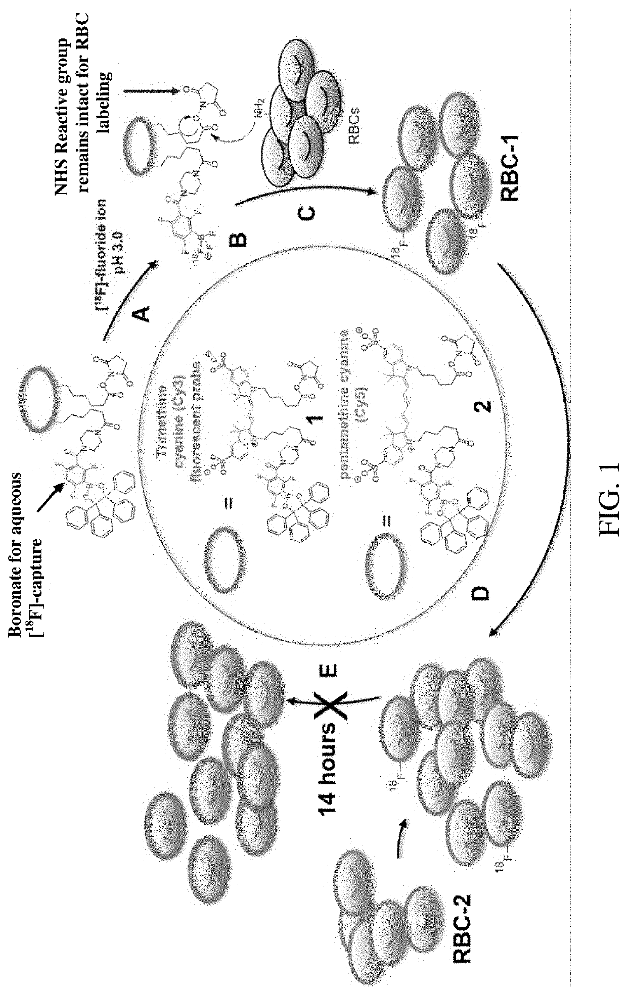
[0083]Synthesis of tri- and penta-methylene N-hydroxy succinimide bearing boronate fluorescent probes (1) and (2)

[0084]The synthesis of the PET / NIRF agents, 1 and 2, was done in a single, two-step reaction. The final yield of 1 was 30%. The final yield of 2 was 15%.

[0085]Synthesis of dioxaborolane bearing trimethine cyanine modified NHS ester (1)

[0086]The following reagents were added to a 1.3 mL glass v-vial in the following order: CY3.18.OH (12 mg, 17 μmols), Piperazin-1-yl(2,4,6-trifluoro-3-(4,4,5,5-tetraphenyl-1,3,2-dioxaborolan-2-yl)phenyl)methanone (12 mg, 19 μmols), hydroxybenzotriazole monohydrate (HOBt, 14 mg, 91 μmols), 800 μL dimethylformamide (DMF), 80 μL pyridine, and N-(3- dimethylaminopropyl)-N′-ethylcarbodiimide hydrochloride (EDC, 150 mg, 785 μmols). The reaction proceeded for 2.5 hours at room temperature before an excess of N-hydroxy succinimide (15 mg, 130 μmols) was added. The reaction was left overnight at room temperature before it was chromatographed by prepa...


PUM

PropertyMeasurementUnit
Structureaaaaaaaaaa

Abstract

A method for internal imaging of biological tissue in a subject by positron emission tomography (PET) or single photon emission computer tomography (SPECT), the method comprising: (i) administering to a subject an imaging agent that includes, at minimum, at least one fluorine-18 radionuclide bound directly or indirectly to a fluorophore, and (ii) imaging internal biological tissue of the subject by PET or SPECT. In further embodiments, the method includes (i) administering to a subject an imaging agent that includes at least one fluorine-18 radionuclide bound directly or indirectly to a fluorophore, and at least one biological entity (e.g., blood cell, peptide, nucleotide, aptamer, targeting agent, antibody, or antibody fragment) bound directly or indirectly to the fluorophore; and (ii) imaging internal biological tissue of the subject by PET or SPECT. In some embodiments, the method further includes simultaneously imaging the internal biological tissue by fluorescence imaging.

Description

CROSS REFERENCE TO RELATED APPLICATIONS[0001]This application claims the benefit of priority from International Patent Application No. PCT / US2019 / 027864, filed on Apr. 17, 2019, and U.S. Provisional Application No. 62 / 658,880, filed on Apr. 17, 2018, which are herein incorporated by reference in their entirety.GOVERNMENT SUPPORT[0002]This invention was made with government support under grant number EB013904 awarded by the National Institutes of Health. The government has certain rights in the invention.FIELD OF THE INVENTION[0003]The present invention generally relates fluorine-18 labeled compositions, and methods of synthesis and use as cell labeling reagents, and more particularly, to fluorophore compositions containing fluorine-18 and methods of using such compositions for cell labeling and biological imaging by positron emission tomography (PET).BACKGROUND OF THE INVENTION[0004]The relationship between imaging and the emergency management of stroke is extremely important given ...

Claims


Application Information

Patent Timeline
IPC IPC(8): A61K51/12A61K49/00
CPCA61K49/0032A61K51/1203A61K49/0043A61B6/037A61B6/508A61B6/481A61B6/4417A61B5/0071A61K51/0497A61K51/0453A61K51/0402A61K49/0021A61K49/0002A61K51/04A61K51/0421A61K49/0097A61K49/0052
Inventor TING, RICHARDARAS, OMERWANG, YE
Owner CORNELL UNIVERSITY
Learn More