Hematopoietic stem and progenitor cell therapy

a hematopoietic stem and progenitor cell technology, applied in the field of cell therapy, can solve the problems of life-threatening, difficult and time-consuming to find the requisite degree of hla donor matched tissue, and allogeneic bone marrow transplants are often associated with a significant incidence of graft-versus-host disease, so as to improve the hematopoietic stem and progenitor cell transplantation method, improve the engraft potential

Pending Publication Date: 2020-10-29
FATE THERAPEUTICS
View PDF2 Cites 0 Cited by
  • Summary
  • Abstract
  • Description
  • Claims
  • Application Information

AI Technical Summary

Benefits of technology

[0013]The invention provides improved hematopoietic stem and progenitor cell transplantation methods. Moreover, the invention provides a superior preparation of hematopoietic stem or progenitor cells that have increased engraftment / engraftment potential and / or increased expansion. In various embodiments, the cells are expanded or proliferated in vivo. In various other embodiments, cells are treated ex vivo, administered to a subject and expanded or proliferated in vivo.
[0078]In particular embodiments, the increase in the engraftment potential of the contacted hematopoietic stem or progenitor cells in the cell population comprises an increase in gene expression of one or more of hyaluronan synthase 1 (HAS1), GTP-binding protein GEM (GEM), dual specificity protein phosphatase 4 (DUSP4), amphiregulin (AREG), Nuclear receptor related 1 protein (NR4A2), renin (REN), cAMP-responsive element modulator (CREM), collagen, type I, alpha 1 (COL1A1), Fos-related antigen 2 (FOSL2), or CXC chemokine receptor 4 (CXCR4) compared to non-contacted hematopoietic stem or progenitor cells, an increase in capacity for self-renewal compared to non-contacted hematopoietic stem or progenitor cells, and no substantial decrease in cell viability compared to non-contacted hematopoietic stem or progenitor cells.

Problems solved by technology

However, bone marrow transplantation is painful for donors and moreover, it is often difficult and time consuming to find the requisite degree of HLA donor matched tissue, especially in particular ethnic populations.
In addition, allogeneic bone marrow transplants are often associated with a significant incidence of graft-versus-host-disease (GVHD).
In many instances, patients receiving hematopoietic stem cell transplants have advanced cancer or other metabolic disorders, which are life threatening.
Thus, any delays in finding a donor having a suitably matched HLA tissue type can compromise the patient outcome, often resulting in fatality.
However, several drawbacks are perceived to exist in using human cord blood transplants, including the risk that hematopoietic stem and progenitor cells from the cord blood transplant may not engraft.
Another drawback of using cord blood transplants is that it takes longer for the cord blood cells to engraft in the patient, which puts the patient at high risk for infection.
Thus, clinicians can be discouraged from using them because they do not have as much information about patients' long-term results after cord blood transplants as they do for marrow transplants.
Moreover, cord blood transplants also have all the same risks as marrow and peripheral blood transplants.
Additionally, a significant barrier to using cord blood as a source of cells for human blood transplants is that there are often not enough blood-forming cells in a single cord blood unit for the size of the patient or to treat the particular indication.
Because the size of a single cord (i.e., the number of blood-forming cells in a single cord) is often insufficient for a blood transplant, two cords may be required, increasing the risks of GVHD and failure to engraft.
Thus, numerous approaches have been tried to expand the number of human hematopoietic stem and progenitor cells in cord blood within isolated grafts in ex vivo settings, which may allow transplantation using a single cord, but these efforts have had limited success.
Thus, the promise of restorative or regenerative hematopoietic stem cell therapies has not been realized, in part, due to difficulties translating promising animal models protocols into human clinical practice, low efficacy of existing clinical protocols, high incidence of complications, e.g., graft-versus-host disease, and relatively few sufficiently matched donors.

Method used


Image

  • Hematopoietic stem and progenitor cell therapy
  • Hematopoietic stem and progenitor cell therapy
  • Hematopoietic stem and progenitor cell therapy

Examples


example 1

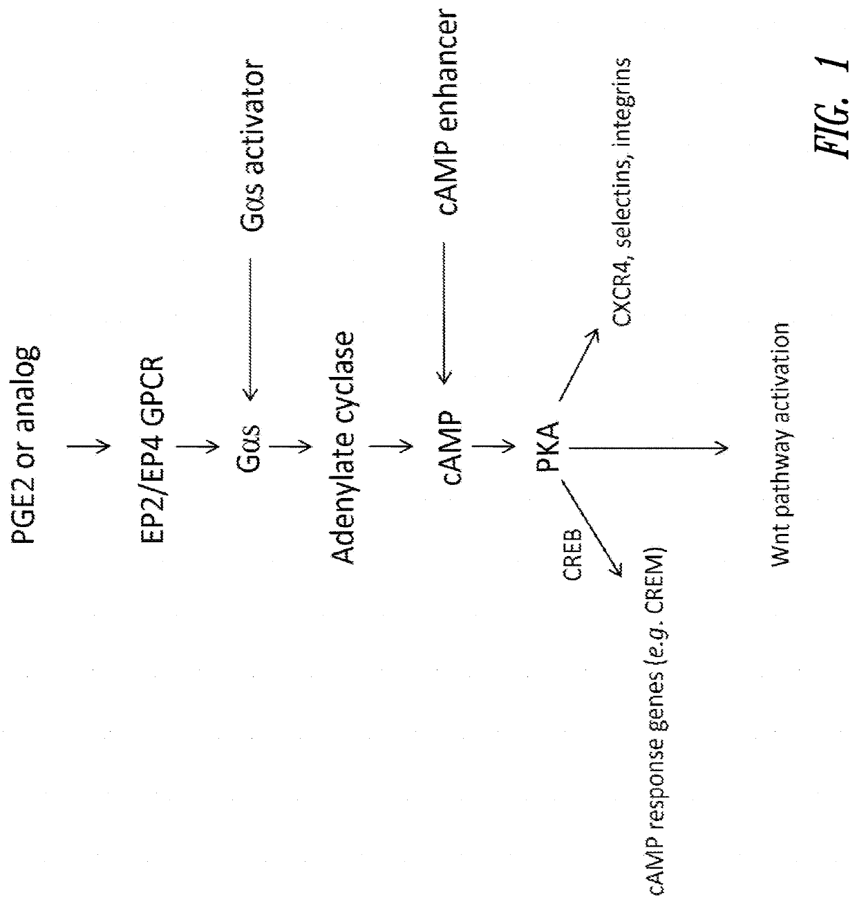
Competitive cAMP Assays

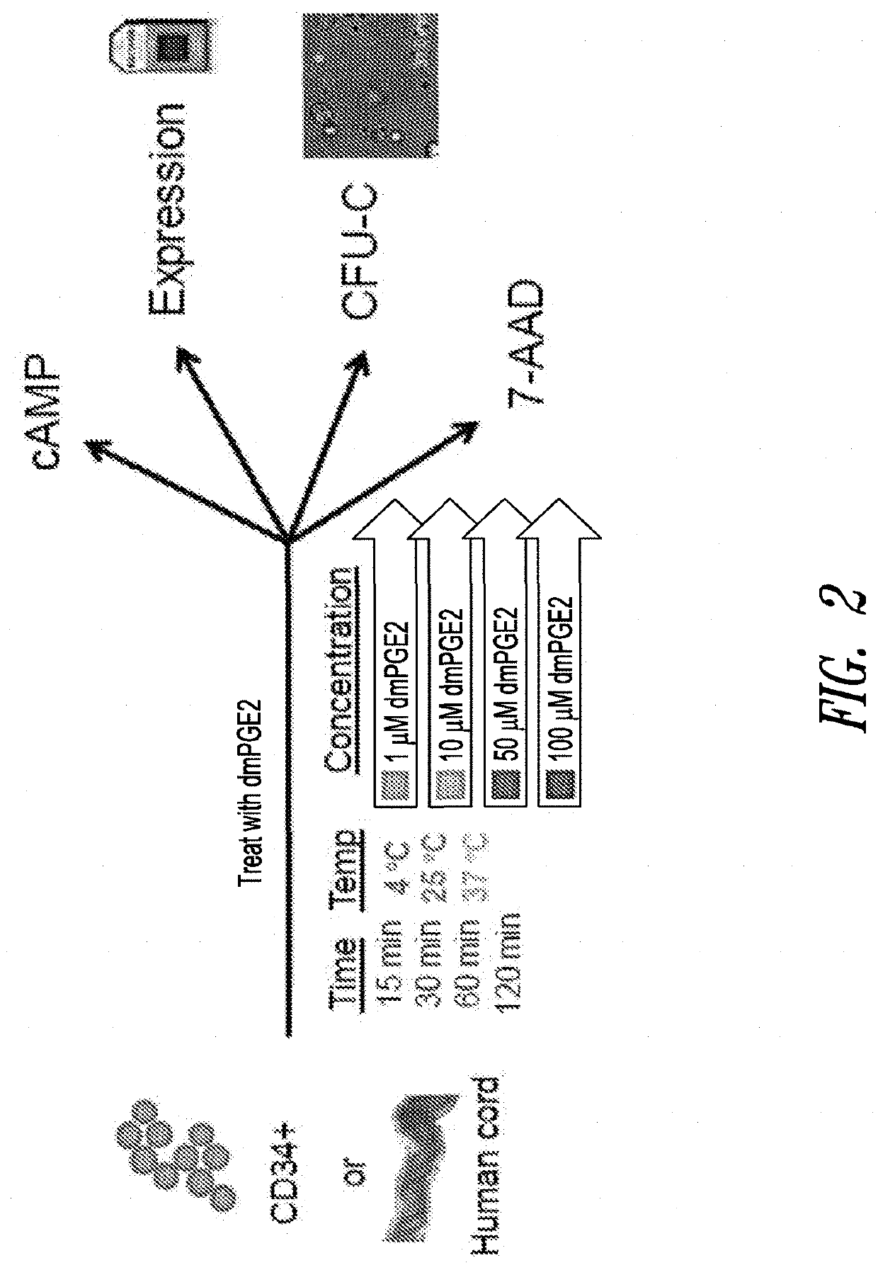
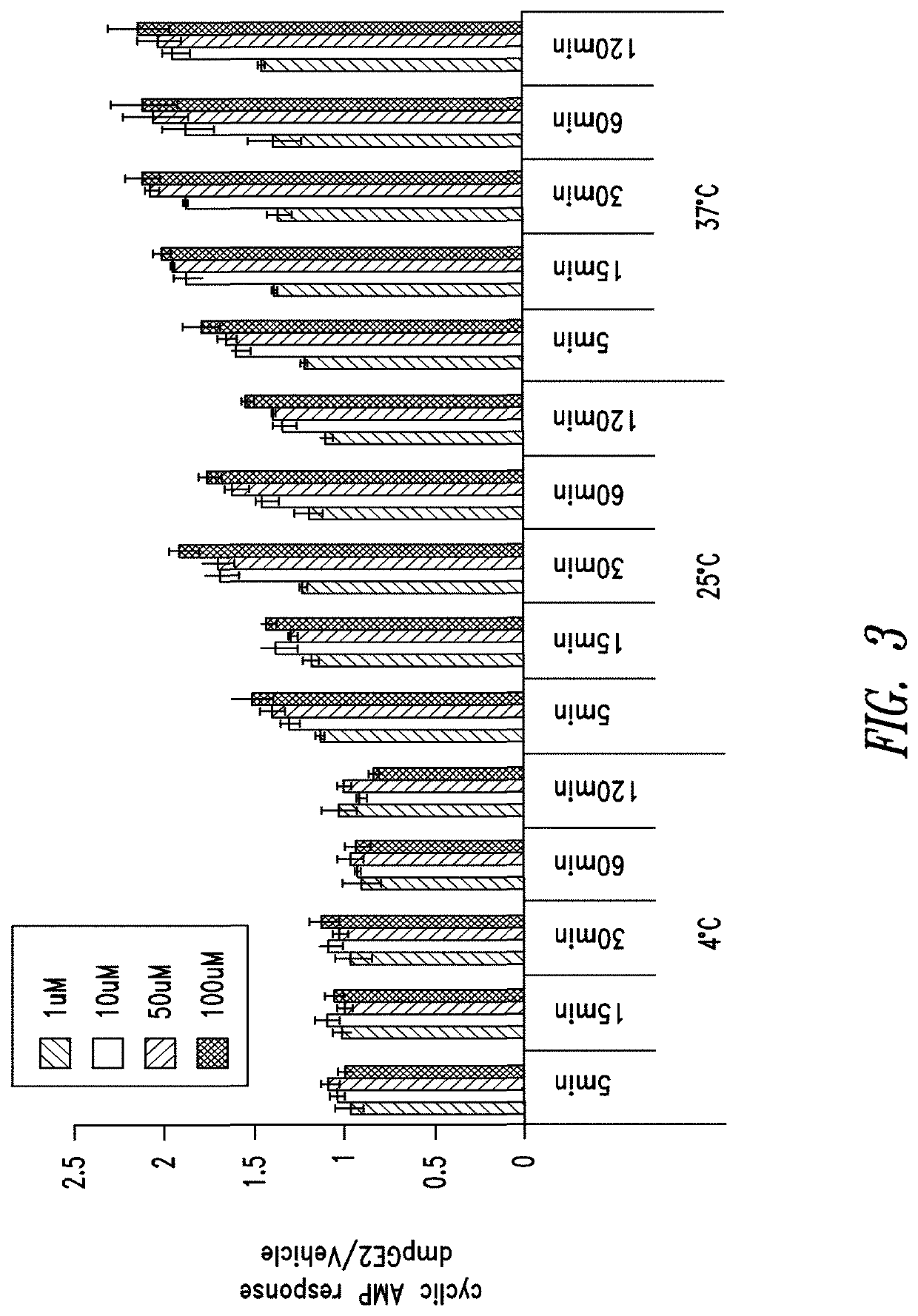
[0351]The cAMP assay was performed on CD34+ cells using the LANCE® cAMP detection kit (Perkin Elmer Inc., Waltham, Mass.) according to the manufacturer's instructions. Briefly, 3,000 CD34+ cells (Stem Cell Technologies, Vancouver, Canada) were aliquoted in each well of a 384-well opaque white plate in the recommended stimulation buffer. Assays were performed in triplicate for all conditions.

[0352]CD34+ cells were placed on ice prior to being stimulated with DMSO or 16,16-dimethyl PGE2 at 4° C. For assays conducted at other temperatures, e.g., 25° C. or 37° C., DMSO or 16,16-dimethyl PGE2 was added to CD34+ cells at room temperature and then the plates were incubated with DMSO or 16,16-dmPGE2 at the experimental temperature (25° C. or 37° C.).

[0353]CD34+ cells were incubated for periods of 5, 15, 30, 60 or 120 minutes. Following the incubation period, detection buffer was added to the stimulated cells and cells were incubated for an additional 1 hour at room te...

example 2

Gene Expression

Whole Genome Expression Arrays

[0357]Human umbilical cord blood and pre-isolated CD34+ cells from human umbilical cord blood were purchased from Stem Cell Technologies Inc. (Vancouver, BC, Canada). Cells were incubated in either low molecular weight dextran with 5% human serum albumin media (LMD / 5% HSA) or Stem Span media (Stem Cells Technology Inc.) for ex vivo treatment with 16,16-dimethyl PGE2. Total RNA was isolated from incubated cells using a Pico Pure RNA Isolation Kit (Molecular Devices, Sunnyvale, Calif.).

[0358]Biotinylated amplified RNA (aRNA) was prepared using the standard protocol for MessageAmp II aRNA Amplification Kit (Applied Biosystems / Ambion, Austin, Tex.) and the optional Second Round Amplification; the copy RNA (cRNA) was transcribed into biotin labeled aRNA using MessageAmp II Biotin Enhanced Kit (Applied Biosystems / Ambion, Austin, Tex.) according to the manufacturer's instructions. Biotin labeled aRNA was purified and fragmented according to the ...

example 3

Cell Viability Assays

[0384]Whole cord blood cells or CD34+ cells obtained from Stem Cell Technologies (Vancouver, Canada) were aliquoted equally in Eppendorf tubes and treated ex vivo at a range of 16,16-dimethyl PGE2 concentrations (10 μM to 100 μM) or DMSO control; temperatures (4° C., 22° C., or 37° C.) for 60 or 120 minutes in LMD / 5% HSA media. After treatment, an aliquot of the incubated cells were assayed using 7-Amino-Actinomycin D (7-AAD) staining as an indicator of cell death. One million whole cord blood were stained with 5 μL of 7AAD staining solution (BD Bioscience, San Jose, Calif.), or 200,000 CD34+ cord blood cells were stained with 1 μL 7-AAD solution. Cells were analyzed on a Guava EasyCyte 8HT System (Millipore) and with the FlowJo software package (Tree Star Inc., Ashland, Oreg.). A separate aliquot of the same cells were also taken for assessment of proliferation potential using CFU-C assays (discussed below in Example 4).

[0385]The results showed that whole cord ...


PUM

PropertyMeasurementUnit
temperatureaaaaaaaaaa
timeaaaaaaaaaa
timeaaaaaaaaaa

Abstract

The invention provides improved methods for cell therapy. In particular, the invention provides therapeutic compositions of modified hematopoietic stem and / or progenitor cells having improved engraftment and homing properties, and methods of making the therapeutic composition. The invention further provides methods of improving the efficacy of hematopoietic stem and progenitor cell transplantation including transplanting the therapeutic composition to subjects in need of hematopoietic system reconstitution.

Description

RELATED APPLICATIONS[0001]This application is a divisional application of U.S. application Ser. No. 13 / 816,723, filed Feb. 12, 2013 and having a 371(c) date of Oct. 8, 2013, which is the U.S. national stage entry of PCT / US2011 / 047657, filed Aug. 12, 2011, which claims the benefit under 35 U.S.C. § 119(e) of U.S. Provisional Patent Application No. 61 / 373,212, filed Aug. 12, 2010, all of which are herein incorporated by reference in their entirety.[0002]This application incorporates by reference in its entirety the Computer Readable Form (“CRF”) of a Sequence Listing in ASCII text format submitted via EFS-Web. The Sequence Listing text file submitted via EFS-Web is entitled “13601-210-999_SEQ_Listings.txt,” was created on Apr. 23, 2020, and is 29,095 bytes in size.BACKGROUNDTechnical Field[0003]The present invention generally relates to cell therapy. Particularly, the present invention relates to improved cell therapies for the hematopoietic system. More particularly, the present inve...

Claims


Application Information

Patent Timeline
Patent Type & Authority Applications(United States)
IPC IPC(8): C12N5/0789A61K35/28A61K35/12
CPCA61K2035/124A61K35/28C12N2501/02C12N5/0647A61P13/02A61P25/00A61P25/28A61P3/00A61P31/18A61P35/00A61P35/02A61P37/02A61P37/04A61P37/06A61P43/00A61P7/00A61P7/04A61P7/06
Inventor SHOEMAKER, DANIELMULTANI, PRATIKDESPONTS, CAROLINEROBBINS, DAVID L.GRAYSON, PAULMENDLEIN, JOHN
Owner FATE THERAPEUTICS
Why Eureka
Try Eureka