

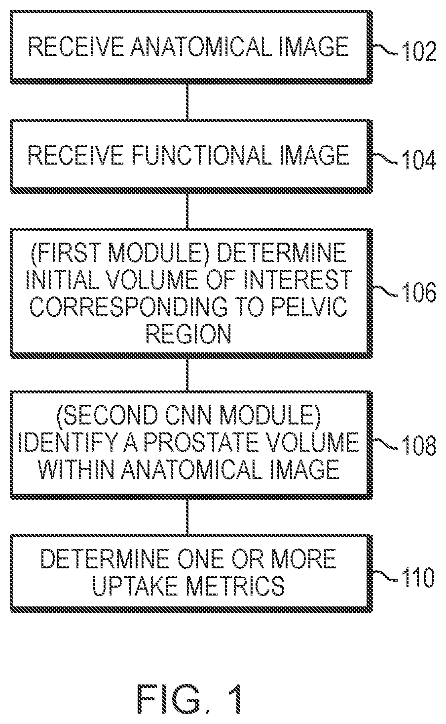
Systems and methods for rapid neural network-based image segmentation and radiopharmaceutical uptake determination
a neural network and image segmentation technology, applied in image enhancement, instruments, applications, etc., can solve problems such as prone to inter- and/or intra-reader variability, and achieve the effects of improving computational efficiency, more computational efficiency, and more computational efficiency
- Summary
- Abstract
- Description
- Claims
- Application Information
AI Technical Summary
Benefits of technology
Problems solved by technology
Method used
Image

Examples
example 4
J. Selection of TBR Threshold for Clinically Significant Findings
[0372]Example 4 is an example showing how a TBR threshold value for partitioning patient prostate cancer pathology into clinically significant and clinically non-significant classifications can be determined.
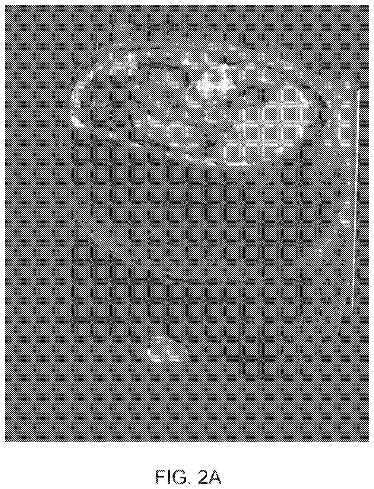
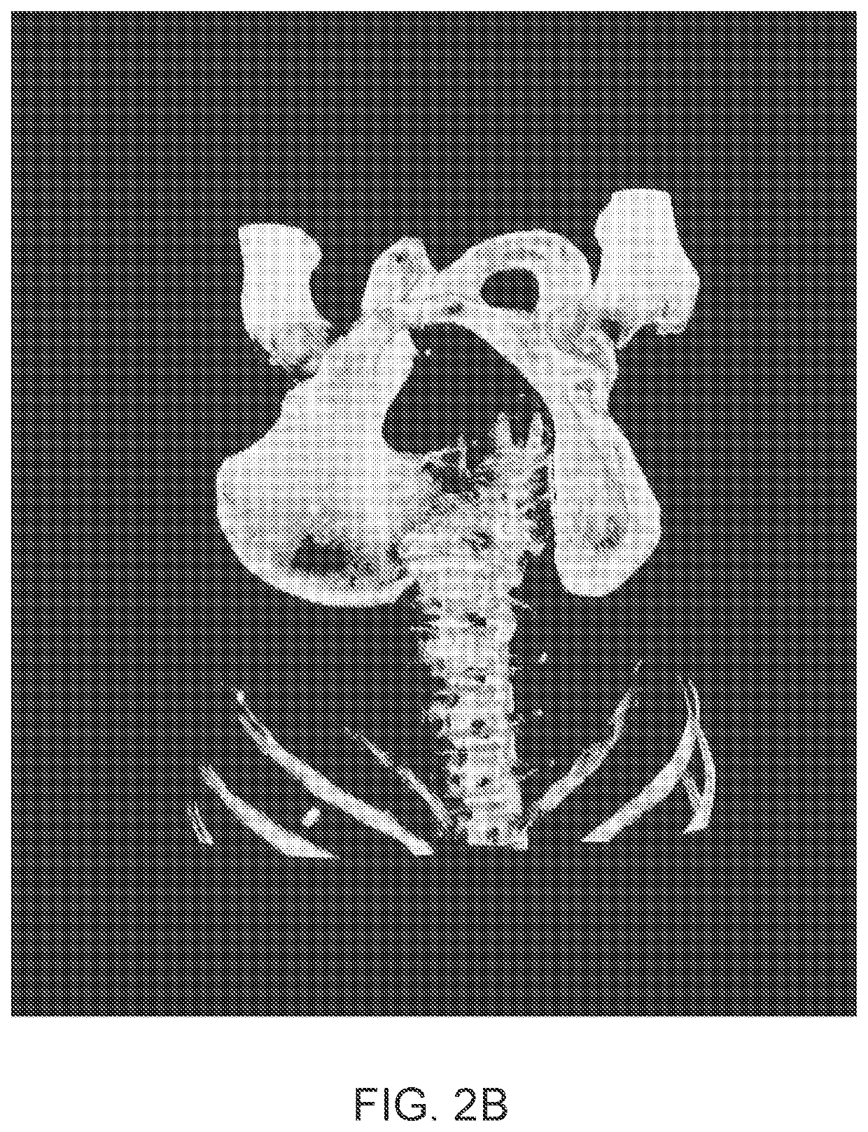
[0373]Two datasets of SPECT / CT images were combined to select an appropriate threshold value. A first dataset comprised images of healthy individuals, taken from a phase I study of the 1404 drug. This dataset contained originally 14 images. Segmentation of a prostate within the images was performed in accordance with the approaches described herein and two images where segmentation of the prostate clearly failed were excluded, resulting in 12 remaining images. A second data set comprised images of individuals with prostate cancer, originating from a phase II study of the 1404 drug. The images were partitioned based on the subject's Gleason grades on histopathology from radical prostatectomy. A total Gleason Score ...
example 6
L. Training and Validation of Convolutional Neural Networks Implemented by the First Machine Learning Module (Localization Machine) and Second Machine Learnigng Module (Segmentation Machine)
[0430]Example 6 is an example showing training and validation of CNN modules used to segment CT images to identify various tissue volumes, including a prostate volume, in accordance with the aspects and embodiments described herein. In this example, the neural networks were defined and trained using the Keras framework with the Tensorflow backend.
[0431]i. Training and validation Data
[0432]The training and validation data comprised CT images coupled with semi-automated segmentations corrected by a radiologist delineating all or some of the following body parts: (i) a prostate; (ii) a urinary bladder; (iii) a rectum; (iv) a left gluteus maximus; (v) a right gluteus maximus; (vi) a left hip bone; (vii) a right hip bone; and (viii) a sacrum and coccyx
[0433]To train a localization CNN, 90 high qualit...
PUM
| Property | Measurement | Unit |
|---|---|---|
| sizes | aaaaa | aaaaa |
| sizes | aaaaa | aaaaa |
| sizes | aaaaa | aaaaa |
Abstract
Description
Claims
Application Information