
System and method for automatic assessment of cancer

a cancer and automatic assessment technology, applied in healthcare informatics, instruments, image enhancement, etc., can solve the problems of difficult early detection and treatment, poor prognosis of patients diagnosed with gbm since the 1980s, etc., to reduce the workload of neuropathologists, accurate data-driven approach, and reduce the effect of diagnosis tim

Active Publication Date: 2020-11-05
KOPPARAPU KAVYA VENKATA KOTA SAI
View PDF0 Cites 1 Cited by
  • Summary
  • Abstract
  • Description
  • Claims
  • Application Information

AI Technical Summary

Benefits of technology

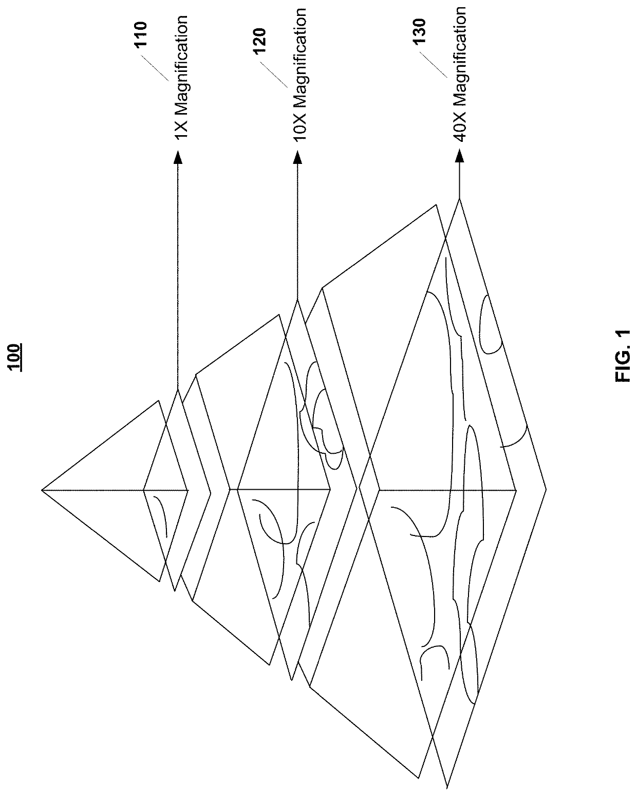
The present patent aims to provide a method to extract tumor information from a single brain biopsy image and use that information to prescribe treatment. It also seeks to enhance the confidence of oncologists and neurologists in making timely treatment decisions. Additionally, it proposes an approach to automatically annotate the biopsy image to highlight tumor features and predict the molecular subtype of the tumor and methylation status of the MGMT gene, which would eliminate the need for expensive and time-consuming genetic testing.

Problems solved by technology

Despite recent advances in neuroimaging, surgery, and drug therapies, the prognosis for patients diagnosed with GBM has not improved since the 1980s.
Currently, treatments for determining GBM are based on time-consuming and costly manual tumor feature segmentation and genetic panel testing to determine molecular subtype and O6-methylguanine methyltransferase (“MGMT”) promoter methylation, both of which are implicated in chemotherapy effectiveness.
Using the approach of the prior art, patients have a mean survival time of 12 months post-diagnosis because the symptoms accompanying GBM are often non-specific, ranging from mild headaches and nausea to personality changes and other stroke-like symptoms, making early detection and treatment difficult.

Method used


Image

  • System and method for automatic assessment of cancer
  • System and method for automatic assessment of cancer
  • System and method for automatic assessment of cancer

Examples


examples

[0161]Following the 20 training epochs, the tumor feature segmentation algorithm achieved a training loss of 0.438, validation loss of 0.427, and validation accuracy of 84.70%. The network achieved an accuracy of 86.02% on the test set of images. Throughout the training process, both the training and validation loss decreased significantly during the first epoch, starting at 3.024 and 3.044 respectively, and gradually decreased before leveling off in Epoch 20. Accordingly, the validation accuracy started at 0.00% and significantly increased during the first epoch before leveling off at 84.70%, as shown in the graphs of FIGS. 11A at 1100 and 11B at 1150.

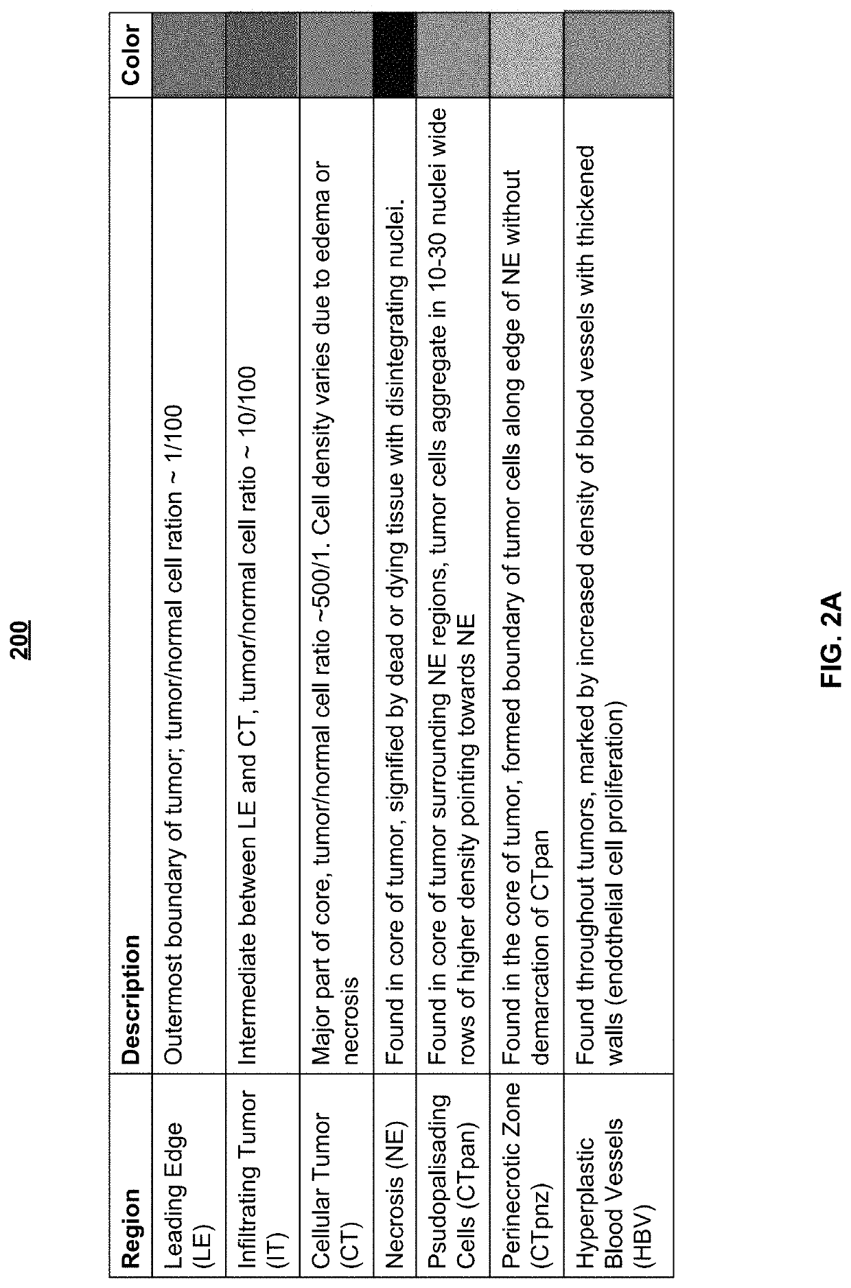
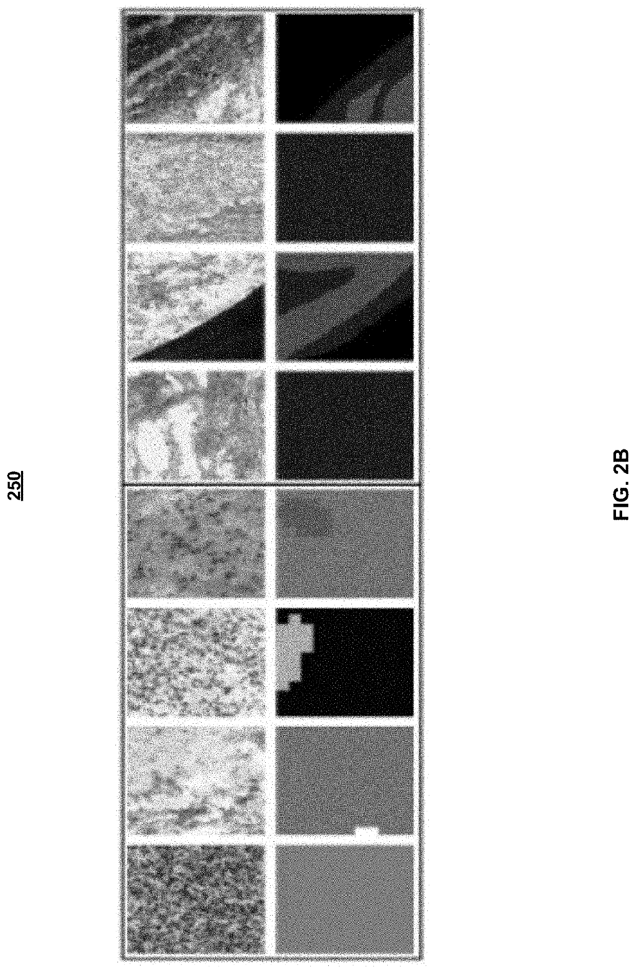
[0162]Throughout the training process, several levels of feature segmentation were achieved, as shown in FIG. 12 at the table 1200. They are as follows:[0163]Stage A (epoch 1): High accuracy was achieved in classification of single-tissue tiles of the necrosis class. From a biological standpoint, this result was expected, as necrosis ...


PUM


Abstract

Cancer can be an aggressive disease. It is critical to determine the most effective patient-specific treatment quickly. Exemplary embodiments use a data-driven approach to extracting tumor information from data obtain from Whole Slide Image that is uploaded through an interface. Exemplary embodiments generate the following information about a tumor from a biopsy slide using neural networks: annotated areas of relevant tissues, molecular subtype, and expression status of an important gene and include three steps: the segmentation of tumor features; prediction of molecular subtype; and prediction of gene methylation status from a WSI.

Description

CROSS-REFERENCE TO RELATED PATENT APPLICATIONS[0001]This application is a Continuation of U.S. patent application Ser. No. 16 / 193,828, filed on Nov. 16, 2018, which claims priority to U.S. Provisional Application No. 62 / 588,651, filed Nov. 20, 2017, entitled “System and Method for Automatic Assessment of Cancer,” the disclosure of which are herein incorporated by reference in their entireties.FIELD OF THE INVENTION[0002]The present disclosure relates generally to a method of treating a patient that has glioblastoma multiforme (“GBM”), also referred to as glioblastoma. Exemplary embodiments integrate multiple biological feature classifiers into a single platform to generate relevant genetic, molecular, and histopathological information from a Whole Slide Image (“WSI”), to detect and treat GBM as soon as possible. Other types of inputs may be used. Specifically, the disclosure relates to a platform that uses trained machine learning architecture, e.g., neural networks, and datasets of...

Claims


Application Information

Patent Timeline
Patent Type & Authority Applications(United States)
IPC IPC(8): G06K9/62G06T7/00G06K9/66G16H30/20G16B25/00G16B40/00G16B20/00G06K9/46G06K9/00G16B40/20
CPCG06T2207/30096G06T2207/20081G06K9/4628G06T2207/30024G06K9/6273G16B40/20G06K9/66G06K9/6269G06T2207/20084G16B25/00G06T7/0012G06T2207/30016G16B40/00G06K9/6267G06K9/00147G06T2207/10056G06K9/6278G16H30/20G16B20/00G06K9/6276G16H50/20G16H30/40G16H20/40G16H20/10G16H40/67G06V20/698G06V10/454G06V10/82G06V30/19173G06F18/24G06F18/2411G06F18/2414G06F18/24147G06F18/24155
Inventor KOPPARAPU, KAVYA VENKATA KOTA SAI
Owner KOPPARAPU KAVYA VENKATA KOTA SAI
Learn More