
Method for detecting cholangiocarcinoma cells

a technology of cholangiocarcinoma and cholangiocarcinoma cells, which is applied in the field of detecting cholangiocarcinoma cells, can solve the problems of difficult diagnosis and detection, poor prognosis, and obstructive jaundice, and achieve the effect of reducing the mixing time and high affinity

Active Publication Date: 2020-11-12
NATIONAL TSING HUA UNIVERSITY +1
View PDF0 Cites 1 Cited by
  • Summary
  • Abstract
  • Description
  • Claims
  • Application Information

AI Technical Summary

Benefits of technology

The invention is a method for capturing and analyzing cholangiocarcinoma cells using a microfluidic chip system. The method has a higher capture rate of cholangiocarcinoma cells than previous methods and reduces the mixing time of the sample and magnetic beads. The detection process involves using special octasaccharides that have high affinity and specificity for capturing cholangiocarcinoma cells. This method can successfully detect cholangiocarcinoma cells without interference from other cells and can also detect low numbers of cells in whole blood. This is the first time an integrated microfluidic system has been developed for detecting circulating tumor cells in a small amount.

Problems solved by technology

The former may cause obstructive jaundice even if the tumor is small.
Regardless of the type of cholangiocarcinoma, the prognosis is very bad.
Because there are no obvious symptoms in cholangiocarcinoma in the early stage, it is difficult to diagnose and detect.
While most patients are diagnosed with cholangiocarcinoma, the disease has progressed to the advanced stage and cannot be cured.
Complete surgical resection is the only hope for cure, but about one-third of patients have tumors that invade the common bile duct, and such tumors cannot be surgically removed, so only a few tumors can be completely resected.
This phenomenon makes it difficult to isolate and purify circulating tumor cells, particularly in a high purity manner.
Although the above-described methods for detecting and isolating circulating tumor cells have been present, the white blood cell contamination in the obtained circulating tumor cells is often unavoidable.
Problems may be caused in subsequent analysis of circulating tumor cells (especially gene expression analysis), which may cause underestimation or misjudgment.
The problem is mainly because the expression level of some white blood cell related genes is still unclear.
Therefore, the presence of these white blood cells can interfere with subsequent analysis.
In addition to the purity of circulating tumor cells, there are some important biological issues that are needed for further consideration.
In this regard, if a conventional method for isolating and detecting circulating tumor cells is used, these circulating tumor cells that are clinically associated with cancer metastasis may be missed, especially the circulating tumor cells in cholangiocarcinoma difficult to detect.

Method used


Image

  • Method for detecting cholangiocarcinoma cells
  • Method for detecting cholangiocarcinoma cells
  • Method for detecting cholangiocarcinoma cells

Examples


example 1

Design and Fabrication of Microfluidic Chip

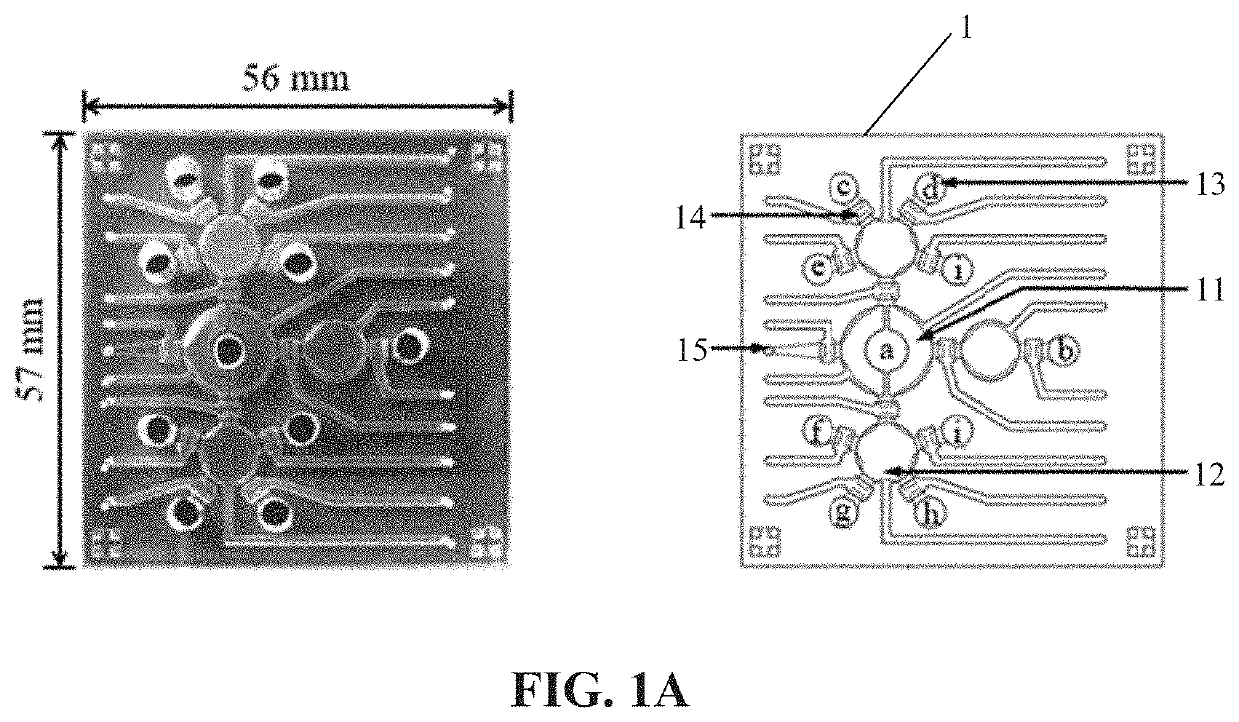
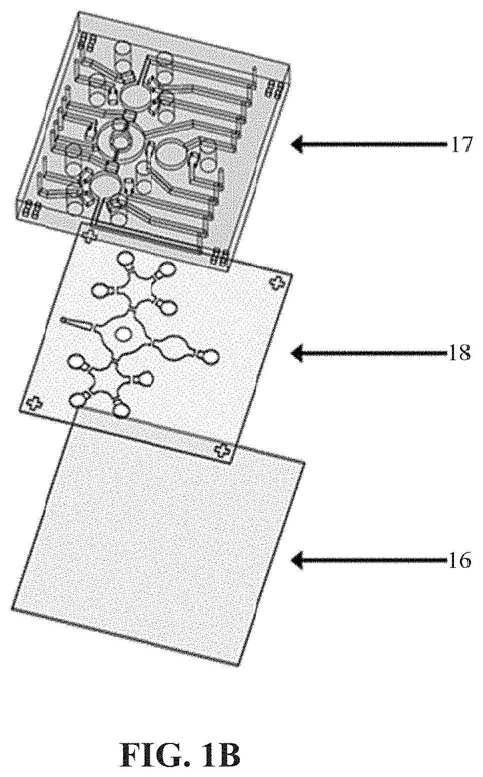
[0041]The microfluidic platform designed in this example includes a blood cell depletion module, a cancer cell isolation module and an IF staining module. The components on the microfluidic chip 1 are an open-type micromixer 11, a plurality of micropumps 12, eight reagent chambers 13 (including the symbols c, d, e, i, f, g, and h in FIG. 1A), a supernatant reservoir b, a plurality of normally-closed valves 14 and a waste outlet 15, shown in FIG. 1A, wherein a indicates blood sample, b indicates supernatant reservoir, c indicates octasaccharide-coated beads, d indicates CD45-coated beads, e indicates 4% paraformaldehyde, f indicates 0.1% Triton X-100, g indicates primary antibodies, h indicates secondary antibodies (Hoechst 33258), and i indicates wash buffer. The open-type micromixer 11, micropumps 12 and normally-closed valves 14 would be controlled by deforming membranes via applying positive and negative gauge pressures using programs an...

example 2

Operating Process on Automatic Microfluidic System

[0043]This example reported an integrated microfluidic system which could automatically perform WBC depletion, CCA cell isolation and IF staining. A whole blood sample (2-3 mL) spiked with 105 human cholangiocarcinoma cell line Huh28 cells (supplied by Division of Surgery, National Cheng Kung University Hospital, Taiwan) was pretreated by RBC lysis buffer for 5 mins first. After centrifuging at 1200 rpm for 5 mins and resuspending in 1×PBS (FIG. 2 (a)), the sample was loaded into the micromixer on microfluidic system to execute WBC depletion by Dynabeads® CD45 three times, wherein the Dynabeads® CD45 are CD45-coated beads. In brief, WBCs in the sample were incubated with Dynabeads® CD45 and the captured WBCs were collected by applying a magnet, the supernatant was transferred to the supernatant reservoir followed by washing out the gathered Dynabeads® CD45 and transferring the supernatant back to the micromixer (FIG. 2 (b)). After pe...

example 3

Performance of the Microfluidic System

[0044]In this example, the microfluidic chip for CCA cell detection consisted of several components such as micromixers, micropumps and microvalves. Before the entire on-chip experiments, the performances of the microfluidic chip including mixing index and shear force was measured, such that the optimum operating conditions could be applied on the microfluidic system.

3.1 Mixing Index of the Micromixer

[0045]In order to effectively incubate cells and octasaccharide-coated beads by the pneumatic micromixer, mixing index which could reflect the mixing efficiency of the micromixer was tested and calculated. The two liquid samples chosen for tests of mixing efficiency were 120 μL DI water and 5 μL blue ink. Gauge pressures from −100 to −500 mmHg were applied at driving frequencies of 1, 2 and 4 Hz to cause different mixing efficiencies. The entire operating process of the microfluidic chip was carried out at a constant gauge pressure of 51.7 mmHg on t...


PUM

PropertyMeasurementUnit
gauge pressureaaaaaaaaaa
pressureaaaaaaaaaa
detection timeaaaaaaaaaa

Abstract

The present disclosure provides a method for detecting cholangiocarcinoma cells. The capture rate of the cholangiocarcinoma cells of the present disclosure is higher than 70%, and a plurality of octasaccharides with high affinity and specificity can be modified on the surface of magnetic beads to capture and analyze cholangiocarcinoma cells under test, wherein the cholangiocarcinoma cells can be circulating tumor cells in cholangiocarcinoma.

Description

CROSS-REFERENCE TO RELATED APPLICATION[0001]This application claims priority of Taiwan patent application No. 108116267, filed on May 10, 2019, the content of which is incorporated herein in its entirety by reference.BACKGROUND OF THE INVENTION1. Field of the Invention[0002]The present invention relates to a method for detecting cholangiocarcinoma cells.2. The Prior Art[0003]Cancer, or malignant tumor, causes millions of deaths each year worldwide, and it is also the number one cause of death in Taiwan in recent years. Among various cancers, cholangiocarcinoma is a rare primary malignant liver tumor, and its clinical symptoms, diagnosis and epidemiological characteristics are all different from those of another primary malignant liver tumor hepatocellular carcinoma. The biliary system is distributed throughout the liver, starting from the bile capillarys formed by the groove on the surface of the hepatic cells, gradually converging into a small bile duct, the left and right intrahep...

Claims


Application Information

Patent Timeline
Patent Type & Authority Applications(United States)
IPC IPC(8): G01N33/574G01N33/543B01L9/00
CPCG01N33/54326B01L9/527G01N33/57438B01L3/5027G01N2400/38B01F31/31B01F33/30
Inventor LEE, GWO BINHUNG, SHANG-CHENGTSAI, WEI-CHUN
Owner NATIONAL TSING HUA UNIVERSITY
Learn More