
METHOD FOR DERIVING CORNEAL ENDOTHELIUM REPLACEMENT CELLS FROM iPS CELLS

Pending Publication Date: 2020-12-24
KEIO UNIV
View PDF0 Cites 0 Cited by
  • Summary
  • Abstract
  • Description
  • Claims
  • Application Information

AI Technical Summary

Benefits of technology

The present invention provides a method for efficiently and safely producing a corneal endothelial substitute cell from an iPS cell. This cell can be used as a medicament for treating diseases caused by functional disorder of corneal endothelial cells, such as corneal transplantation and cellular therapy. The technical effect of this patent is to provide a more efficient and safe method for producing a useful cell for eye treatment.

Problems solved by technology

The above-mentioned function of corneal endothelial cell is impaired by a damage to the corneal endothelium such as a decrease in the corneal endothelial cell and the like, thus resulting in the edema of the corneal stroma.
This decreases transparency of the cornea, and reduces the visual acuity.
While the penetrating keratoplasty is an established technique, the supply of cornea is short in Japan as the situation stands, and the rejection reaction poses a problem.
Since the number of donor of cornea is limited, the problem of donor shortage cannot be overcome, like penetrating keratoplasty.
Furthermore, since corneal endothelial cell is difficult to culture, preparation of cultured cells in a number sufficient for transplantation places a large burden in terms of time and cost.
However, the mechanism of induction from stem cells to the corneal endothelium has not been elucidated because both rely on additives made of animal-derived components such as bovine serum.
In addition, they are not production methods of a level that can be used in clinical researches since they use biological materials.

Method used


Image

  • METHOD FOR DERIVING CORNEAL ENDOTHELIUM REPLACEMENT CELLS FROM iPS CELLS
  • METHOD FOR DERIVING CORNEAL ENDOTHELIUM REPLACEMENT CELLS FROM iPS CELLS
  • METHOD FOR DERIVING CORNEAL ENDOTHELIUM REPLACEMENT CELLS FROM iPS CELLS

Examples


example 1

e in Gene Expression Between iPS Cell-Derived Neural Crest Cell and Human Corneal Endothelium by DNA Microarray

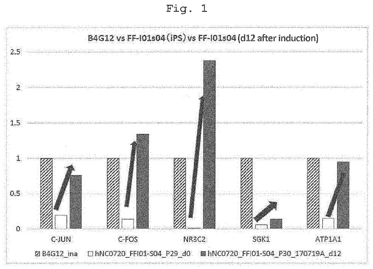
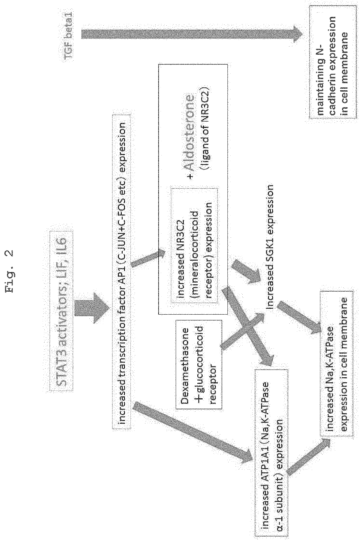
[0082]Using two kinds of cells of (a) genuine human corneal endothelial cells collected from the cornea for overseas donor research and (b) iPS cell-derived neural crest cells (iPS-NCC) induced from iPS cell (FF-I01s01 line; the Center for iPS Cell Research and Application, Kyoto University) by a modified method of the neural crest cell induction method of a previous report (Bajpai et al., Nature. 2010 Feb. 18; 463(7283):958-62), Agilent Technology DNA microarray analysis was outsourced to Hokkaido System Science Co., Ltd., and gene expression profiles were compared by DNA microarray. iPS-NCC (also indicated as iNCC) was confirmed by the expression of neural crest cell markers P75NTR and CD49D. It was newly found that corneal endothelial cells highly express KGF, IGF2, LIF, and IL6 (Tables 2-1, 2-2). It was also found that AP1 (C-FOS+C-JUN), which is considered to be locate...

example 2

n of ATP1A1, ZO-1, N-Cadherin and PITX2 11 Days after CECSi Cell Induction

[0093]According to the induction protocol used in Example 1, iPS cells (FF-I01s04 line) were induced to differentiate. The expression of ATP1A1, ZO-1, N-cadherin and PITX2 was examined using cells on day 11 from the differentiation induction.

(Immunostaining)

[0094]CECSi cells on a 35 mm dish were fixed with ice-cooled 4% paraformaldehyde after removing the medium. After washing the dish twice with PBS, a blocking solution (PBST containing 10% Normal Donkey Serum (10% NDS / PBST)) was added and blocking was performed for 30 min. Then, the primary antibody reaction was performed at room temperature for 1 hr with the following antibody amounts.

(i) ZO1, Na,K,ATPase α1 double staining

Rabbit anti human ZO-1; Invitrogen, 40-2200, 1:500, 1.5 μL / dish

Mouse anti human Na,K-ATPase α1; Novus, NB300-146, 1:500, 1.5 μL / dish

blocking solution (10% NDS / PBST), 0.75 mL / dish

(ii) N-cadherin, PITX2 double staining

Rabbit anti human PITX...

example 3

n of Induction Efficiency into CECSi Cells

(Verification by FACS)

[0099]Furthermore, induction efficiency was quantitatively evaluated by performing flow cytometry using integrin alpha-3 (CD49c) expressed on corneal endothelial cells as a surface antigen.

(Flow Cytometry)

[0100]After collecting CECSi cells on 10 cm dish with Accutase, the cells were washed by adding HESS and centrifuging and discarding the supernatant. After suspending the cells in HBSS, 2 μL of each of the following antibody reagents was added to 1×10⁶ cells / 100 μL of cell suspension.

PE anti-human CD49c, BioLegend #343803

PE anti-human CD49d, Biolegend #304304

[0101]Alternatively, 2 μL of rBC2LCN-FITC (Wako Pure Chemical Industries, Ltd. #80-02991) was added. The antibody reaction was performed at room temperature for 1 hr.

[0102]The cells were washed by adding HBSS and centrifuging and discarding the supernatant, and flow cytometry was performed using SH800 manufactured by SONY.

[0103]The results are shown in FIG. 6. The ...


PUM


Abstract

Using the medium of the present invention for inducing a cornea endothelial substitute cell from an iPS cell, the medium containing an insulin-like growth factor and a STAT3 activator, and not containing a basic fibroblast growth factor or a ROCK inhibitor, and the method of the present invention for inducing corneal endothelial substitute cells from iPS cells by using the medium, it becomes possible to efficiently produce corneal endothelial substitute cells, particularly to efficiently produce corneal endothelial substitute cells from iPS cells. Furthermore, it becomes possible to stably produce large amounts of corneal endothelial substitute cells by inducing differentiation of iPS cells into corneal endothelial substitute cells more efficiently.

Description

TECHNICAL FIELD[0001]The present invention relates to a method for inducing a corneal endothelial substitute cell from an iPS cell. Furthermore, the present invention relates to a corneal endothelial substitute cell obtained by the method, a medicament using the same and the like.BACKGROUND ART[0002]The above-mentioned function of corneal endothelial cell is impaired by a damage to the corneal endothelium such as a decrease in the corneal endothelial cell and the like, thus resulting in the edema of the corneal stroma. This decreases transparency of the cornea, and reduces the visual acuity. Such condition is called bullous keratopathy. In the meantime, it is known that human corneal endothelial cell once injured scarcely shows an ability to regenerate. When the corneal endothelial cells have decreased due to certain injury, an effective and sole treatment thereof is corneal transplantation. In fact, about half the number of applicable cases of corneal transplantation is for bullous...

Claims


Application Information

Patent Timeline
IPC IPC(8): A61K35/30C12N5/079A61P27/02
CPCA61K35/30C12N5/0621A61P27/02C12N2501/117C12N2501/15C12N2501/2306C12N2501/105C12N2506/45C12N2500/38A61K35/545A61L27/3808A61L27/3834A61L27/3886A61L27/3895A61L2430/16
Inventor SHIMMURA, SHIGETOHATOU, SHINTSUBOTA, KAZUO
Owner KEIO UNIV
Learn More