Method for isolating cell nuclei having enhanced antigenicity from fixed cells or ffpe tissue section, and antigen activator and kit therefor

a cell nucleus and antigenicity technology, which is applied in the field of isolating cell nuclei having enhanced antigenicity from fixed cells or ffpe tissue sections, can solve the problems of limited useable antibodies, achieve enhanced antigenicity, high objectivity, reproducibility and universality, and detect quickly

Pending Publication Date: 2020-12-31
NITTO BOSEIKI CO LTD
View PDF1 Cites 1 Cited by
  • Summary
  • Abstract
  • Description
  • Claims
  • Application Information

AI Technical Summary

Benefits of technology

[0101]According to the present invention, established is a method for detecting (quantitatively determining) Ki-67-positive cells with high objectivity, reproducibility and universality.
[0102]The present invention provides a protocol (including a pretreatment reagent and a process) to isolate cell nuclei containing a target antigen with enhanced antigenicity from a FFPE tissue section. The recovered dispersion can be analyzed at a single cell nucleus level. For example, a protein, a nucleic acid or the like on a nuclear membrane or in a nucleus can be an object to be detected and can be detected by staining them with a labeling substance (fluorescent substance, chemiluminescent subst...

Problems solved by technology

However, digestion of the matrix with an enzyme may cause an epitope...

Method used


Image

  • Method for isolating cell nuclei having enhanced antigenicity from fixed cells or ffpe tissue section, and antigen activator and kit therefor
  • Method for isolating cell nuclei having enhanced antigenicity from fixed cells or ffpe tissue section, and antigen activator and kit therefor
  • Method for isolating cell nuclei having enhanced antigenicity from fixed cells or ffpe tissue section, and antigen activator and kit therefor

Examples


reference example 1

Observation of Isolation of Cell Nuclei by Water Flow Shear

1. Materials and Method

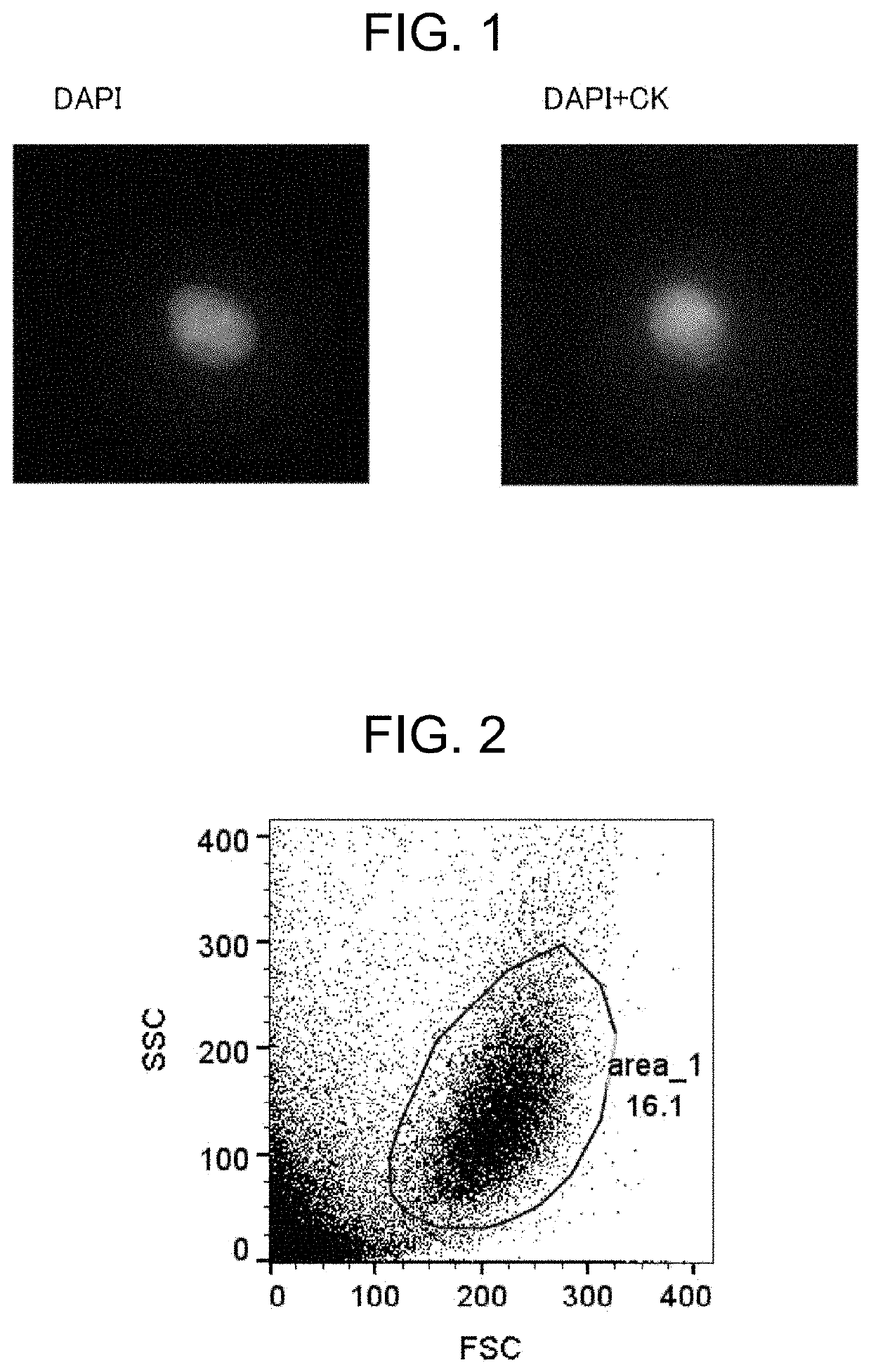
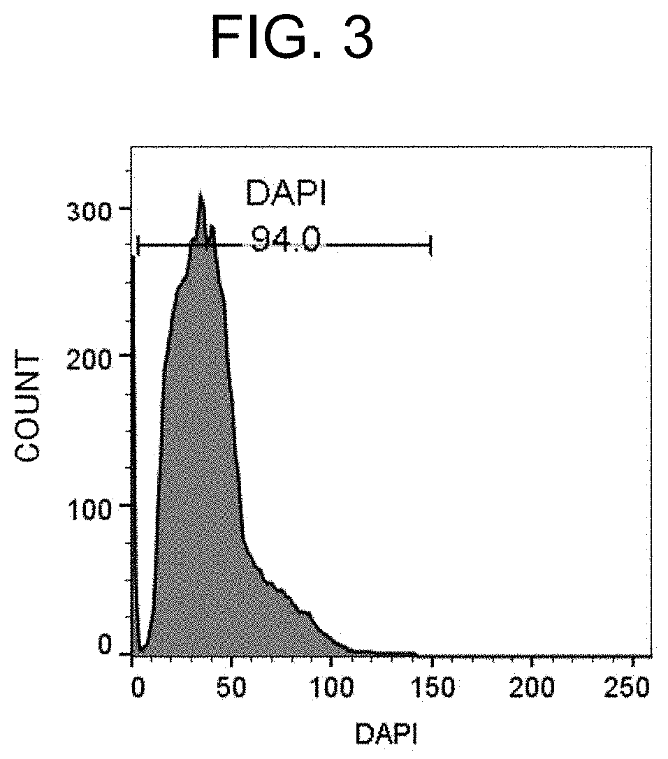
[0193]A FFPE tissue section of a breast cancer tissue was deparaffinized / hydrophilized, subjected to antigen retrieval with heat treatment, and dispersed by a water flow shear apparatus. The recovered product was then immunofluorescently stained, and observed with a microscope.

1-1. FFPE Tissue Block

[0194]A FFPE tissue block of a breast cancer tissue was purchased from ProteoGenex, Inc.

1-2. Preparation of FFPE Section

[0195]A FFPE section with a thickness of 20 μm was prepared by slicing a FFPE tissue block with a sliding microtome manufactured by Thermo Fisher Scientific.

1-3. Deparaffinization / Hydrophilization

[0196]A sufficient amount of xylene was added to the FFPE section, the section was placed still for 10 minutes, and xylene was then removed. These steps were carried out again to completely remove paraffin. Subsequently, the section was immersed in 100% ethanol, 95% ethanol, 70% ethanol, 50% ethano...

reference example 2

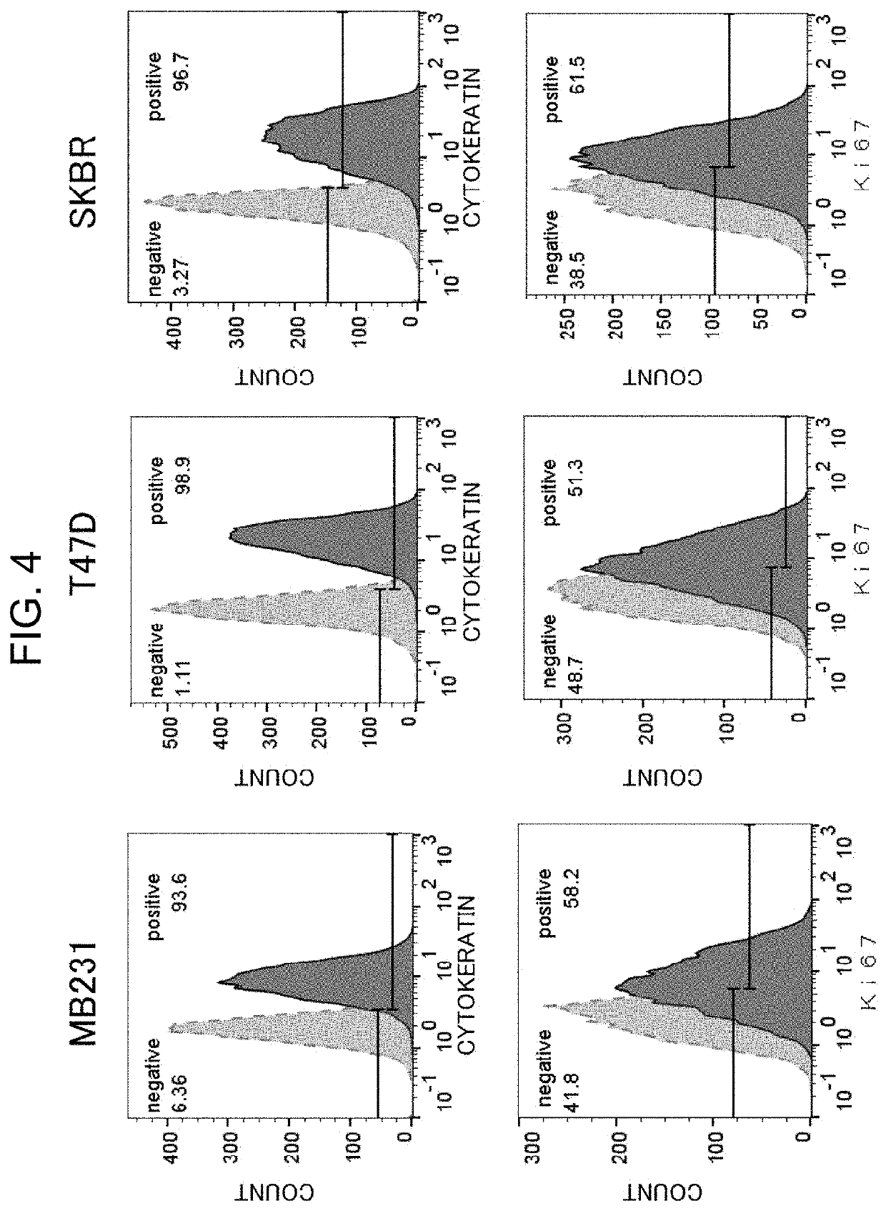
[0202]Examination of Signals Derived from Cytokeratin and Ki-67 in Formalin-Fixed Breast Cancer Cells

1. Materials and Method

[0203]Formalin-fixed breast cancer cells were subjected to antigen retrieval with heat treatment and immunofluorescently stained, and signals from cytokeratin and Ki-67 were examined by a flow cytometer.

1-1. Cells

[0204]Three breast cancer cell lines: T47D, MDA-MB-231 (described as MB231 or 231 in the figures) and SKBr3 (described as SKBR in the figures) were acquired from ATCC (American Type Culture Collection).

1-2. Cell Culture and Formalin Fixation

[0205]The T47D cell line was cultured in RPMI-1640 medium, the MDA-MB-231 cell line was cultured in Leibovitz's L-15 medium, SKBr3 cells were cultured in McCoy's 5A medium. These mediums are supplemented with 10% fetal bovine serum (FBS). After sufficient proliferation of the cells, a culture solution was drawn in, and the cells were washed with PBS, followed by adding TrypLE Express (Thermo Fisher Scientific). The ...

example 1

[0211]Enhancement of Ki-67 Signals in Formalin-Fixed Cells by Antigen Activation with Thrombin

1. Materials and Method

[0212]With formalin-fixed breast cancer cells, signal intensities with and without thrombin treatment were compared.

1-1. Cells

[0213]The three breast cancer cell lines used in Reference Example 2 were used.

1-2. Cell Culture and Formalin Fixation

[0214]The same procedure as in Reference Example 2 was carried out.

1-4. Antigen Retrieval with Heat Treatment

[0215]The same treatment as in Reference Example 1 was carried out.

1-5. Antigen Activation with Enzyme

[0216]A thrombin reagent (25 mM Tris-HCl pH 7.4, 150 mM NaCl, 1000 KU / L thrombin, 10 mM CaCl2)) was added to the cells after the antigen retrieval with heat treatment, and the cells were heated on a heat block at 37° C. for 20 minutes.

1-7. Immunofluorescent Staining

[0217]The same procedure as in Reference Example 2 was carried out. Here, a Ki-67 antibody (Dako, clone: MIB-1, mouse monoclonal antibody) was used as a primar...


PUM

PropertyMeasurementUnit
Fractionaaaaaaaaaa
Fractionaaaaaaaaaa
Fractionaaaaaaaaaa

Abstract

Provided are a pretreatment method for detecting the number of cells containing Ki-67 protein-positive nuclei using Ki-67 antibody; a method for the detection; a kit to be used in the detection method; and determination of a therapy regimen using the aforesaid method. Attempts were made to activate Ki-67 antigen with the use of an enzyme having been considered as inappropriate for the activation thereof. By pretreating a sample with an enzyme not recognizing the epitope of MIB-1 (a rare cutter enzyme), the antigen activation of Ki-67 antigen was enhanced, while enhancing the antigenicity of other antigens including a cytokeratin too. As a result, a method for more objectively and more universally quantifying Ki-67-positive cells at higher reproducibility, said method comprising isolating cell nuclei from an FFPE section while enhancing the antigenicity and performing a reaction between Ki-67 protein, i.e., the target, existing in the cell nuclei with a fluorescently labeled antibody, has been completed.

Description

TECHNICAL FIELD[0001]The present invention relates to a pretreatment method for use in detecting cells containing Ki-67 protein-positive cell nuclei with a Ki-67 antibody, a method for detecting the cells, a kit for use in the pretreatment method or the detection method, and selection of a therapeutic regimen with the detection method.BACKGROUND[0002]Formalin fixation of a surgically removed tissue is the most common method for preserving a cancer tissue over the world, and serves as a standard pathological technique. The most common method for preserving a tissue includes immersing the entire tissue in an aqueous formalin solution for a long time (8 to 48 hours), and then embedding the fixed tissue in paraffin wax for long-term preservation at room temperature. Accordingly, molecular analysis methods with a formalin-fixed cancer tissue are most commonly used for analysis of, for example, a tissue from a cancer subject.[0003]The immunohistological staining, which is one of diagnosti...

Claims


Application Information

Patent Timeline
IPC IPC(8): G01N33/574G01N1/30C12Q1/6886
CPCG01N33/57496G01N2333/723G01N2333/47C12Q1/6886G01N1/30C12Q2600/158C12Q2600/106C12Q2600/118G01N2333/4742C07K14/47C12N9/50C12Q1/37G01N33/6875G01N2800/52G01N33/56966G01N15/14G01N33/57484C07K16/18C12N9/6424C12N9/2474C12N5/0693C12Q1/6813G01N2015/1486C12N2509/10G01N2333/435
Inventor SATO, NATSUKIKUROIWA, MIKANAKATSUJI, MASATOSHIISHIHARA, HIDEKI
Owner NITTO BOSEIKI CO LTD
Why Eureka
Try Eureka