Alk5 inhibitors as skeletal muscle hypertrophy inducers
- Summary
- Abstract
- Description
- Claims
- Application Information
AI Technical Summary
Benefits of technology
Problems solved by technology
Method used
Image
Examples
example 1
[0181]Materials and Methods
[0182]Cell Source and Cell Culture Healthy donor primary skeletal cells (Donor 1, Donor 3 and Donor 5) were Clonetics™ Human Skeletal Muscle Myoblasts (HSMM). In addition, cells from three DMD (Duchenne muscular dystrophy) donors (Donor Z, W and D1) and from a healthy donor (Donor A) were sourced.
[0183]Donor characteristics are detailed in table 1 below.
TABLE 1Donor characteristicsDonor 1Donor 3Donor 5SourceLonzaLonzaLonzaDonor162120age(years)DonorFemaleMaleFemalesexDonorCausasianCausasianCausasianraceStatusHealthyHealthyHealthyDesmin>90%>90%>90%positivecellsDonor ZDonor WDonor D1Donor ASourceHospitalHospitalHospitalHospitalDonor3111age(years)DonorMaleMaleMaleFemalesexDonorCausasianCaucasianCausasianCausasianraceStatusDMDDMDDMDHealthydeletion ofdeletion ofduplicationexons 48 toexons 46-of exons 85252and 9Desmin10%90%50%>90%positivecells
[0184]Muscle cells were maintained in culture following the supplier instructions with supplements and fetal bovine serum ...
example 2
line Receptor Clustering Assay
[0215]Materials and Methods
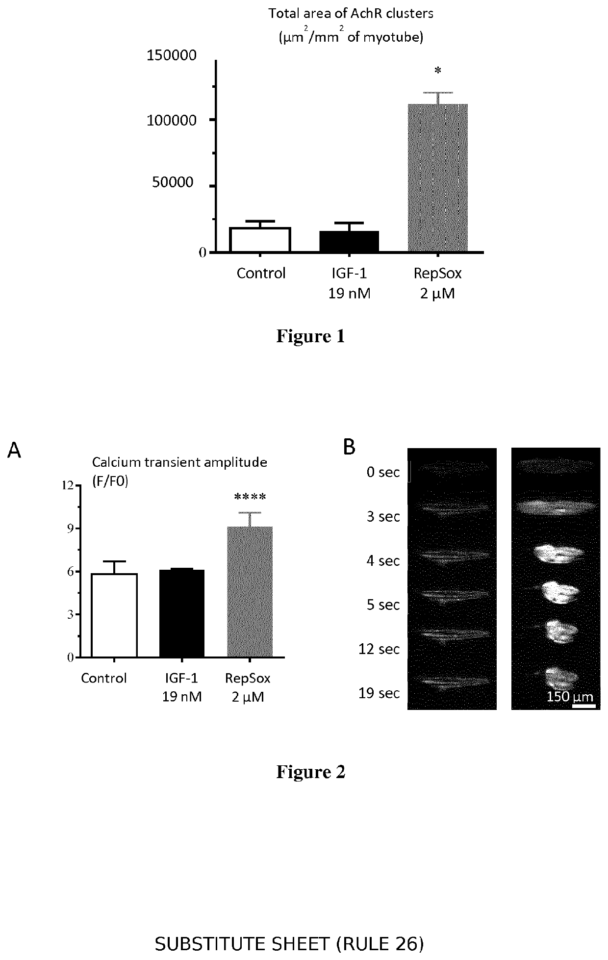
[0216]The MyoScreen™ protocol was performed as described in the Hypertrophy and Atrophy rescue assay described in Example 1 but was stopped at Day 9 instead of Day 6. At the end of the assay, AchR were immunostained using a specific antibody in addition to Troponin T and nuclei. Images were acquired at ×20 with an Operetta High Content Imaging System from Perkin Elmer. Image processing and analyses were performed with a dedicated algorithm developed on the Acapella High Content Imaging Software (Perkin Elmer). Specific readouts were calculated in each well: nuclei count and myotube fusion index, number of AchR, AchR mean area, AchR total area normalized by the myotube total area.
[0217]Results
[0218]Remarkably, when labeled with a specific anti-AChR antibody, MyoScreen myotubes at 6 days post-differentiation display AChR clusters punctuated along the sarcolemma membrane in the middle of the myotube fibre and in distinct regions ...
example 3
[0219]Materials and Methods
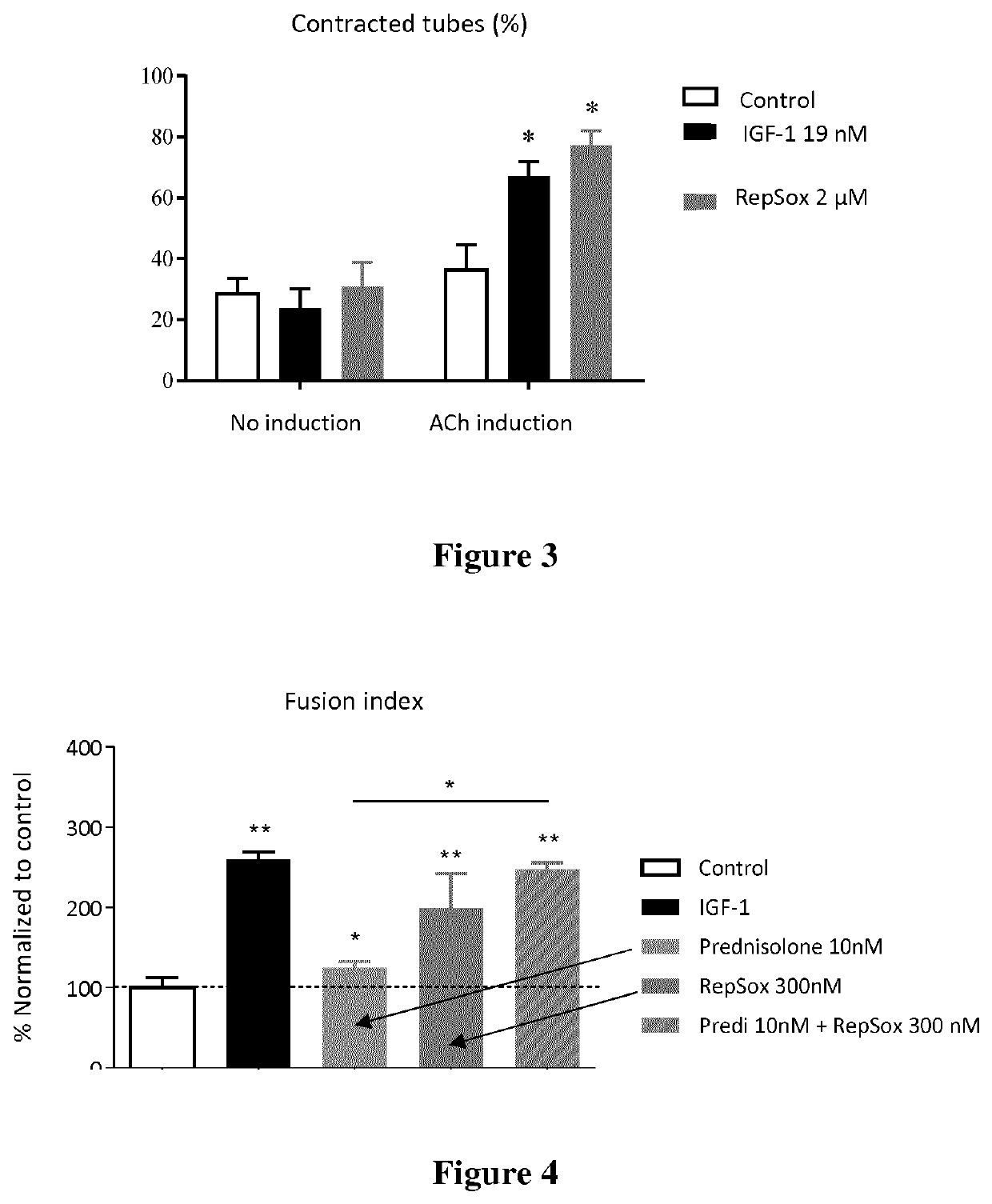
[0220]Cells were washed with calcium buffer containing (in mM): 130 NaCl, 5.4 KCl, 1.8 CaCl2, 0.8 MgCl2, 5.6 D-glucose, 10 Hepes, pH 7.4. Cells were then incubated for 1 h with 2 μM Fluo-4 AM (F14201-Invitrogen) in an incubator (37° C., 5% CO2). After washing to remove unloaded dye, myotube responses to 20 μM ACh (A6625-Sigma-Aldrich), were videoimaged. Stream acquisitions were set at 1 sec intervals and one field of view was acquired per well. Images were processed and analyzed automatically using a dedicated software developed in-house integrating the open-source ImageJ framework. The image analysis tool segments the myotubes in images acquired in the same well. The Fluo-4 total fluorescence intensity was measured inside each myotube and corrected for background. The total intensity was normalized to the myotube area to obtain the mean fluorescence intensity readout. For data presentation, fluorescence intensities (F) were normalized to the mean...
PUM
| Property | Measurement | Unit |
|---|---|---|
| Mass | aaaaa | aaaaa |
| Molar density | aaaaa | aaaaa |
| Molar density | aaaaa | aaaaa |
Abstract
Description
Claims
Application Information
 - R&D
- Intellectual Property
- Life Sciences
- Materials
- Tech Scout
- Unparalleled Data Quality
- Higher Quality Content
- 60% Fewer Hallucinations
Browse by: Latest US Patents, China's latest patents, Technical Efficacy Thesaurus, Application Domain, Technology Topic, Popular Technical Reports.
© 2025 PatSnap. All rights reserved.Legal|Privacy policy|Modern Slavery Act Transparency Statement|Sitemap|About US| Contact US: help@patsnap.com


