

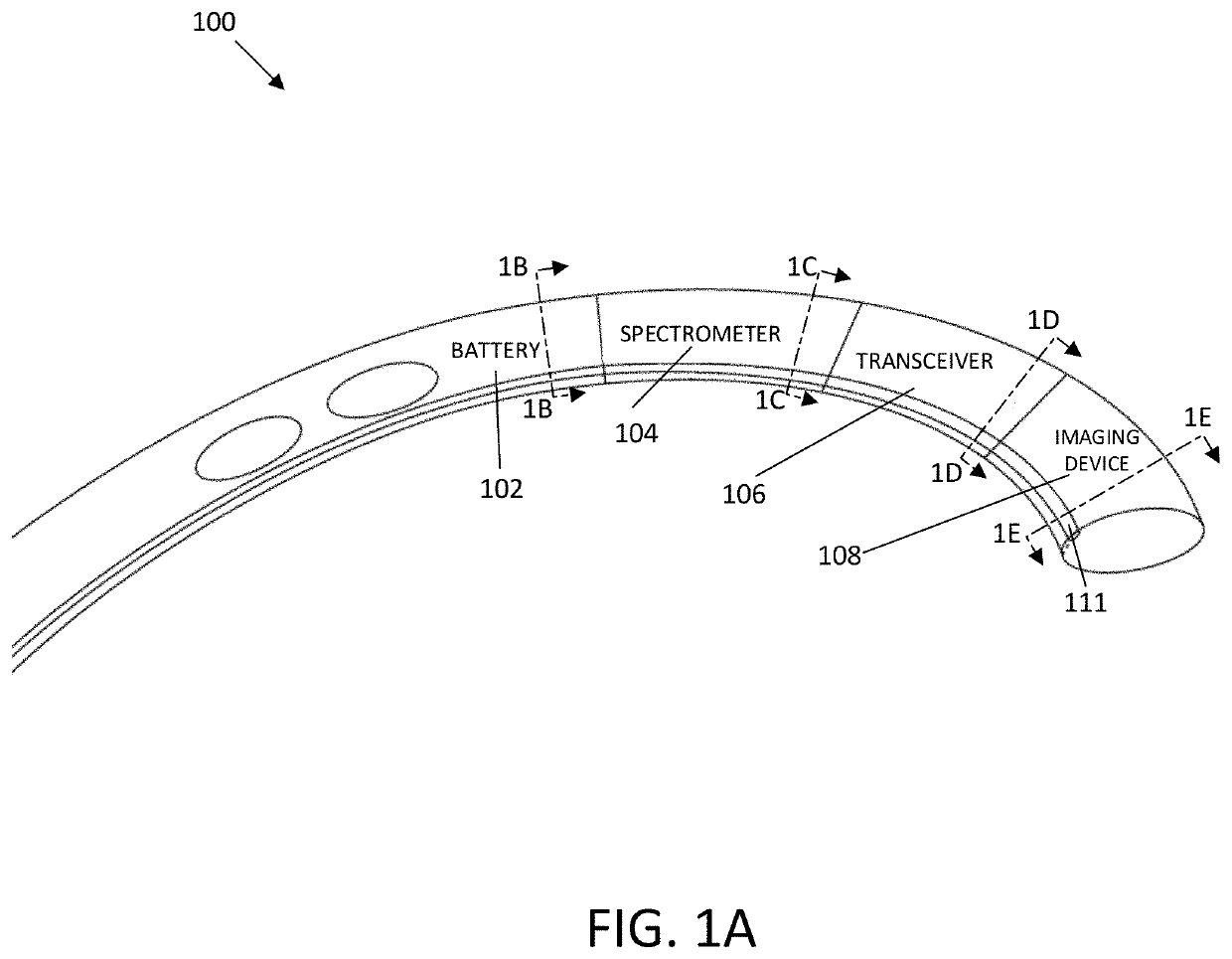
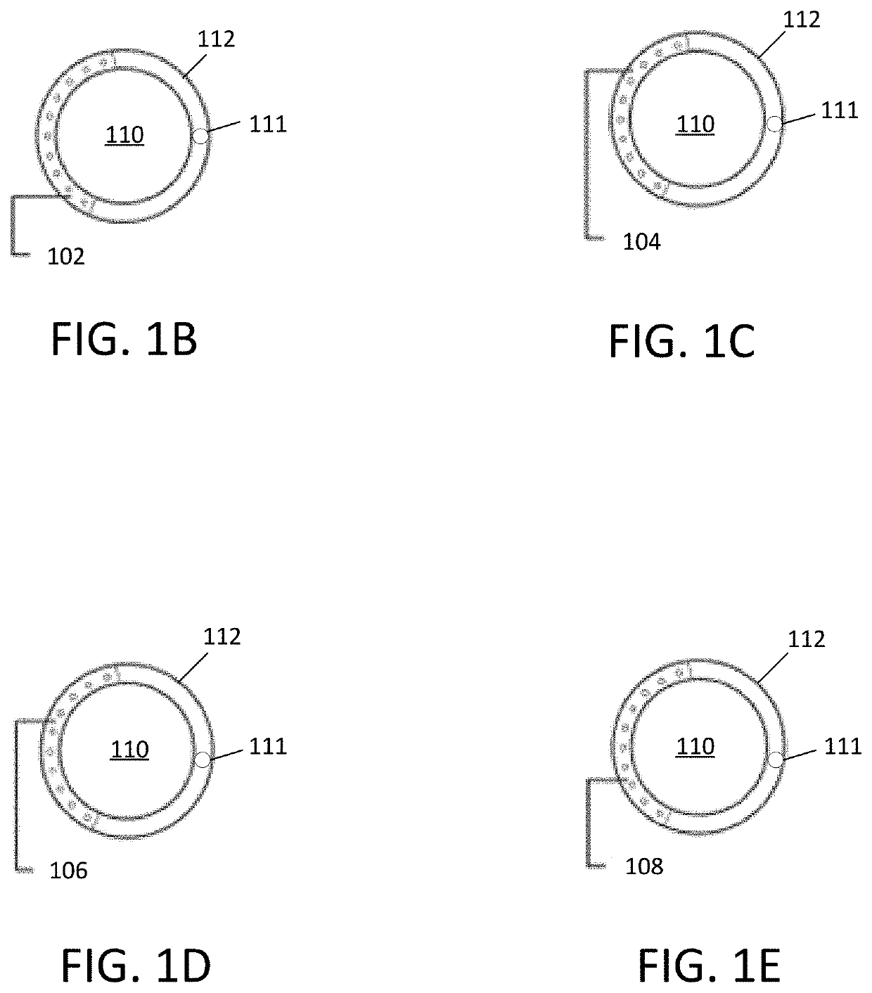
Robotic artificial intelligence nasal/oral/rectal enteric tube
a robotic intelligence and enteric tube technology, applied in the field of robotic artificial intelligence nasal/oral/rectal enteric tubes, can solve the problems of difficult tube placement, affecting the patient's ability to achieve the effect of pulmonary access, and affecting the patient's ability to achieve respiratory tract access,
- Summary
- Abstract
- Description
- Claims
- Application Information
AI Technical Summary
Benefits of technology
Problems solved by technology
Method used
Image

Examples
example
[0153]The following is a non-limiting example in accordance with embodiments of the invention.
[0154]Experimental Data Support
[0155]The process of the autonomous endotracheal tube insertion was validated by dividing it into two individual parts: an object detection functionality that guides the robot and an integrated system that controls the robot individually.
[0156]Image Dataset (for Robot Guidance Experiment) and Phantom Model (for Robot Control Experiment)
[0157]A commercially available training model, Koken Model for Suction and Tube Feeding Simulator: LM-097B (Koken Co Ltd, Bunkyo-ku, Japan), was purchased for experimentation. The images used for training of the model in tracheal detection were obtained utilizing this phantom. These images were obtained by manual control and automatic control of the robot during image / data gathering.
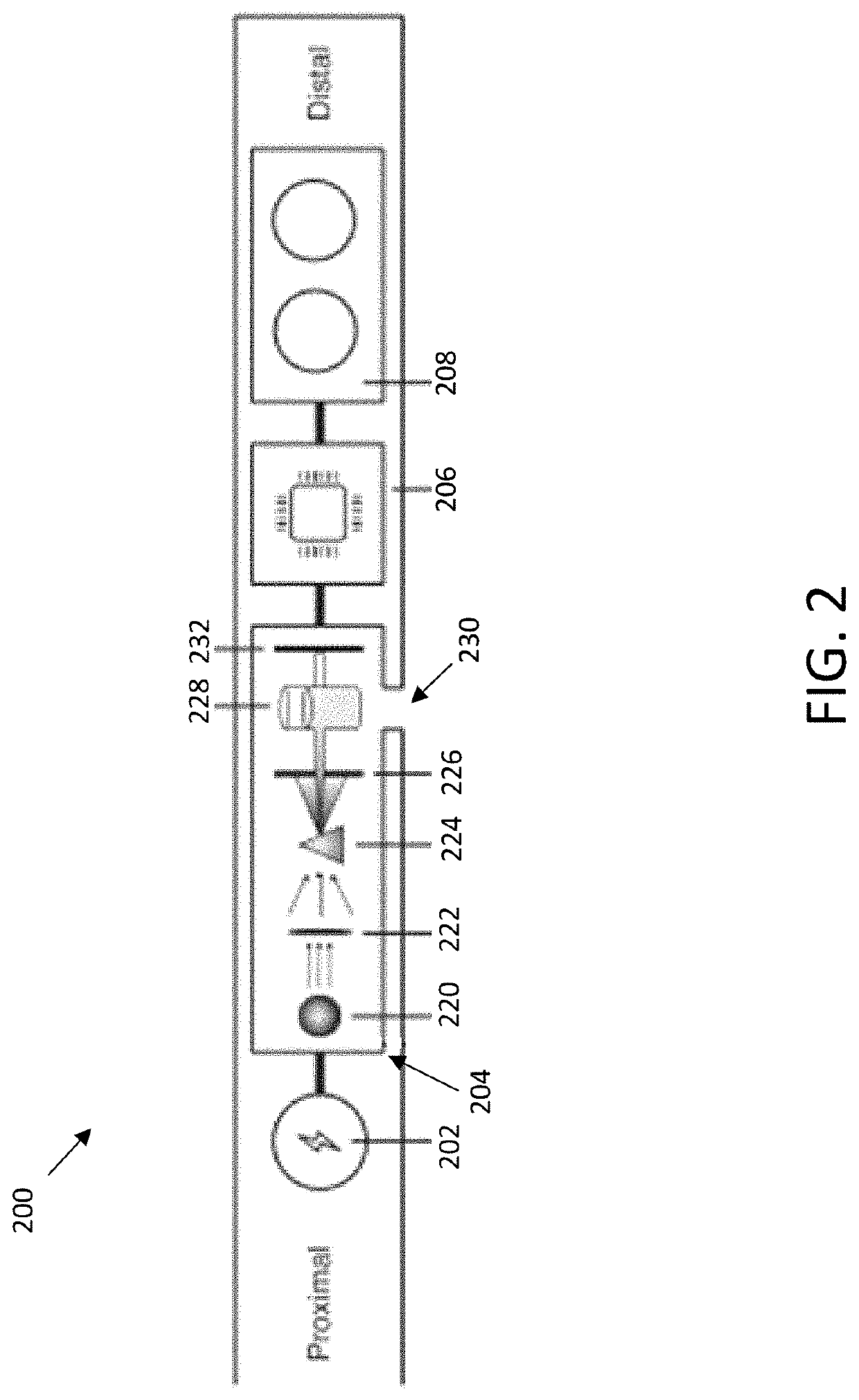
[0158]System Configuration
[0159]A macroscopically one-way closed loop system was built, consisting of 1) robot, 2) robot controlling computer, and 3...
PUM
 Abstract
Description
Claims
Application Information