Ultrasound imaging system and method

a technology of ultrasound imaging and ultrasound, applied in the field of ultrasound imaging system and method, can solve the problems of reversing pulsus paradoxus, the process of obtaining time trace recordings of blood pressure near the heart is limited to patients with invasive radial or intra-cardiac catheters, and the estimating of stroke volume is limited by pulse contour analysis

Active Publication Date: 2021-07-01
KONINKLJIJKE PHILIPS NV
View PDF0 Cites 3 Cited by
  • Summary
  • Abstract
  • Description
  • Claims
  • Application Information

AI Technical Summary

Benefits of technology

[0067]The ECG signal permits temporal tracking of the phases of the heart cycle, so that data collection can be timed to coincide with the beginning and end of each heart cycle for instance. For the first imaging procedure, the ECG input signal permits for instance identification of the end-systolic and end-diastolic phases, so that ultrasound data collection can be timed to coincide with these time points only. For the second imaging procedure, the start and end of each cardiac cycle can be identified, so that data collection can be timed to extend continuously throughout each given cycle.

Problems solved by technology

As a result, the hemodynamic effect is reversed during mechanical ventilation, resulting in reversed pulsus paradoxus.
While this indirect method is functional, the process of obtaining time trace recordings of blood pressure near the heart is limited to patients with invasive radial or intra-cardiac catheters in place.
Furthermore, pulse contour analysis has its own limitations with regard to estimating stroke volume, such as variability of the pressure transducer location relative to the phlebostatic axis.
Together, these parameters provide a relatively accurate estimate of CO, but require a trained user and are difficult to repeat.
In addition, it is subject to considerable variability due to the requirement to maintain the probe positioning unchanged over multiple cardiac cycles and the need to perform an estimation of LVOT diameter.
It is not possible for this method to be performed without a trained sonographer or cardiologist.

Method used


Image

  • Ultrasound imaging system and method
  • Ultrasound imaging system and method
  • Ultrasound imaging system and method

Examples


Embodiment Construction

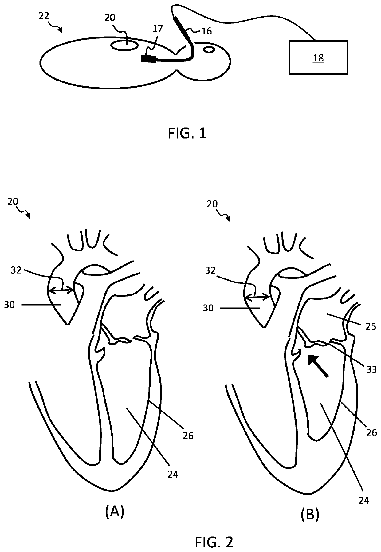
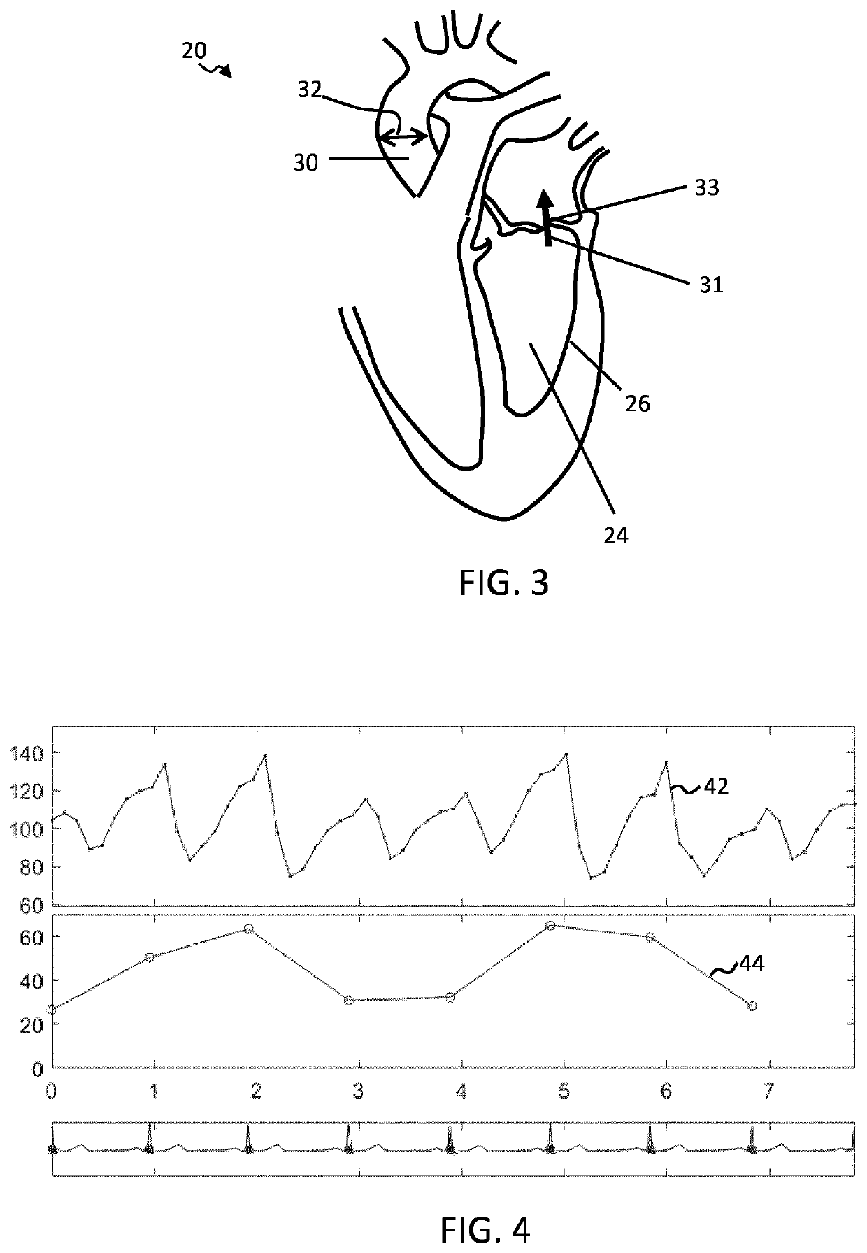
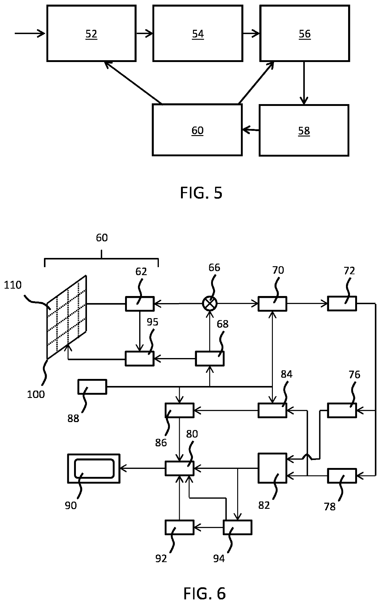
[0099]The invention provides an ultrasound imaging system for determining stroke volume and / or cardiac output. The imaging system includes a transducer unit for acquiring ultrasound data of a heart of a subject, and a controller. Alternatively the imaging system may include an input for receiving the acquired ultrasound data by a transducer unit rather than the unit itself. The controller is adapted to implement a two-step procedure, the first step being an initial assessment step, and the second being an imaging step having two possible modes depending upon the outcome of the assessment. In the initial assessment procedure, it is determined whether regurgitant ventricular flow is present. This is performed using Doppler processing techniques applied to an initial ultrasound data set. If regurgitant flow does not exist, stroke volume is determined using segmentation of 3D ultrasound image data to identify and measure the volume of a ventricle at each of end systole and end-diastole,...


PUM


Abstract

An ultrasound imaging system is for determining stroke volume and/or cardiac output. The imaging system may include a transducer unit for acquiring ultrasound data of a heart of a subject (or an input for receiving the acquired ultrasound data), and a controller. The controller is adapted to implement a two-step procedure, the first step being an initial assessment step, and the second being an imaging step having two possible modes depending upon the outcome of the assessment. In the initial assessment procedure, it is determined whether regurgitant ventricular flow is present. This is performed using Doppler processing techniques applied to an initial ultrasound data set. If regurgitant flow does not exist, stroke volume is determined using segmentation of 3D ultrasound image data to identify and measure the volume of the left or right ventricle at each of end systole and end-diastole, the difference between them giving a measure of stroke volume. If regurgitant flow does exist, stroke volume is determined using Doppler techniques applied to ultrasound data continuously collected throughout a cardiac cycle.

Description

FIELD OF THE INVENTION[0001]This invention relates to an ultrasound imaging system and method, in particular for determining stroke volume.BACKGROUND OF THE INVENTION[0002]Outflow of blood from the left or right ventricle of the heart is a parameter used by clinicians in a range of diagnostic and therapeutic applications. Total outflow of blood during a single cardiac cycle (a single heartbeat) is known as the (left-ventricular) stroke volume and outflow of blood per minute is known as cardiac output. Both are key parameters used in a range of clinical applications.[0003]One important use of stroke volume is in assessing intravenous fluid responsiveness during hemodynamic fluid intervention.[0004]When breathing, the lungs expand and contract. These volume changes lead to changes in intrathoracic pressure, the pressure being lower during inspiration than during expiration. The pressure changes have an impact upon the hemodynamic function of the heart; in particular the effective forc...

Claims


Application Information

Patent Timeline
Patent Type & Authority Applications(United States)
IPC IPC(8): A61B8/06G06T7/33A61B5/318A61B8/00A61B8/14A61B8/12
CPCA61B8/065G06T7/344A61B5/318A61B8/466G06T2207/30104A61B8/12G06T2207/10136G06T2207/20121A61B8/14A61B8/0883A61B8/4405A61B8/4472A61B8/469A61B8/483A61B8/486A61B8/488A61B8/5276A61B8/543A61B8/06
Inventor RAJU, BALASUNDAR IYYAVUBINGLEY, PETERWEBER, FRANK MICHAELSUTTON, JONATHAN THOMASWEKEL, TILMANBOUWMAN, ARTHURKORSTEN, ERIK
Owner KONINKLJIJKE PHILIPS NV
Why Eureka
Try Eureka