
Particles functionalized with imageable radioisotopes and methods of making and use thereof

Pending Publication Date: 2021-08-05
DREHER MATTHEW ROBERT +1
View PDF1 Cites 0 Cited by
  • Summary
  • Abstract
  • Description
  • Claims
  • Application Information

AI Technical Summary

Benefits of technology

The patent text describes the problem of determining the distribution of therapeutic microparticles in the body, which makes it difficult to predict the effects of the treatment. The text proposes the use of imageable radioisotopic particles as surrogates to approximate the distribution of the microparticles and predict the radiation dosing more accurately. This approach can enhance the effectiveness of the treatment and reduce side effects for patients.

Problems solved by technology

However, because therapeutic microparticles are not readily imageable, their distribution in the body is difficult to determine.
The properties of these therapeutic microspheres make them difficult and impractical to predict distributions of therapeutic doses within the body.
In turn, it is difficult to track and accurately assess where therapeutic microspheres have ultimately lodged.
Without the ability to accurately determine where the microparticles reside in the body, the dose of radiation at a target location for therapeutic microspheres is difficult to predict or measure.
Moreover, the dose of harmful radiation to healthy areas of the body is also difficult to determine.

Method used


Image

  • Particles functionalized with imageable radioisotopes and methods of making and use thereof
  • Particles functionalized with imageable radioisotopes and methods of making and use thereof
  • Particles functionalized with imageable radioisotopes and methods of making and use thereof

Examples


example 1

Preparation of Zirconium-89 Coupled Yttrium Aluminum Silicon Oxide (YAS) Microspheres

[0691]A 4 mL glass vial was charged with yttrium aluminum silicon oxide (YAS) glass beads (TheraSphere®, Biocompatibles UK Ltd, provided as non-radioactive spheres which had not been exposed to neutron bombardment. Y2O3 was therefore in the form of naturally occurring 89Y) and suspended in 300 μL of deionized water. Next, a 2 μL aliquot of Zirconium-89 in 1 M oxalic acid (3D imaging, Little Rock, Ark.)) (about 100 microcuries (μCi)) was added to the reactor vial, followed by 2 μL of 2 M sodium carbonate, followed by a Teflon®-coated magnetic stir bar. The reaction mixture was stirred and heated for 2 hours on a 120° C. aluminum heating block, then removed from the block and allowed to cool to room temperature. The microspheres were suspended in 3.0 mL of deionized water and passed through a 0.22 μm syringe filter to collect the microspheres for labeling analysis. The glass vial was rinsed with an ad...

example 2

Preparation of 99mTc Coupled Yttrium Aluminum Silicon Oxide Microspheres

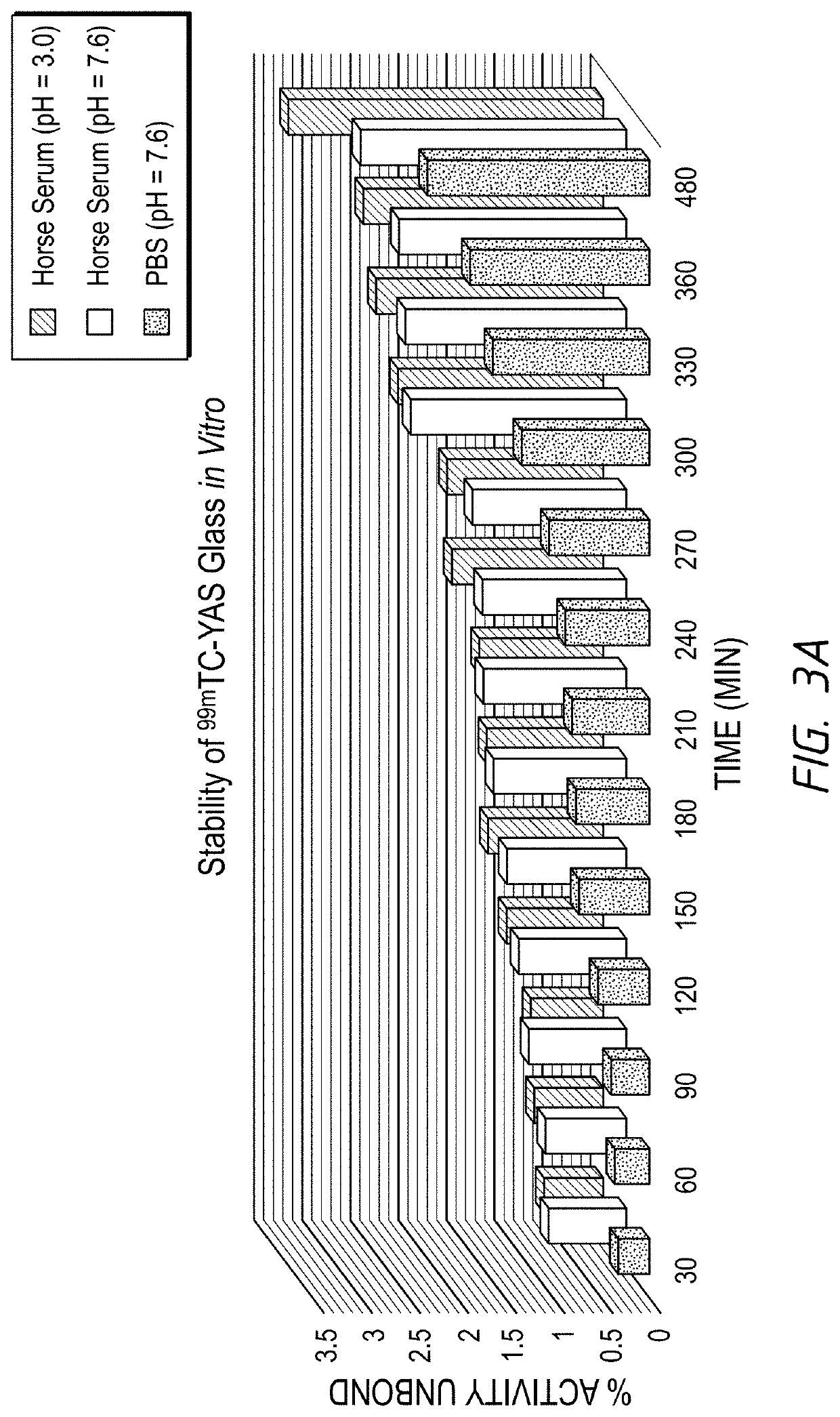
[0692]Tin(II) chloride dihydrate (1 mg) was added to a 1 dram vial and dissolved in 400 μL of deionized water. In a separate 1 dram vial, yttrium aluminum silicon oxide glass beads (89Y, non-radioactive TheraSphere—TS) were added, followed by 100 μL of [99mTc]sodium pertechnetate (>30 mCi / mL). The tin(II) chloride dihydrate solution was added to the TheraSphere vial, briefly mixed (approximately 3 seconds) and the vial was capped and allowed to react at room temperature for 60 minutes. The microspheres were suspended in 3.0 mL of deionized water and passed through a 0.22 μm syringe filter to collect the microspheres for labeling analysis. The glass vial was rinsed with an additional 4 mL of deionized water, which was then passed through the syringe filter. The syringe filter (containing the labeled microspheres), the glass vial, and the deionized water filtrate were analyzed by a gamma well counter and the resul...

example 3

[89Zr] Microsphere Ligand Challenge with DFO Chelate

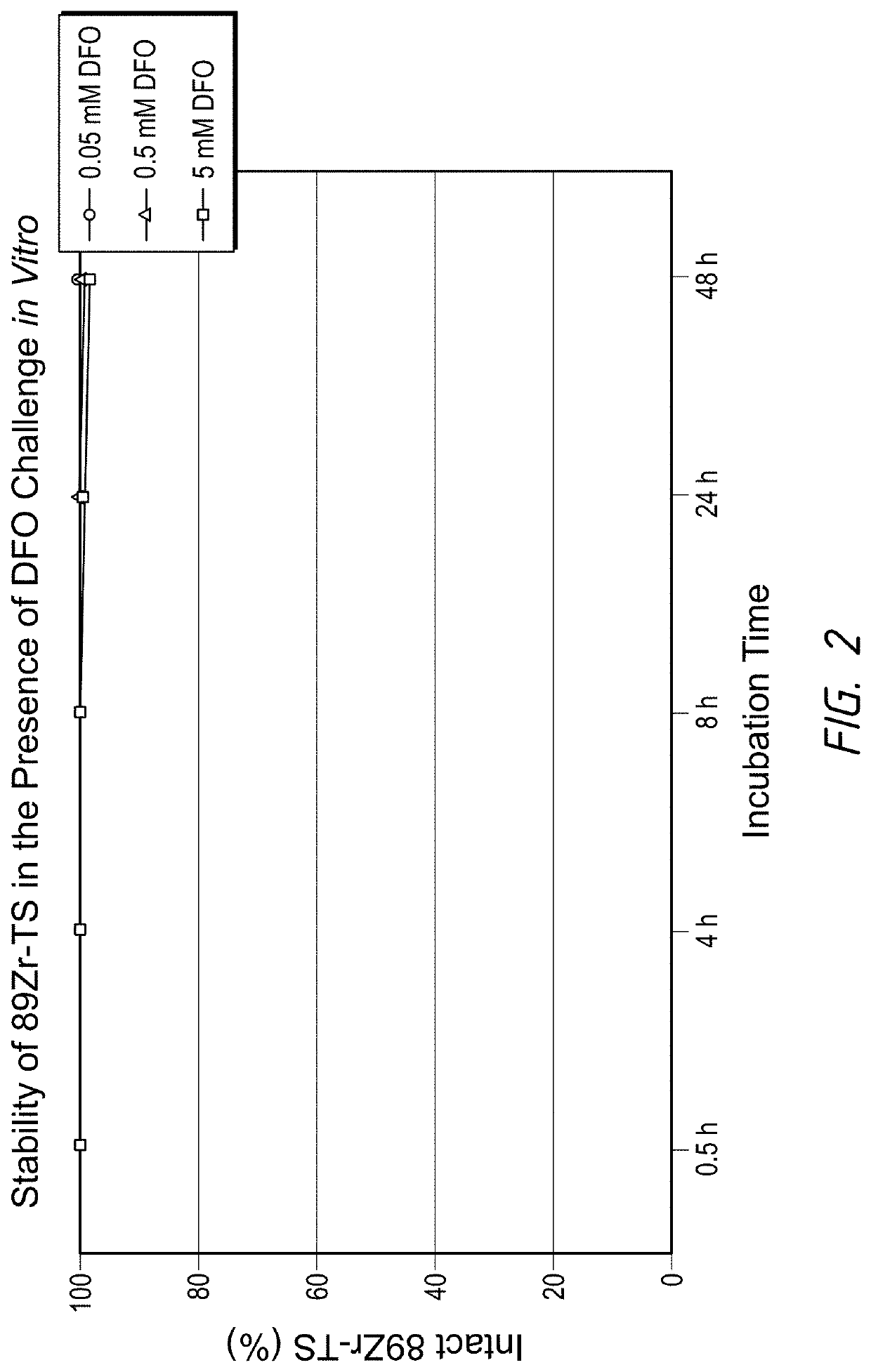
[0693]YAS microspheres (e.g., Therasphere) (suspended in 200 μL sterile saline, pH 7-8) were mixed with increasing concentrations of desferrioxamine (DFO), ranging from 0.05 to 5 mM, and incubated at 37° C. under constant stirring. At each time point, 5 μL of solution was removed, added to a 0.45 μm spin filter, and diluted with 100μL of deionized water. The spin filter was centrifuged at 13,200× G for 60 seconds, 100 μL of deionized water was added back to the spin filter and centrifugation repeated. The spin filter was removed from the microcentrifuge tube and the supernatant was analyzed by gamma spectroscopy, then counted in a gamma counter, to detect formation of possible 89Zr-DFO. Negligible 89Zr detachment was detected for 89Zr-TS within 48 h, demonstrating a strong binding affinity of 89Zr to TS. Results are shown in FIG. 2.


PUM


Abstract

Some embodiments relate to imageable radioisotopic microspheres. In some embodiments, the imageable microspheres are radiolabeled with imageable radioisotopes. In some embodiments, the imageable radioisotope is directly coupled to a surface of a substrate of the microsphere. In some embodiments, the imageable microspheres can be used as surrogate particles to predict the distribution of therapeutic microspheres comprising radiotherapeutic isotopes.

Description

CROSS REFERENCE TO RELATED APPLICATIONS[0001]The present application claims the benefit of priority to U.S. Provisional Patent Application No. 62 / 970,587, filed Feb. 5, 2020, the entirety of which is hereby incorporated by reference herein.BACKGROUNDField[0002]The present disclosure pertains to particles functionalized with imageable radioisotopes, their use as surrogates for therapeutic particles, methods of making such imageable particles, and methods of using the particles for biological imaging and dosimetry.Description of the Related Art[0003]One approach to the treatment of patients with certain cancers is to introduce radioactive isotopes into the patient's circulatory system. A measured amount of radioactive isotopes are injected into the patient such that they accumulate at the site of the cancer sufficiently to treat the cancer.SUMMARY[0004]In one method for the radio-treatment of cancer, radioactive microspheres are delivered to a point in the vasculature of a patient suc...

Claims


Application Information

Patent Timeline
IPC IPC(8): A61K51/12A61N5/10A61B6/03A61K51/02
CPCA61K51/1244A61N5/1001A61N2005/1021A61K51/02A61B6/037A61N2005/1052A61N5/1071A61K51/1251
Inventor STONE, JAMES RADFORDNEUMANN, KIEL DOUGLASDREHER, MATTHEW ROBERT
Owner DREHER MATTHEW ROBERT
Learn More