

Methods of measuring extracellular vesicles and nanoparticles in complex matrices by light scattering
a technology of light scattering and nanoparticles, applied in the field of light scattering detection methods, can solve the problems of inability to accurately and rapidly measure the presence of nanoparticles, the purity, concentration and absolute number of complex matrices, and the current approaches for the detection, isolation and purification of biological nanoparticles such as extracellular vesicles derived from cell culture or other biological samples require laborious and time-consuming methods, and the current ultra-centrifugation protocols
- Summary
- Abstract
- Description
- Claims
- Application Information
AI Technical Summary
Benefits of technology
Problems solved by technology
Method used
Image

Examples
example 1
and Exosomes have Detectable Fluorescent Signal at Excitation and Emission Wavelengths with Similar Values
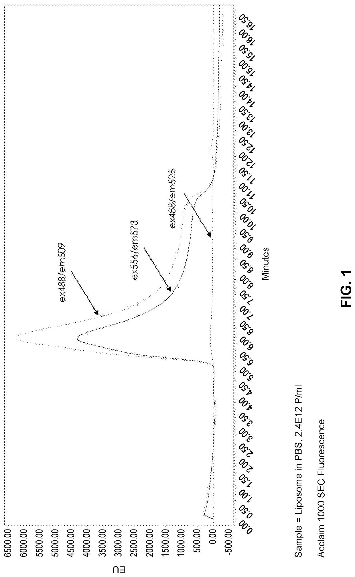
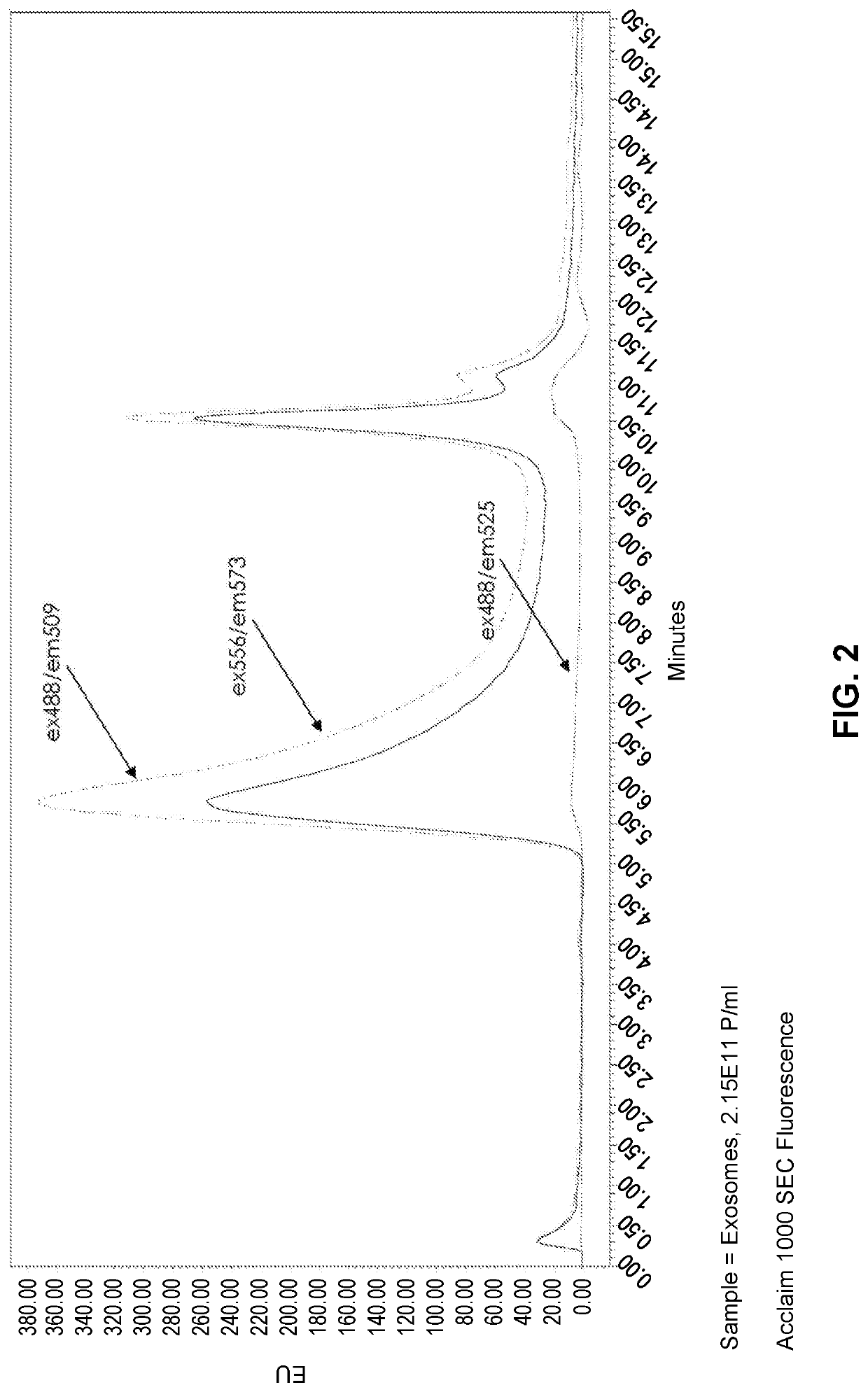
[0152]Purified liposomes lacking a fluorescent marker (FormuMax Scientific Inc., Cat # F10104) were analyzed by size exclusion chromatography using an ACCLAIM™ SEC-1000 column. The SEC column was operated under isocratic flow at 0.5 mL / min using an Agilent 1100 HPLC system with PDA and fluorescence detectors. Samples were excited at UV wavelengths 488 nm and 556 nm and detected at fluorescence excitation and emission wavelength pairs 488 / 509, 556 / 573, and 488 / 525. The liposomes were detected at 488 / 509 and 556 / 573 but not at 488 / 525, despite very similar excitation and emission profiles for 488 / 509 and 488 / 525 runs (FIG. 1). Purified exosomes were analyzed by the same method and a similar trend was found, where the samples were detectable at the excitation / emission wavelength pairs that were separated by ˜20 nm, but not in the 488 / 525 pair, which is separated by 37 nm (FIG. 2). ...
example 2
Scanning of Purified Exosome Samples Across a Broad Frequency Range
[0153]To determine whether the light scattering phenomenon described above persisted across a range of spectral values, purified exosomes were analyzed by SEC with excitation ranging from 400 nm to 690 nm, and emission detected at 10 nm above the excitation wavelength. For this experiment an ACCLAIM™-1000 SEC column (ThermoFisher Scientific) was operated under a 0.5 mL / min isocratic flow in a mobile phase of 100 mM Na-phosphate, 200 mM NaCl, pH 7.2 with the Waters 2695 ALLIANCE® HPLC system. A W2998 UV / Vis detector was used to monitor 210 nm, 254 nm, 280 nm wavelengths; and the W2475 fluorescence detector was used for 3D spectral scanning from the 400 nm to 700 nm range.
[0154]The fluorescence excitation wavelength was fixed in 10 nm increments through multiple injections, and the emission was read from 10 nm greater than excitation, up to 700 nm. For example, the first injection had a fixed excitation 400 nm, and the...
example 3
Properties of Additional Nano-Sized Biological Substrates
[0161]To understand whether the light scattering observations shown in the previous examples were specific to spherical particles, a similar experiment was carried out by analyzing purified E. coli ribosomes (˜25 nm diameter near-spherical particles; New England Biolabs; #P07635) at UV280, ex280 / em350, ex460 / em470, ex556 / em573, and ex488 / em525. As shown in FIG. 15, ex280 / em350, which detects protein tryptophan fluorescence, and the ex / em pairs separated by 10-20 nm reliably detected the same peak of purified ribosomes. Monitoring at ex488 / em525 failed to detect any signal. These results are consistent with the observations shown above, wherein excitation and emission pairs separated by less than 20 nm can detect nano-sized particles by SEC, whereas ex / em pairs of a greater separation, such as ex488 / em525 fail to detect the particles. Notably, ex460 / em470 yielded a much greater signal than even ex280 / em350. Normalizing maximum ...
PUM
| Property | Measurement | Unit |
|---|---|---|
| excitation wavelength | aaaaa | aaaaa |
| excitation wavelength | aaaaa | aaaaa |
| emission wavelength | aaaaa | aaaaa |
Abstract
Description
Claims
Application Information