Eureka AIR delivers breakthrough ideas for toughest innovation challenges, trusted by R&D personnel around the world.

Real-time methods to enable precision-guided cpr to improve neurological outcome and predict brain damage after ischemic injury and reperfusion

Pending Publication Date: 2022-06-23
RGT UNIV OF CALIFORNIA
View PDF0 Cites 0 Cited by
  • Summary
  • Abstract
  • Description
  • Claims
  • Application Information

AI Technical Summary

Benefits of technology

The present invention provides a way to measure brain function quickly and continuously after resuscitation from a cardiac arrest. This is important because brain damage can occur quickly after resuscitation, and the invention can help doctors assess the severity of the damage and prescribe appropriate treatment. The invention uses optical signals from the body, which are non-invasive and can be measured at different time windows after resuscitation. This technology can be used in a variety of clinical scenarios, including cardiac arrest, stroke, and drug overdose. By monitoring cerebral blood flow and metabolism, the invention can provide information about brain damage and help doctors make informed treatment decisions.

Problems solved by technology

Survivors of CA typically sustain significant brain damage due to cerebral ischemia, reperfusion injury, and compromised cerebral autoregulation.
After hypoxic-ischemic injury (e.g., CA), reperfusion (e.g., CPR) can deliver oxygenated blood to the brain to support energy production, but this massive influx of oxygen can also cause neuronal injury if it is not metabolized efficiently.
As a result, relying on only perfusion data is insufficient to accurately assess the hemodynamic response of CA patients, and a complementary measure of cerebral metabolism is required for correct assessment of injury severity and prognostication of recovery.
Early intervention is critical for improving outcome for patients suffering cerebral ischemia, but there are no reliable quantitative methods for rapidly diagnosing severity of injury, prognosing outcome, and informing treatment in real time.
However, these measurements occur distant from the brain and are often not informative of cerebral hemodynamic processes.
Thus, the prior art attempts to quantify cerebral perfusion and metabolism struggle to do so sufficiently and quickly enough to enable the clinician to take early action.
Further still, methods for direct measurement of oxygen consumption in the brain are typically invasive and do not provide any information about perfusion.
Techniques for non-invasive CBF measurement do not have the temporal resolution required to monitor rapid changes in cerebral perfusion and metabolism.
As such, it remains difficult to obtain accurate real-time feedback on CBF and metabolism concurrently to guide emergent clinical intervention to optimize neurological outcome for CA patients.

Method used


Image

  • Real-time methods to enable precision-guided cpr to improve neurological outcome and predict brain damage after ischemic injury and reperfusion
  • Real-time methods to enable precision-guided cpr to improve neurological outcome and predict brain damage after ischemic injury and reperfusion
  • Real-time methods to enable precision-guided cpr to improve neurological outcome and predict brain damage after ischemic injury and reperfusion

Examples


example 1

[0102]The following is a non-limiting example of the present invention. It is to be understood that said example is not intended to limit the present invention in any way. Equivalents or substitutes are within the scope of the present invention.

Methods

Animal Preparation

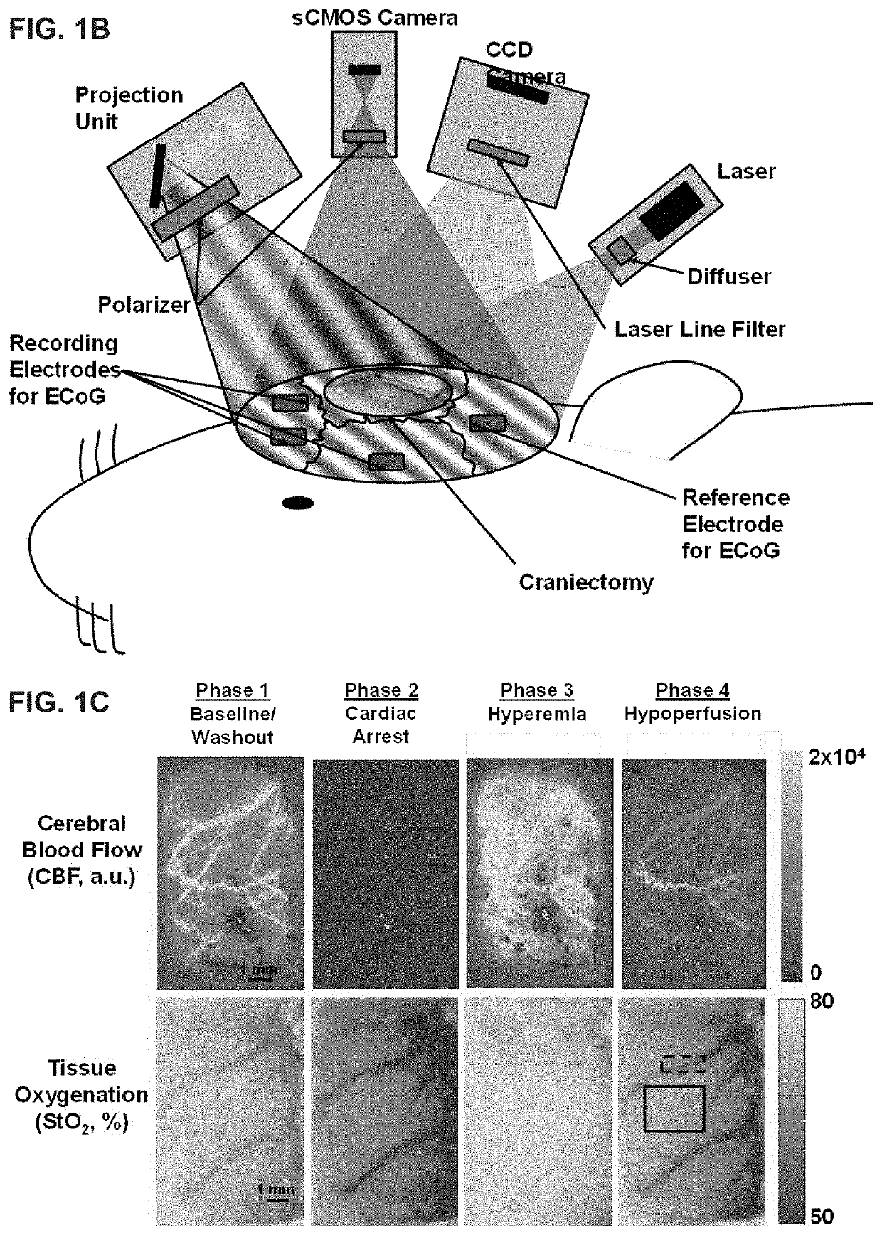
[0103]All procedures described in this protocol were approved by the Institutional Animal Care and Use Committee (IACUC) at the University of California, Irvine (protocol number 2013-3098-01). Ten male Wistar rats (weight ˜300-400 g) were used in this study. In the animal preparation procedures, rats were fasted with three pellets the night before the experiment as standard procedure for the CA experiments. On the day of the experiment, rats were anesthetized with isoflurane and endotracheally intubated to enable controlled breathing with a ventilator. Then, epidural screw electrodes were implanted for ECoG, and a partial craniectomy (4 mm right-to-left×6 mm anterior-to-posterior) was performed to expose a portion of ...

example 2

[0131]The following is a non-limiting example of the present invention. It is to be understood that said example is not intended to limit the present invention in any way. Equivalents or substitutes are within the scope of the present invention.

Introduction

[0132]There is a significant clinical need for quantitative methods to measure cerebral metabolism in vivo to assess damage and recovery in brain-injured patients. Current techniques for quantifying brain metabolism are typically expensive, bulky, and unable to provide high temporal resolution. Diffuse optical spectroscopy and imaging have the potential to rapidly and portably monitor cerebral blood flow (CBF) and brain oxygenation (StO2) simultaneously. Combining these measurements can quantify the cerebral metabolic rate of oxygen (CMRO2) to measure brain metabolism on an absolute physiological scale (units of μM O2 consumed / min) without the need for physiological perturbations (e.g., gas challenges). The present example introdu...


PUM


Abstract

A multimodal optical imaging platform is used to obtain cerebral perfusion-metabolism mismatch metrics for rapid assessment of acute brain injury, ongoing (real-time) feedback to optimize cardiopulmonary resuscitation to improve neurological outcome, and rapid prognosis of recovery. Light of several wavelengths and types is delivered to the tissue, which is then absorbed and scattered by tissue components such as blood and cellular components. Some of this light scatters back to the surface, where it is captured by a detector. The resulting data are processed to obtain blood flow and oxygenation parameters, as well as tissue scattering. These parameters are then combined to calculate metabolism and flow-metabolism coupling / decoupling metrics, which are used to determine ischemic damage, ongoing need for optimal blood flow and oxygenation, and to predict cerebral recovery in patients with acute brain injury during and immediately after cardiac arrest, stroke, traumatic brain injury, etc.

Description

CROSS-REFERENCES TO RELATED APPLICATIONS[0001]This application is a continuation-in-part and claims benefit of U.S. patent application Ser. No. 17 / 534,986 filed Nov. 24, 2021, the specification of which is incorporated herein in its entirety by reference.[0002]U.S. patent application Ser. No. 17 / 534,986 is a continuation-in-part and claims benefit of PCT Application No. PCT / US2020 / 035440 filed May 29, 2020, which claims benefit of U.S. Provisional Application No. 62 / 854,215, filed May 29, 2019, the specification(s) of which is / are incorporated herein in their entirety by reference.[0003]U.S. patent application Ser. No. 17 / 534,986 is also a continuation-in-part and claims benefit of U.S. patent application Ser. No. 17 / 377,123 filed Jul. 15, 2021, which is continuation-in-part and claims benefit of U.S. patent application Ser. No. 16 / 985,113 filed Aug. 4, 2020, now abandoned, which is a continuation-in-part and claims benefit of U.S. patent application Ser. No. 16 / 837,478 filed Apr. 1...

Claims


Application Information

Patent Timeline
IPC IPC(8): A61H31/00A61B5/1455A61B5/0205A61B5/026
CPCA61H31/005A61B5/14553A61B5/0205A61N1/39044A61B5/14552A61H2230/207A61B5/0261A61B5/02028A61B5/14546A61B5/369A61B5/4836A61B5/4866A61B5/4875A61B5/6814A61B5/6841A61H2230/25A61B5/0075A61B5/0086A61B5/14542
Inventor WILSON, ROBERT H.CROUZET, CHRISTIANAKBARI, YAMACHOI, BERNARDTROMBERG, BRUCE J.
Owner RGT UNIV OF CALIFORNIA
Why Eureka
Eureka Blog
Learn More