

Tunable leukocyte-based biomimetic nanoparticles and methods of use
a biomimetic nanoparticle and leukocyte technology, applied in the field of medicine, can solve problems such as thwarting the ability of leukocyte-based biomimetic nanoparticles to deliver their payload to the target tissu
- Summary
- Abstract
- Description
- Claims
- Application Information
AI Technical Summary
Benefits of technology
Problems solved by technology
Method used
Image

Examples
example
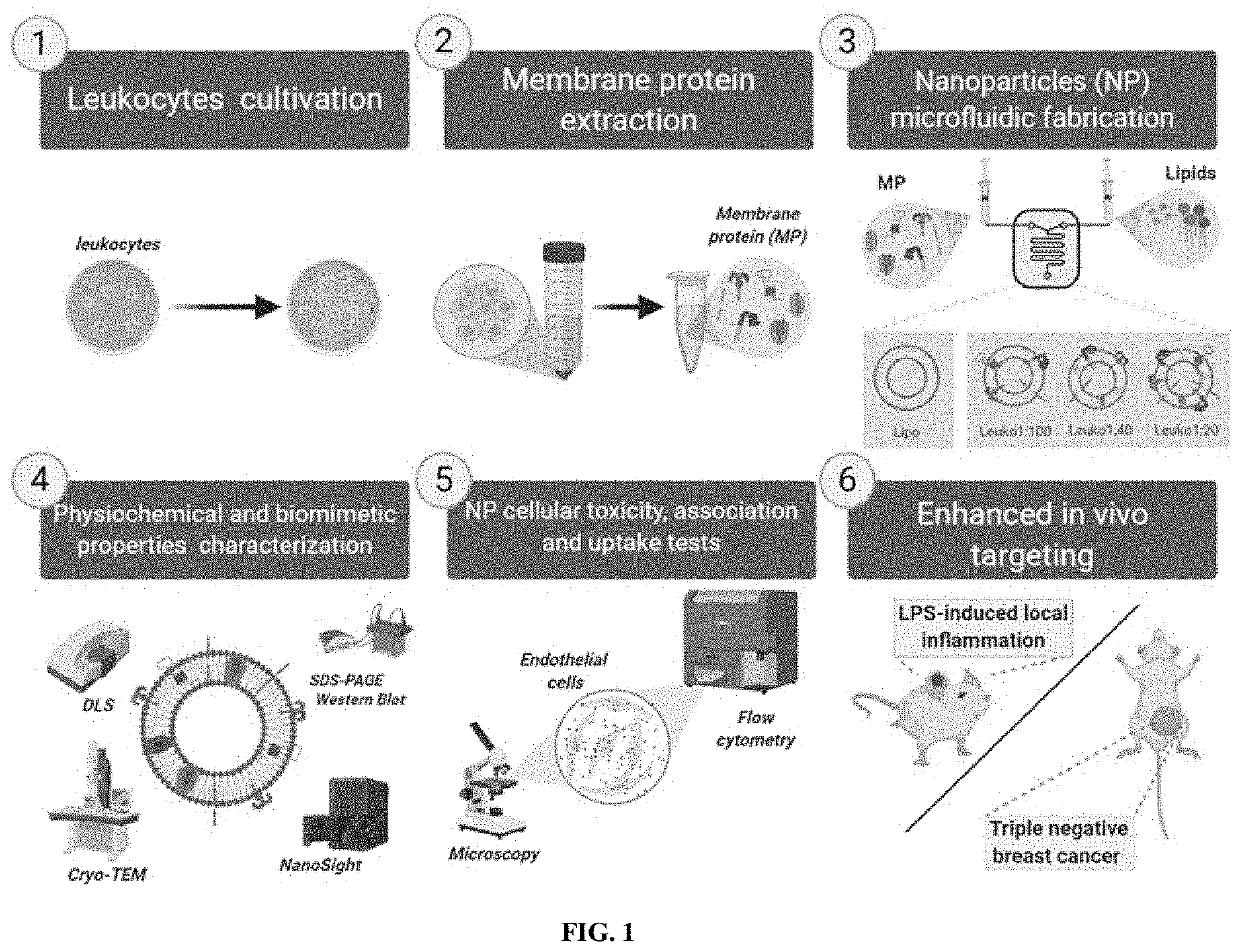
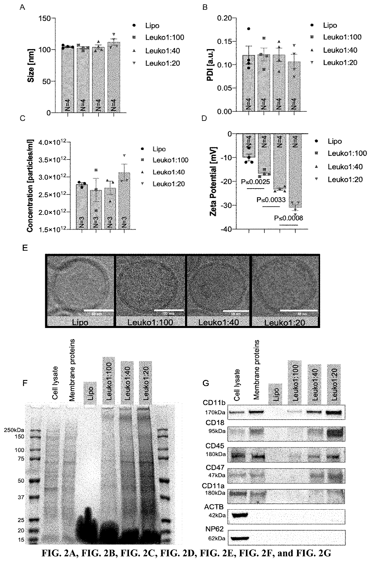
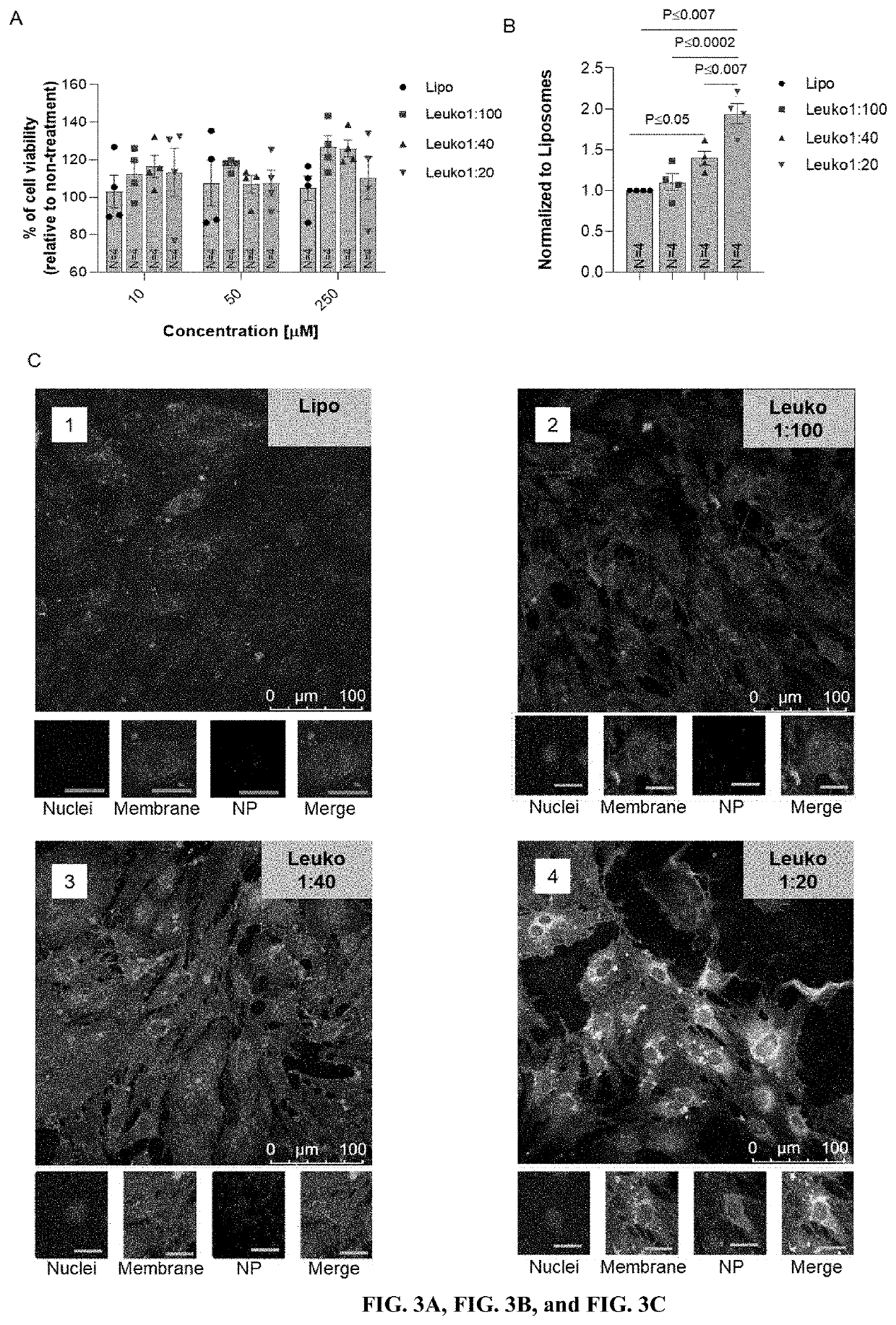
[0072]The following example is included to demonstrate preferred embodiments of the invention. It should be appreciated by those of skill in the art that the techniques disclosed in the example that follows represent techniques discovered by the inventors to function well in the practice of the invention, and thus can be considered to constitute preferred modes for its practice. However, those of skill in the art should, in light of the present disclosure, appreciate that many changes can be made in the specific embodiments which are disclosed and still obtain a like or similar result without departing from the spirit and scope of the invention.
Enhancing Inflammation Targeting Using Tunable Leukocyte-Based Biomimetic Nanoparticle Compositions
Materials and Methods
Reagents
[0073]Membrane protein extraction kit, chloroform, methanol, Tween 20 and 2-Mercaptoethanol (Sigma Aldrich, St. Louis, Mo., USA). Dipalmitoylphosphatidylcholine (DPPC), 1,2-dioleoyl-sn-glycerol-3-phosphocholine (DOPC...
PUM
| Property | Measurement | Unit |
|---|---|---|
| temperature | aaaaa | aaaaa |
| diameter | aaaaa | aaaaa |
| diameter | aaaaa | aaaaa |
Abstract
Description
Claims
Application Information