Method for introducing pharmaceutical drugs and nucleic acids into skeletal muscle

Inactive Publication Date: 2000-08-29
INOVIO
View PDF13 Cites 278 Cited by
  • Summary
  • Abstract
  • Description
  • Claims
  • Application Information

AI Technical Summary

Benefits of technology

Conventional electroporation, however, employs high field strengths from 0.4 to several kV / cm. In contrast to conventional electroporation, the field strength used in the present invention ranges from about 25 V / cm to 250 V / cm. These lower field strengths are thought to cause less muscle damage without sacrificing, and indeed increasing, transfection efficiencies. Furthermore, using the method of the present invention, transfection efficiencies can be tightly regulated by altering such parameters as frequency, pulse duration and pulse number.

Problems solved by technology

Despite these new discoveries, a major obstacle facing the medical profession is how to safely deliver effective quantities of these agents to patients to treat disease or for genetic immunization.
First, a large percent of orally or intravenously delivered drugs are degraded by the body before arriving at the target organ or cells.
Second, oral and intravenous drug and gene delivery is non-specific.
Currently, however, there is no non-viral method for effectively delivering pharmaceutical drugs and DNA into skeletal muscle in vivo.
The clinical applicability of direct muscle injection, however, is limited mainly because of low transfection efficiency, typically less than 1% transfection efficiency.
While injection in regenerating muscles induced by Bivucain show higher efficiency, the method has limited applicability in humans because of the severe damage caused to the muscle.

Method used


Image

  • Method for introducing pharmaceutical drugs and nucleic acids into skeletal muscle
  • Method for introducing pharmaceutical drugs and nucleic acids into skeletal muscle
  • Method for introducing pharmaceutical drugs and nucleic acids into skeletal muscle

Examples


example 1

Stimulated Versus Unsimulated Muscles

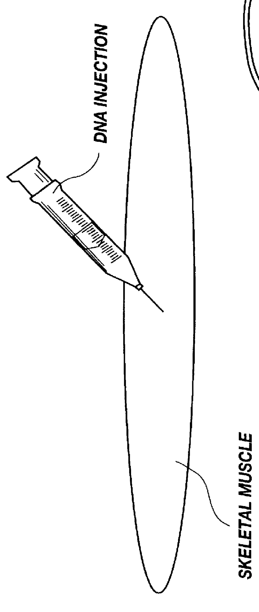
Transfection efficiencies were determined by injecting skeletal muscles with the pSV40-luc reporter construct into the soleus muscle. Three days after injection, the muscles were removed and luciferase activity was measured using the Promega Luciferase Assay System (Madison, Wis.) according to manufacturer's protocols. Unstimulated EDL muscles from the same rats were used as control. The data are shown below in Table 1.

example 2

Transfection Efficiency Versus Frequency

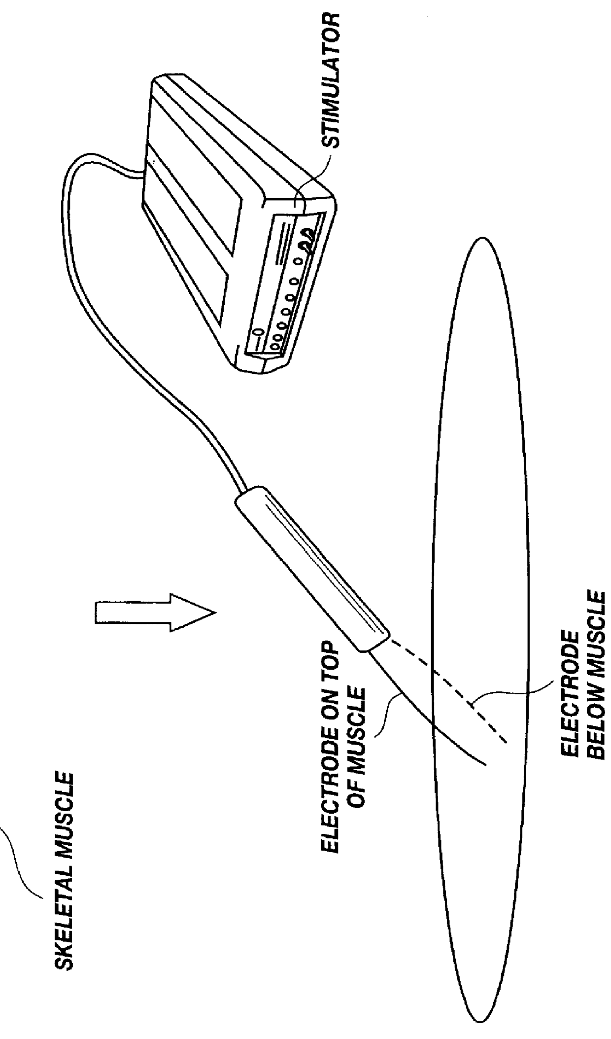
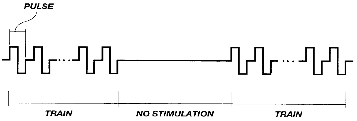
Rats were injected with 50 .mu.l of 1 mg / .mu.l of a plasmid carrying lac Z gene. Immediately following injection, electrodes were placed between 2-3 mm apart and the muscle was stimulated with the following stimulation parameters: voltage=30 volts; pulse duration=0.2 ms (total 0.4 ms, bipolar); trains=30, 1 second on 1 second off for 1 minute. Transfected fibers were counted from a 1 mm slice from middle of muscle. The number of transfected fibers is shown below in Table 2 and illustrated in FIG. 7. These data also illustrate that the method of the present invention transfects more than just surface muscle fibers; muscle fibers several cell layers deep are also transfected.

example 3

Transfection Efficiency Versus Pulses

Soleus muscles of Wistar rats (200-270 grams) were injected with 50 .mu.g of RSV luciferase DNA plasmid in 50 .mu.l 0.9% NaCl. Shortly after injection, the muscles were electrically stimulated using the following parameters: 1000 Hz, between 0-1000 bipolar pulses of 200 .mu.l duration in each train were applied to the muscle 30 times over a period of 1 minute. Muscles were removed 3 days after transfection and frozen in liquid nitrogen. Cryostat sections were taken from the of the muscles and stained with Hematoxolin, Eosin and Safran (see Example 9). The remaining pieces were homogenized as described in Example 4 below. As illustrated in FIGS. 10-12, transfection efficiency increased with the number of pulses delivered to the muscle.


PUM

PropertyMeasurementUnit
Timeaaaaaaaaaa
Timeaaaaaaaaaa
Dielectric strengthaaaaaaaaaa

Abstract

A method is disclosed for delivering molecules such as pharmaceutical drugs and nucleic acids into skeletal muscle in vivo. The pharmaceutical drug or nucleic acid is first injected into the muscle at one or multiple sites. Immediately, or shortly after, injection, electrodes are placed flanking the injection site and a specific amount of electrical current is passed through the muscle. The electrical current makes the muscle permeable, thus allowing the pharmaceutical drug or nucleic acid to enter the cell. The efficiency of transfer permits robust immune responses using DNA vaccines and produces sufficient secreted proteins for systemic biological activity to be observed.

Description

2. FIELD OF THE INVENTIONThe present invention is related to a method for making skeletal muscle semipermeable to pharmaceutical drugs and nucleic acids. More specifically, skeletal muscle is made semipermeable by electrically stimulating the muscle at low field strengths following pharmaceutical drugs and nucleic acids injection.3. TECHNICAL BACKGROUNDScientists are continually discovering genes which are responsible for many human diseases, such as genes responsible for some forms of breast cancer, colon cancer, muscular dystrophy and cystic fibrosis. In addition, scientists are continually discovering genes that code for bacterial and viral antigens (e.g., viral capsid proteins). Despite these new discoveries, a major obstacle facing the medical profession is how to safely deliver effective quantities of these agents to patients to treat disease or for genetic immunization.Currently, most pharmaceutical agents are taken orally or intravenously. Oral and intravenous drug and gene ...

Claims


Application Information

Patent Timeline
IPC IPC(8): A61K41/00A61K39/04A61N1/32A61N1/36A61N1/30
CPCA61K39/04A61K41/00A61N1/325A61N1/205A61K2039/54A61K2039/545A61N1/36017A61N5/00
Inventor MATHIESEN, IACOBLOMO, TERJE
Owner INOVIO
Why Eureka
Try Eureka