
Particle-mediated bombardment of DNA sequences into tissue to induce an immune response

Inactive Publication Date: 2001-02-27
DUKE UNIV +2
View PDF12 Cites 175 Cited by
  • Summary
  • Abstract
  • Description
  • Claims
  • Application Information

AI Technical Summary

Benefits of technology

Also disclosed herein is a method of administering a protein or peptide to a vertebrate subject. This method is based in part on our finding that vertebrate tissue transformed by particle bombardment is surprisingly free of callus formation, inflammation, and other defensive responses. Thus, proteins and peptides released from the transformed cells (by virtue of their being transformed) can circulate throughout the animal subject in which the cells reside, and cells which circulate in the animal subject (e.g., lymphocytes) have access to the transformed cells. In this method, target vertebrate tissue (preferably dermis or hypodermis tissue) is selected and microprojectiles provided. The microprojectiles carry polynucleic acid sequences, the sequences comprising, in the 5' to 3' direction, a

Problems solved by technology

The utility of transforming nematodes is, however, comparatively limited.

Method used


Image

  • Particle-mediated bombardment of DNA sequences into tissue to induce an immune response
  • Particle-mediated bombardment of DNA sequences into tissue to induce an immune response
  • Particle-mediated bombardment of DNA sequences into tissue to induce an immune response

Examples


example 1

Particle-Mediated Transformation of Terminally Differentiated Skeletal Myotubes

This example demonstrates that primary cultures of fully differentiated, non-dividing skeletal myotubes can be transformed in vitro using a DNA-particle accelerator. The introduced genes are not rapidly degraded, but remain transcriptionally active over the life of the culture (twelve days).

Myoblast cultures (4.times.10.sup.5 cells) were established from breast muscles of eleven-day chick embryos in gelatin-coated 60 mm plastic dishes in Dulbecco's Minimum Essential Medium (DMEM) supplemented with 10% horse serum and 5% embryo extract. After five days without fresh media, cultures consisted almost entirely of multinucleated myotubes, some of which showed cross-striations. Some cultures were also treated with cytosine arabinoside (ara-C: 1.5 to 3.0 .mu.g / ml) to inhibit growth of residual undifferentiated myoblasts or non-myogenic cells. See G. Paulath et al., Nature 337, 570 (1989). At this stage (five day...

example 2

Fate Over Time of DNA Introduced in Terminally Differentiated Skeletal Myotubules by Particle Bombardment

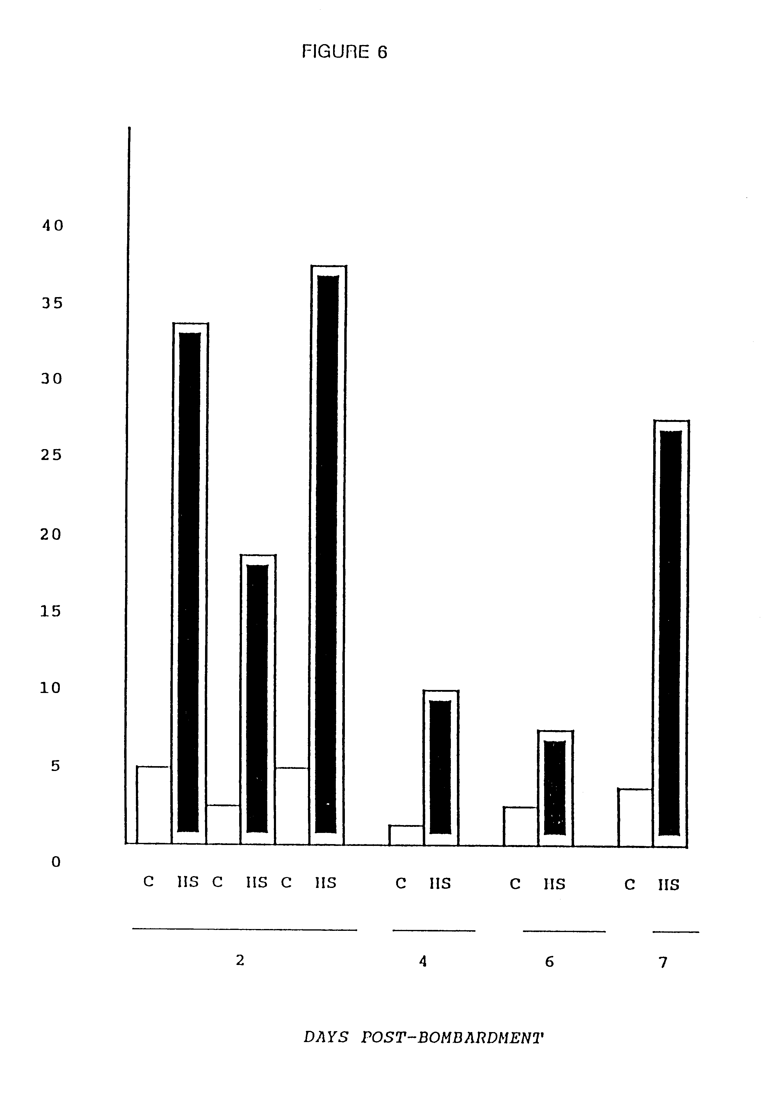
This Example addressed the fate of the introduced DNA over time by measuring the response of an inducible promoter at varying intervals after bombardment. Myotubes were transformed as described in Example 1 above, but with the firefly luciferase gene under the control of the human HSP70 promoter. See B. Wu et al., Proc. Nat. Acad. Sci. USA 83, 629 (1986). On days 2-7 following bombardment, luciferase activity was measured in sister cultures that were either maintained at 37.degree. C. (Control=C) or placed at 45.degree. C. for 90 minutes followed by recovery at 37.degree. C. for three hours (Heat Shock=HS).

The cultures maintained for 6-7 days following bombardment were re-fed at two day intervals with conditioned media (depleted of mitogenic growth factors) from untransfected myotube cultures. FIG. 6 demonstrates that inducible expression of the introduced plasmid was maintained ...

example 3

Site of Transgene Expression in Cultures of Terminally Differentiated Myotubules

This Example was conducted to determine whether trans-gene expression was occurring within the fully differentiated myotubes, as distinguished from mononuclear cells that remain within these primary cultures. Cultures of differentiated myotubes were transfected, by microprojectile bombardment as described in Example 1, with a plasmid construct containing the Drosophila Alcohol Dehydrogenase (ADH) gene under the control of the Rous Sarcoma Virus long terminal repeat (pRSV-ADH). On the following day, cells were fixed and stained according to the method described by C. Ordahl et al., Molecular Biology of Muscle Development 547 (C. Emerson et al. Eds. 1986), and photographed under phase contrast with misaligned phase rings.

Although Drosophila ADH was detectable in mononuclear cells, the large majority of the activity was found within multinucleated fused myotubes. Interestingly, two patterns of myotube stain...


PUM

PropertyMeasurementUnit
Diameteraaaaaaaaaa
Diameteraaaaaaaaaa
Concentrationaaaaaaaaaa

Abstract

A method of transferring a gene to vertebrate cells is disclosed. The method comprises the steps of: (a) providing microprojectiles, the microprojectiles carrying polynucleic acid sequences, the sequences comprising, in the 5' to 3' direction, a regulatory sequence operable in the tissue cells and a gene positioned downstream of the regulatory sequence and under the transcriptional control thereof; and (b) accelerating the microprojectiles at the cells, with the microprojectiles contacting the cells at a speed sufficient to penetrate the cells and deposit the polynucleic acid sequences therein. Preferably, the target cells reside in situ in the animal subject when they are transformed. Preferred target cells are dermis or hypodermis cells, and preferred genes for insertion into the target cells are genes which code for proteins or peptides which produce a physiological response in the animal subject.

Description

SUMMARY OF THE INVENTIONThis invention relates to the transformation of animal cells and tissue with heterologous DNA by microprojectile bombardment.BACKGROUND OF THE INVENTIONThe transformation of living cells by propelling microprojectiles at those cells at high velocity, with the microprojectiles carrying exogenous DNA or RNA, was originally proposed by T. Klein, E. Wolf, R. Wu and J. Sanford, Nature 327, 70 (1987). See also J. Sanford et al., Particulate Science and Technology 5, 27 (1987). The original work involved the tranformation of onion epidermal cells with RNA derived from tobacco mosaic virus. The findings with onion epidermal cells have been extended to other plants. For example, the transformation of soybean callus by particle bombardment is described by P. Christou et al., Plant Physiol. 87, 671 (1988), and the transformation of soybean meristem is described by D. McCabe et al., Bio / Technology 6, 923 (1988). European Patent Application Publication No. 0 301 749 to P....

Claims


Application Information

Patent Timeline
IPC IPC(8): C07K14/435C07K14/61C12N15/87C12N15/89A61K48/00C12N15/09C12N5/10C12N9/04
CPCC07K14/61C12M35/04C12N9/0006C12N15/87C12N15/895A61K48/00A61K2039/53A61K2039/54A61P17/00A61P37/04
Inventor JOHNSTON, STEPHEN A.WILLIAMS, R. SANDERSSANFORD, JOHN C.MCELLIGOTT, SANDRA G.
Owner DUKE UNIV
Learn More