

Activated ras interaction assay
a technology of lysates and assays, applied in the field of activated ras interaction assays, can solve the problems of not detecting all, type of assays suffer the drawbacks, and reactions using lysates from cell cultures
- Summary
- Abstract
- Description
- Claims
- Application Information
AI Technical Summary
Problems solved by technology
Method used
Image

Examples
example 1
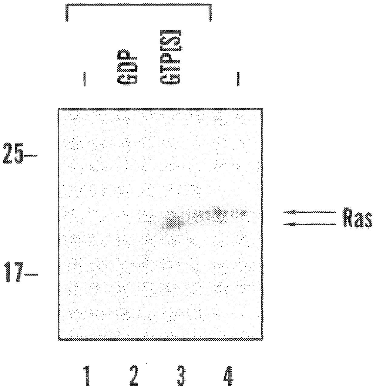
Assay for detection of activated Ras
To create an expression vector for production of GST-RBD, a BamHI-HinDIII fragment of plasmid pKScRaf1 was blunt-ended and ligated into the SmaI site of pGEX-2T. pGEX-RBD encodes amino acids 1-149 of cRaf-1 fused to GST. Plasmid pGEX-RBD has been deposited pursuant to, and in satisfaction of, the requirements of the Budapest Treaty on the International Recognition of the Deposit of Microorganisms for the Purposes of Patent Procedure with the American Type Culture Collection ("A.T.C.C.") at 10801 University Boulevard, Manassas, Va. 20110-2209. Plasmid pGEX-RBD was deposited on Sept. 21, 1999, and received A.T.C.C. Designation Number PTA-738. GST-RBD expression in transformed E. coli was induced with 1 mM isopropyl-beta-D-thiogalacto pyranoside ("IPTG") for 1-2 hours and the fusion protein was purified on glutathione Sepharose beads. The beads were washed in 20 mM HEPES, pH 7.5, 120 mM NaCl, 10% glycerol, 0.5% NP-40, 2 mM EDTA, 10 .mu.g / ml leupeptin...
example 2
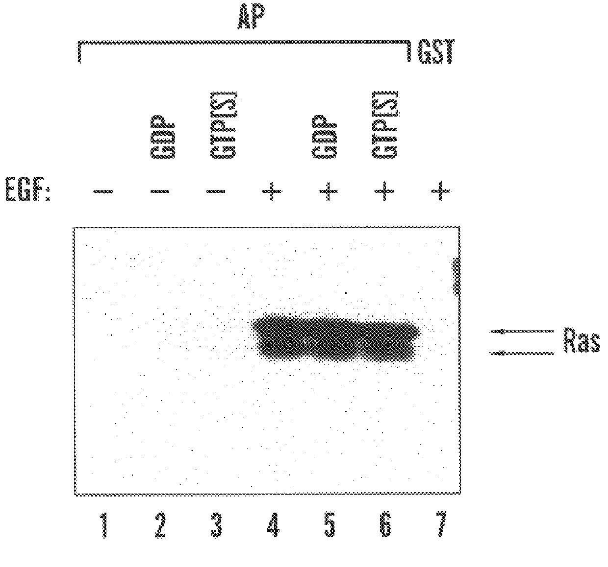
Ras is activated during G1 progression in HeLa cells
To evaluate whether Ras activity may be modulated during progression through G1 phase, HeLa cells were released from a mitotic arrest by means of a thymidine-nocodazole double block and released into G1 phase (in suspension culture unless otherwise indicated) or unsynchronized cells were serum-starved and treated with agonists. Shown are immunoblols probed with anti-Ras antibody. In FIG. 2(a), lanes 1-9 show GST-RBD affinity precipitates from cell lysates prepared at the indicated times after release from mitotic arrest, lanes 10-12 show affinity precipitates from unsynchronized (lane 10) or serum-starved cells treated without (lane 11) or with (lane 12) EGF (100 ng / ml for 10 minutes), lanes 13 and 14 show anti-Ras immunoprecipitate (lane 13) or whole cell lysate (lane 14) from 50% or 5% (relative to affinity precipitated) unsynchronized cell lysate. In FIG. 2(b), lanes 1-4 show affinity precipitates from cells released for 1 hours...
example 3
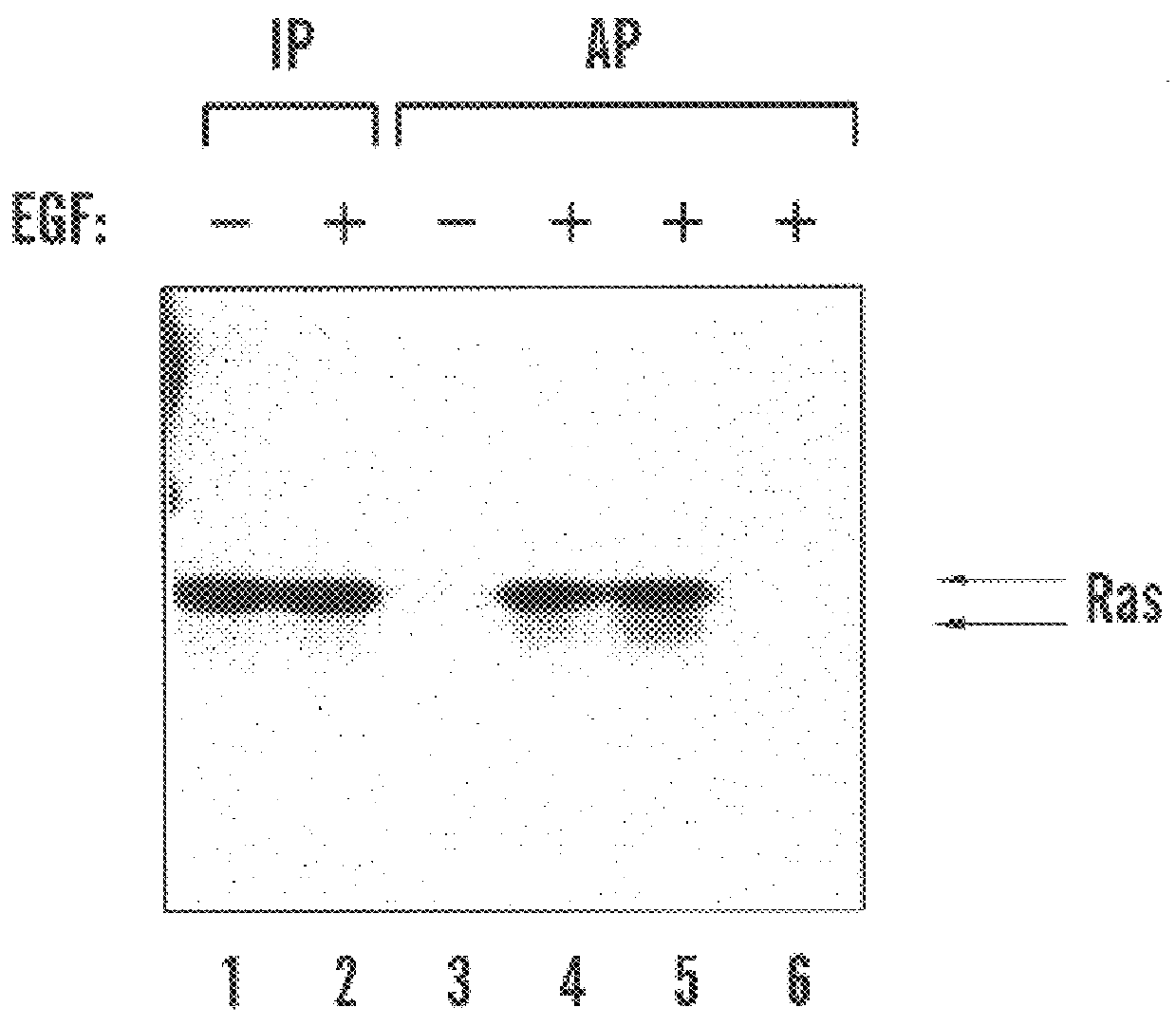
Activation of Ras in mid-G1 phase is uncoupled from Erk2 activation
Activation of Ras by growth factors leads to a rapid activation of the MAP kinases Erk1 and Erk2. Activated RasL61 has been shown to strongly activate co-expressed Erk2 in HeLa cells (Minden et al., "Selective Activation of the JNK Signalling Cascade and c-Jun Transcriptional Activity by the Small GTPases Rac and Cdc42Hs," Cell, 81:1147-57 (1995), which is hereby incorporated by reference). To determine whether Erk2 activity was stimulated in response to Ras activation during G1 progression, HeLa cell extracts were analyzed for Ras and Erk2 activity following mitotic release.
In FIG. 3(a), lanes 1-10 show anti-Erk2 immunoprecipitates from cells treated as described in lanes 1-10 in FIG. 2(c). Immunoprecipitates were subjected to in-gel kinase assay using myelin basic protein as substrate (upper panel) or immunoblotted with anti-Erk2 monoclonal antibody (lower panel). In FIG. 3(b), lanes 1-9 show GST-RBD affinity preci...
PUM
 Abstract
Description
Claims
Application Information