STAT function-regulatory protein
a stat-regulatory protein and function-regulatory technology, applied in the field of stat-induced stat inhibitors, can solve the problem of little known about genes (targets) and achieve the effect of improving the efficiency of the stat-regulatory protein
- Summary
- Abstract
- Description
- Claims
- Application Information
AI Technical Summary
Problems solved by technology
Method used
Image
Examples
example 1
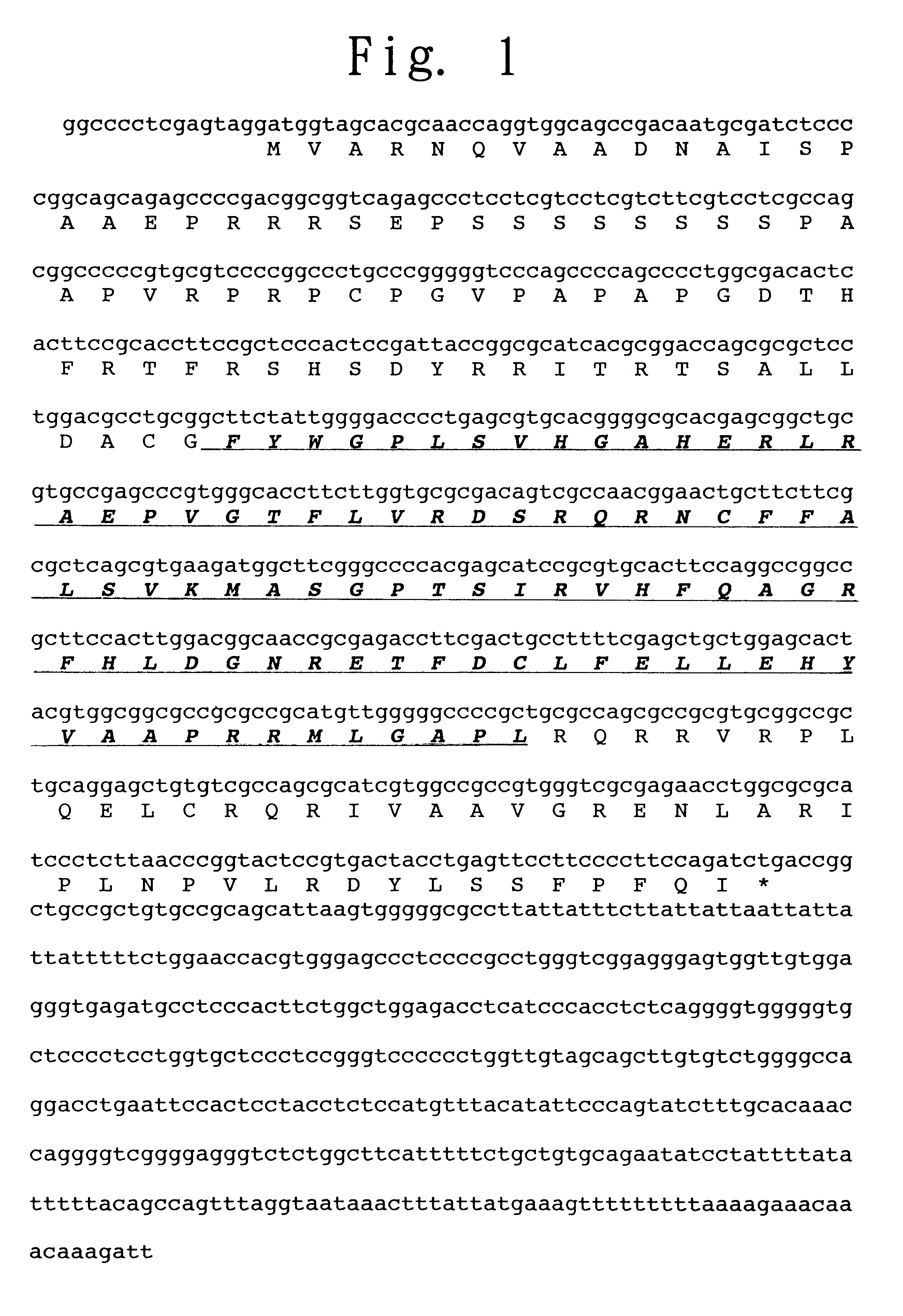
Preparation of SIIS-1, the Novel Protein of the Present Invention
(1) Preparation of a Monoclonal Antibody Against the SH2 Domain of a STAT:
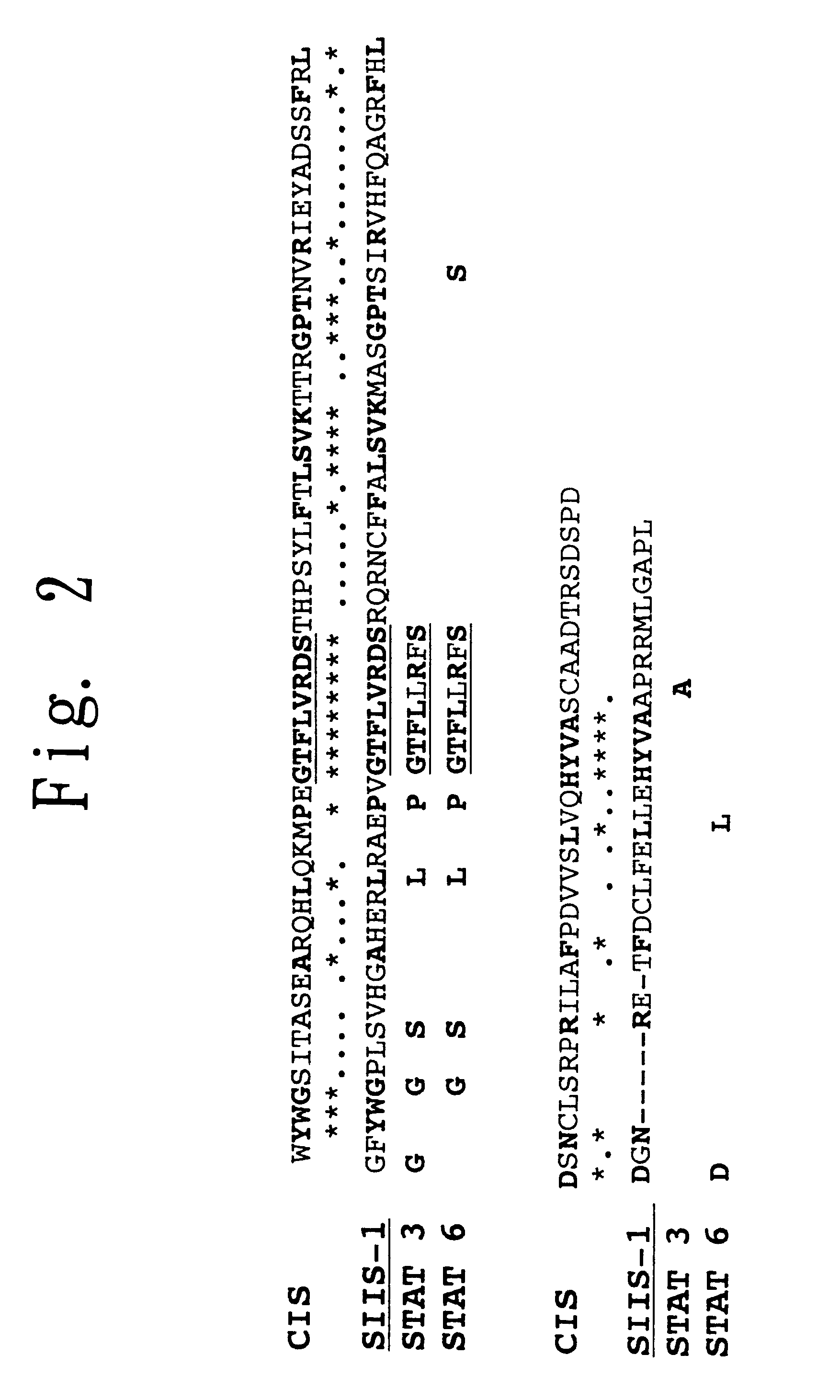
A monoclonal antibody against the amino acid sequence GTFLLRFS (Gly-Thr-Phe-Leu-Leu-Arg-Phe-Ser in 3-letter abbreviation) which is found in the SH2 domain and is highly conserved in proteins of the STAT family was produced as follows. The synthetic oligopeptide TKPPGTFLLRFSESSKEG (SEQ ID NO: 6) (Thr-Lys-Pro-Pro-Gly-Thr-Phe-Leu-Leu-Arg-Phe-Ser-Glu-Ser-Ser-Lys-Glu-Gly in 3-letter abbreviation), corresponding to the 600th to 617th amino acids in the SH2 domain of STAT3, was coupled to keyhole limpet hemocyanin, and BALB / c mice were immunized therewith. Spleen cells from each of the immunized BALB / c mice were fused with mouse myeloma cells P3-X63-Ag8-653 to thereby establish a hybridoma clone FL-238, which produces a monoclonal antibody against the synthetic oligopeptide GTFLLRFS in which the N-terminus thereof is protected by Fmoc. The monoclonal an...
example 2
SIIS-1 Expression in Various Murine Tissues
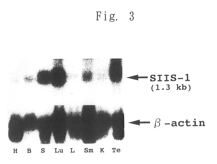
A radiolabeled full-length cDNA of SIIS-1 as a probe was hybridized to mouse NTN blot membrane (manufactured and sold by CLONTECH, USA). As a control, a commercially available .beta.-actin probe was used instead of the SIIS-1 probe. The results are shown in FIG. 3, wherein H indicates heart, B indicates brain, S indicates spleen, Lu indicates lung, L indicates liver, Sm indicates skeletal muscle, K indicates kidney and Te indicates testis.
The results show that SIIS-1 mRNA was ubiquitously expressed, and the expression was strong in lung, spleen and testis, and weak in all other tissues.
example 3
Analysis of the Possible Induction of SIIS-1 in Factor-dependent Cell Lines
In the following analysis, cells were stimulated with the following cytokines: MH60 cells and M1 cells were stimulated with IL-6 (50 ng / ml) plus sIL-6R (5 ng / ml); IL-4-dependent CT4S cells were stimulated with IL-4 (10 U / ml) plus IL-2 (10 ng / ml); and G-CSF-dependent NFS60 cells were stimulated with G-CSF (20 ng / ml) plus IL-3 (5 ng / ml).
With respect to all of the cell lines excluding M1 cells, the cells were factor-depleted for 4 hours in RPM1 1640 medium containing 1% BSA, and subsequently, the cells were stimulated with the above-mentioned cytokines by culturing in a cytokine-containing medium for various periods of time (min.) shown in FIG. 4. With respect to M1 cells, the cells were cultured in the same manner as described in Reference Example 1, and subsequently, the cells were stimulated with the above-mentioned cytokines by culturing the cells in a cytokine-containing medium for various periods of time (...
PUM
| Property | Measurement | Unit |
|---|---|---|
| pH | aaaaa | aaaaa |
| northern blot | aaaaa | aaaaa |
| resistance | aaaaa | aaaaa |
Abstract
Description
Claims
Application Information
 - R&D
- Intellectual Property
- Life Sciences
- Materials
- Tech Scout
- Unparalleled Data Quality
- Higher Quality Content
- 60% Fewer Hallucinations
Browse by: Latest US Patents, China's latest patents, Technical Efficacy Thesaurus, Application Domain, Technology Topic, Popular Technical Reports.
© 2025 PatSnap. All rights reserved.Legal|Privacy policy|Modern Slavery Act Transparency Statement|Sitemap|About US| Contact US: help@patsnap.com


