Detection of HLA-G

a technology of placenta and hla, which is applied in the field of detection of hla, can solve the problems of not being able to quickly detect in a clinical setting, unable to accurately predict currently no good predictive test for the development of pre-eclampsia, etc., and achieves accurate measurement, increased likelihood of causing pregnancy, and limited detection time

Inactive Publication Date: 2003-09-02
LIBRACH CLIFFORD L +1
View PDF3 Cites 6 Cited by
  • Summary
  • Abstract
  • Description
  • Claims
  • Application Information

AI Technical Summary

Benefits of technology

Another object of the invention is to provide a miniaturized detection assay, conducive to obtaining rapid results in a clinical setting.
The inventive method may be used to diagnose or detect an HLA-G indicative condition. In this embodiment, a control value for an HLA-G indicative condition can be compared with the quantity of HLA-G found in the sample. Certain conditions may be indicated if HLA-G is low or absent, while others may be indicated by increased levels of HLA-G. One of skill in the art could easily determine the indicative levels useful in diagnosing a condition. Such HLA-G indicative conditions may include, but are not limited to pre-eclampsia, increased risk of pre-eclampsia, adverse fetal outcome, increased risk of adverse fetal outcome, cancer, or increased risk of cancer development.
Currently, in vitro fertility (IVF) laboratories are able to select pre-embryos only on the basis of their morphology and rate of in vitro cleavage during the first 48 to 72 hours after fertilization. These criteria are useful, but not always good indicators of developmental potential. In most cases, 3 or 4 embryos are chosen based on these relatively crude indicators and then transferred into the uterine cavity. If additional, more stringent pre-embryo selection criteria were available, based on biochemical, genetic or developmental parameters, it would be possible to transfer one or two healthy pre-embryos, which have the highest chance of survival, without exposing patients to the psychological trauma caused by recurrent embryo implantation failure, spontaneous abortions, multiple IVF trials or the risk of multiple pregnancy. Therefore, a more predictive test for successful implantation would be invaluable.
Coating Microplates. In this assay system an indirect coating method was used. A comparison of assay sensitivity between direct and indirect coating antibody showed that indirect coating increased assay sensitivity.
Miniaturization of the HLA-G Assay. Pregnancy rates from transferred embryos having positive HLA-G secretion were significantly higher than those with undetectable HLA-G. Thus, to ascertain whether an embryo to be transferred has an increased likelihood of leading to pregnancy, a HLA-G detection assay can be conducted. However, the time required for detection is extremely limited, for example during an IVF procedure, and an accurate measurement taken within a clinical setting is desirable. A miniaturized HLA-G detection method can be used in a clinical setting so as to provide a rapid quantitative measurement of HLA-G in the medium in which an embryo is incubated, without requiring that a sample be sent off for laboratory analysis. Such a rapid detection assay advantageously involves either a visually distinguishable end-point (ie--colorimetric) or may involve a reader which is either hand-held, or bench-top sized, using an enzymatic, fluorometric or colorimetric end-point.
A miniaturized assay according to the invention may involve any device or method for analyzing a biological sample, such as is known in the art. The assay may be present on a support, such as on a strip or a chip, formed of plastic, cellulose, or any other acceptable material. By conducting the assay according to the invention in a miniaturized fashion, there is a reduced requirement for larger pieces of laboratory equipment. Further, there is less waste, as individual samples can be assessed with small amounts of biological fluids ranging from levels as low as 2 to 50 .mu.l. A miniaturized assay, requiring a small sample volume allows detection even when sample size is severely reduced.

Problems solved by technology

Elucidating the exact role played by the placenta in this disease has been difficult because fundamental aspects of the normal biology of this interesting organ are not well understood.
This suggests that an inappropriate response to some paternally derived antigen, possibly HLA-G, may result in pre-eclampsia.
However, there is currently no good predictive test for the development of pre-eclampsia, even though shallow invasion in the first trimester sets up conditions for the clinical signs that manifest later in pregnancy, most commonly late in the third trimester.
Conventional methods for diagnosing pre-eclampsia or for detecting antigens in a biological sample require such a sample to be sent away to a laboratory for analysis, and do not allow rapid detection in a clinical setting.

Method used


Image

  • Detection of HLA-G
  • Detection of HLA-G
  • Detection of HLA-G

Examples


example 2

Serum and Placental HLA-G is Decreased in Women with Pre-eclampsia

HLA-G levels in serum and placenta tissues from women with pre-eclampsia at term were compared with levels in normal pregnant women. HLA-G levels were determined by using an ELISA according to the invention in 20 subjects with pre-eclampsia and 14 control pregnant women. Both serum and placental HLA-G levels were significantly decreased in the pre-eclampsia group (median 0.026 .mu.g / ml in serum and median 0.026 .mu.g / mg protein in placenta), in comparison with normal pregnant women (median 0.093 .mu.g / ml in serum and median 0.088 .mu.g / mg protein in placenta, p=0.0112 and p=0.0406, respectively). There was a significant correlation between serum and placental HLA-G levels (r=0.603, p=0.002). The impaired expression of HLA-G suggested that there was an alternation of the maternal-fetal immune relationship in pre-eclampsia and decreased HLA-G plays a role in etiology of the disorder.

Pre-eclampsia is a disease that affec...

example 3

Differences in Serum Human Leukocyte Antigen-G (HLA-G) in Normal and Pre-Eclamptic Women at During Different Trimesters of Pregnancy

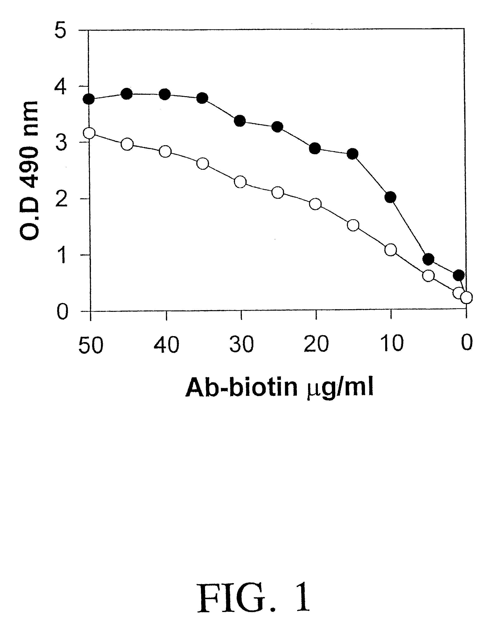
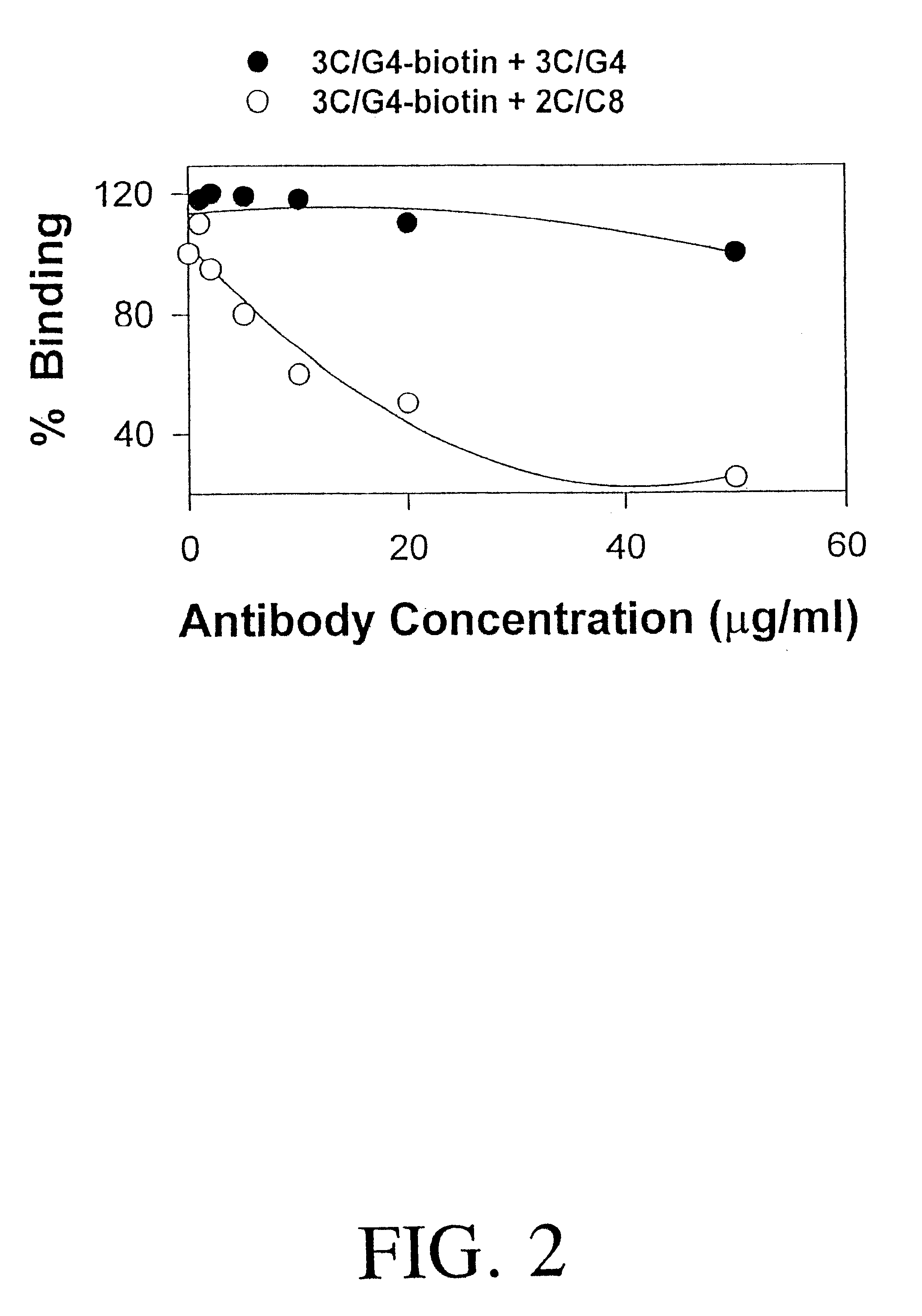
Serum samples were collected from normal pregnant women or from pre-eclamptic women at first trimester, second trimester and third trimester (N=12). An ELISA assay according to the invention was conducted, using 96-well plates prepared with immobilized 3C / G4 antibody, in which samples were incubated. HRP-labeled 2C / C8 binding was detected with OPD.

FIG. 13 illustrates the data points and median values for HLA-G levels in serum taken from women in the first trimester of their pregnancy either during a normal pregnancy, shown in closed circles (.circle-solid.), or during pre-eclampsia, shown by open circles (.smallcircle.).

FIG. 14 illustrates the data points and median values for HLA-G levels in serum taken from women in the second trimester of their pregnancy either during a normal pregnancy, shown in closed circles (.circle-solid.), or during pre-eclamps...

example 4

Progesterone Increases HLA-G Expression in vitro

HLA-G is expressed on placental tissues at the maternal-fetal interface. This restricted pattern of expression suggests that HLA-G may play a crucial role in maternal immune tolerance to the fetus. It has been reported that HLA-G expression is developmentally regulated and changes with stage of gestation. There are conflicting reports that the cytokine gamma IFN has an effect on HLA-G expression in vitro. Since, some studies suggest that progesterone is important in suppressing the maternal immunological response to the fetus, it was hypothesized that this steroid hormone may play a role in regulating HLA-G expression. The present study was performed to explore potential effects of progesterone on HLA-G expression in cytotrophoblasts and in the JEG-3 choriocarcinoma cell line in vitro.

Methods. Purified cytotrophoblasts were prepared from first trimester placental tissue by standard techniques. JEG-3 choriocarcinoma cells were cultured ...


PUM

PropertyMeasurementUnit
molecular massaaaaaaaaaa
volumeaaaaaaaaaa
concentrationsaaaaaaaaaa

Abstract

The present invention relates to the detection of HLA-G. Antibodies to both soluble and membrane bound HLA-G are disclosed. Exemplary antibodies include 2C / C8, 3C / G4, and 4H84. 2C / C8 and 4H84 antibodies bind to the same region of HLA-G, which is a different region than that bound by 3C / G4. Methods of detection and diagnosis are disclosed as well as kits, including a miniaturized assay suitable for a clinical setting. Further, a method of selecting an embryos for in vitro fertilization is disclosed. A sandwich ELISA test is provided using two antibodies that bind to HLA-G at different regions. The HLA-G test according to the invention is over 1000 times more selective in binding to HLA-G than to antigens HLA-A2, HLA-B4, HLA-C, or mixed WBC preparations.

Description

The present invention relates to the detection of HLA-G. The present invention provides antigens for generating specific antibodies to both soluble and membrane bound HLA-G, as well as exemplary antibodies.A central question in pregnancy is how the fetal-placental unit avoids maternal immune rejection. Although fetal and maternal cells interact throughout pregnancy, the fetus typically remains a privileged site, not subject to rejection. It is likely that the particular nature of the cells at the fetal-maternal interface and their products help prevent rejection of the fetus by the maternal immune system.Implantation and placental development physically connect the mammalian embryo to the maternal uterus. Establishing this connection is essential for subsequent development. The initial developmental events which occur in the embryo set aside unique extraembryonic cellular lineages which are the precursors of the placenta. The first differentiation event gives rise to trophoblasts, w...

Claims


Application Information

Patent Timeline
Patent Type & Authority Patents(United States)
IPC IPC(8): C07K16/18C07K16/28G01N33/569G01N33/68C12N5/20
CPCC07K16/2833G01N33/56977G01N33/689C07K2317/34Y10S436/906Y10S530/853
Inventor LIBRACH, CLIFFORD L.YIE, SHANG-MIAN
Owner LIBRACH CLIFFORD L
Why Eureka
Try Eureka