
Diagnostic procedures using 129Xe spectroscopy characteristic chemical shift to detect pathology in vivo

a technology of characteristic chemical shift and spectroscopy, which is applied in the direction of magnetic variable regulation, measurement using nmr, instruments, etc., can solve the problems of insufficient dose delivery to more distant (away from the pulmonary vasculature) target areas of interest, insufficient inhalation or ventilation delivery, and insufficient quantifiable information from experiments, so as to reduce the impact of certain potentially data-corrupting parameters

Inactive Publication Date: 2004-02-24
POLAREAN
View PDF25 Cites 23 Cited by
  • Summary
  • Abstract
  • Description
  • Claims
  • Application Information

AI Technical Summary

Benefits of technology

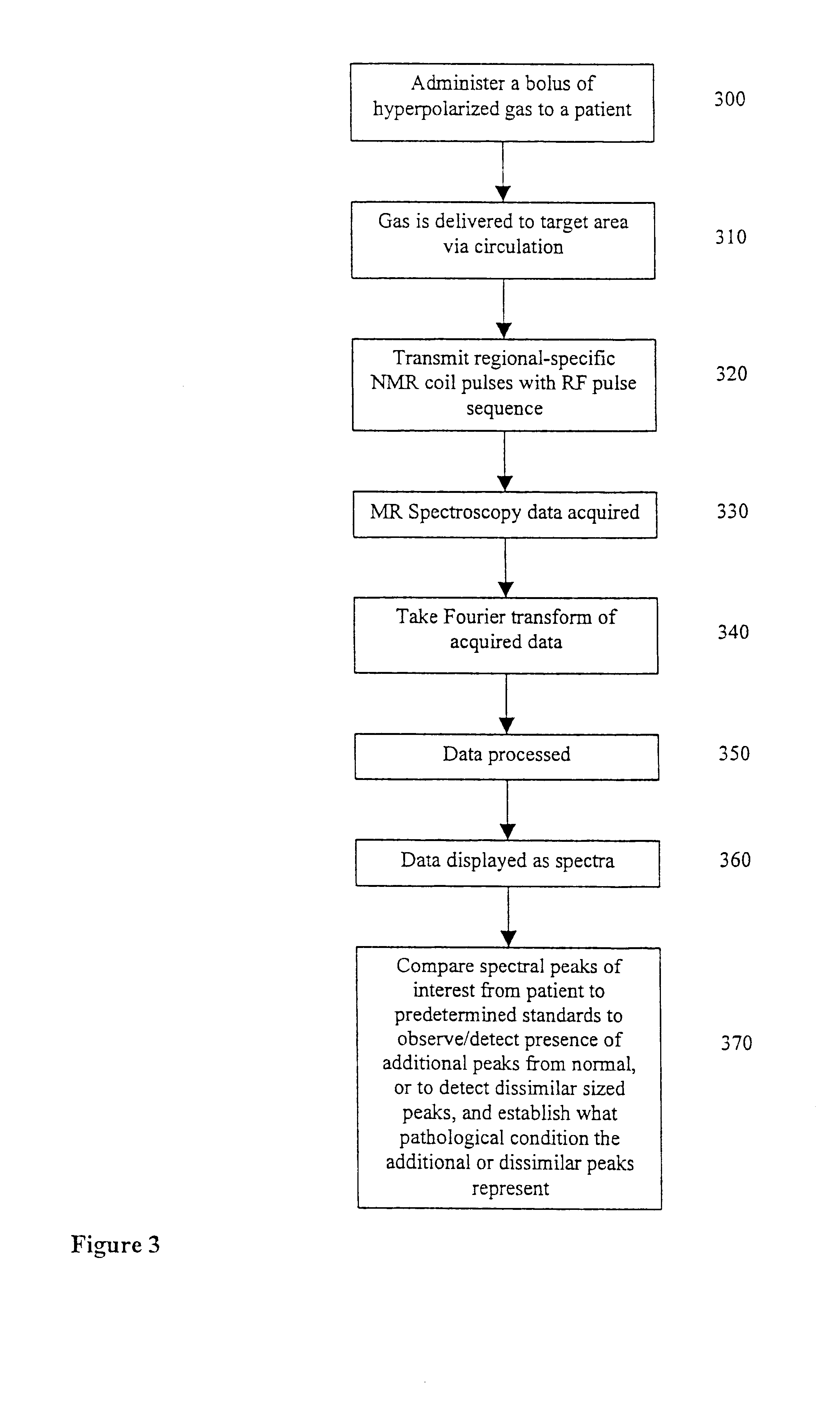
"The present invention is a method for detecting and diagnosing pathological conditions in vivo using hyperpolarized .sup.129 Xe gas. The method involves administering the gas to a patient and analyzing the signals received from the gas in a region of interest. The method can account for variations in polarization and volume of the gas, as well as filtering out background information. The method can also be used to monitor the progression of a disease and the efficacy of treatments. The invention provides a non-invasive or minimally invasive way to probe tissues in vivo to detect pathological or abnormal conditions."

Problems solved by technology

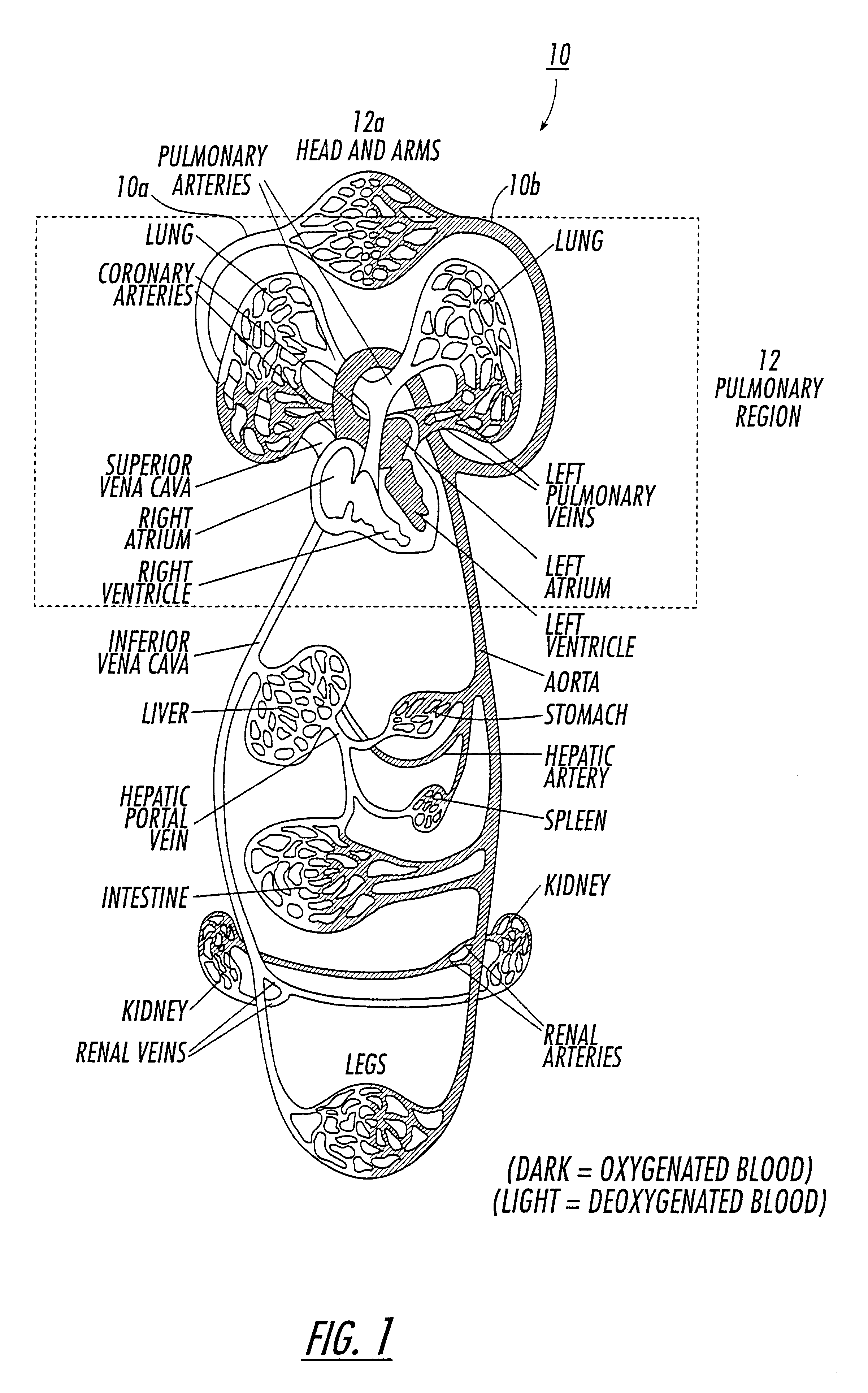
While inhaled delivery .sup.129 Xe methods are suitable, and indeed, preferable, for many MR applications for several reasons such as the relatively non-invasive characteristics attendant with such a delivery to a human subject, inhalation or ventilation-based deliveries may not be the most efficient method to deliver a sufficiently large dose to more distant (away from the pulmonary vasculature) target areas of interest.
Since MR imaging requires high signal strength to achieve a clinically useful spatial resolution in the resulting image, inhalation delivery may not produce clinically desirable images of distal or remote target organs or regions.
However, these experiments provided very little in the way of quantifiable information.
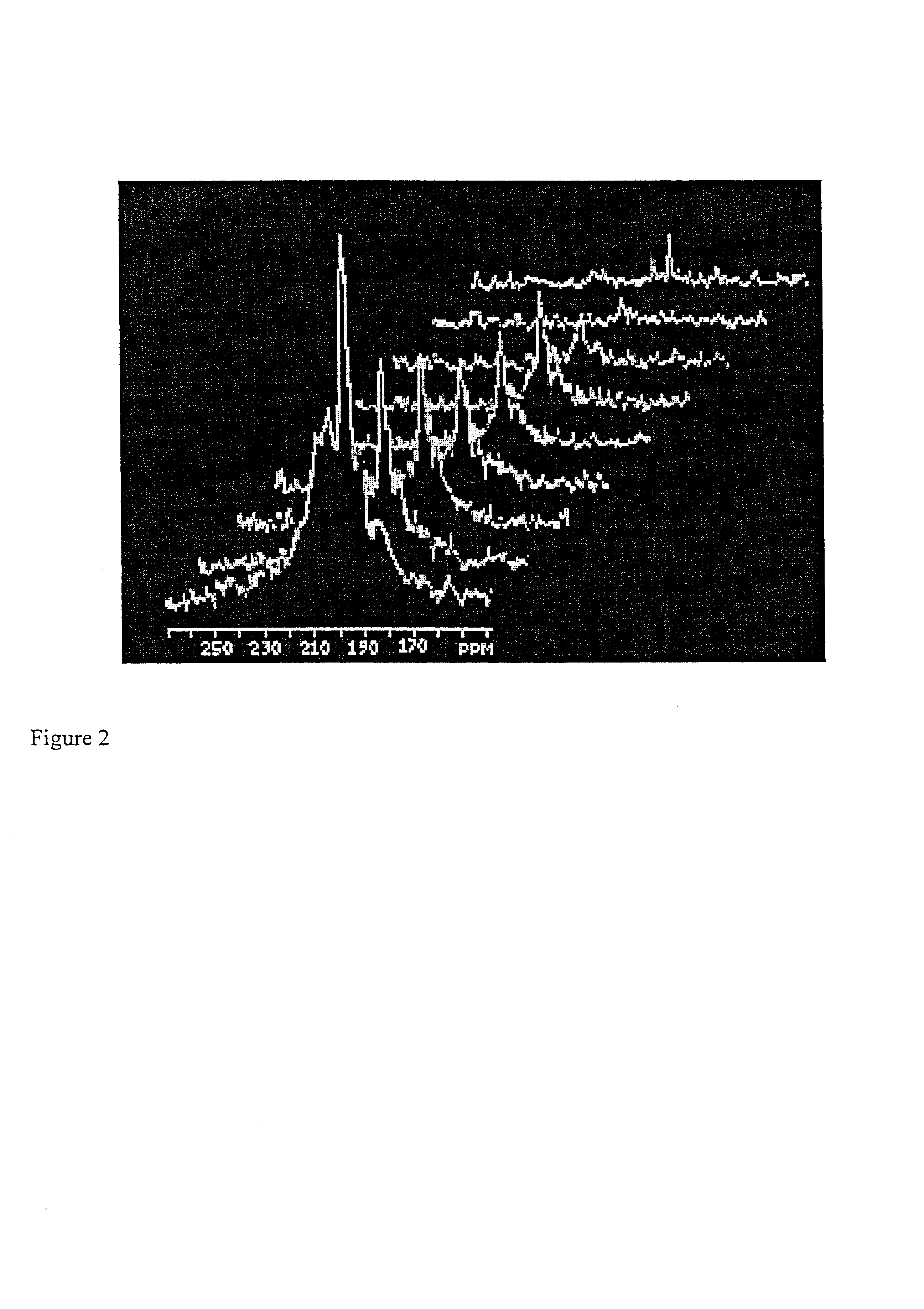
Diseases of interest often cannot be diagnosed merely by the appearance of peaks denoting characteristic chemical shifts, since healthy tissues may also exhibit the same characteristics (e.g., some lipid is expected, but an excess or reduced amount of lipid may be problematic).

Method used


Image

  • Diagnostic procedures using 129Xe spectroscopy characteristic chemical shift to detect pathology in vivo
  • Diagnostic procedures using 129Xe spectroscopy characteristic chemical shift to detect pathology in vivo
  • Diagnostic procedures using 129Xe spectroscopy characteristic chemical shift to detect pathology in vivo

Examples


example

Xenon-129 NMR Spectroscopy in Human Aortic Tissue

Background

The well-established property of the .sup.129 Xe nucleus to respond to changes in the environment with a change in NMR-resonance frequency led to a proposal that the lipid-rich atherosclerotic plaques in humans could be detected by measuring the chemical shift of the xenon signal. .sup.129 Xe spectra have been recorded in human tissue at 7 T and 25 bars of pressure. The .sup.129 Xe was thermally polarized for this experiment.

Method

Samples of aortic wall were taken from recently deceased humans, unfrozen but kept on ice. The innermost layer of the aortic wall, the intima, was dissected by lifting by tweezers and gently cutting it free with a sharp scalpel. The tissue was sliced into pieces of a few mm. Then, for each patient, a control part of "healthy" vessel wall was taken and compared to plaques cut out the same way. The plaques were scored on a graded scale ranging from 1-5 with grade 1 representing totally soft, fatty de...


PUM


Abstract

An in vivo non-invasive method for detecting and / or diagnosing a pathological condition using hyperpolarized <129>Xe spectroscopy is disclosed. Generally stated, the method includes determining the magnitude of spectral peaks which represent particular chemical shifts and comparing the observed magnitudes to those of healthy individuals. Preferably, the method includes subtracting substantial backgrounds and accounting for secondary conditions such as the polarization of hyperpolarized gas administered. Additionally, a quantitative analysis of hyperpolarized <129>Xe spectra advantageously allows a physician to establish the extent of disease progression. Advantageously, this method can be used regardless of the method of hyperpolarized <129>Xe administration.

Description

The present invention relates to magnetic resonance spectroscopy methods utilizing chemical shifts of hyperpolarized .sup.129 Xe.MRI using hyperpolarized noble gases has been demonstrated as a viable imaging modality. See e.g., U.S. Pat. No. 5,545,396 to Albert et al. The contents of this patent are hereby incorporated by reference as if recited in full herein. Albert et al. proposed several techniques of introducing the hyperpolarized gas (either alone or in combination with another substance) to a subject, such as via direct injection, intravenous injection, and inhalation. See also "Biological magnetic resonance imaging using laser-polarized .sup.129 Xe," Nature, pp. 199-201 (Jul. 21, 1994). Other researchers have since obtained relatively high-quality images of the lung using pulmonary ventilation of the lung with both hyperpolarized .sup.3 He and .sup.129 Xe. See J. R. MacFall et al., "Human lung air spaces: Potential for MR imaging with hyperpolarized He-3," Radiology 200, 553...

Claims


Application Information

Patent Timeline
Patent Type & Authority Patents(United States)
IPC IPC(8): A61B5/0275A61B5/055A61B5/026G01R33/28G01R33/44G01R33/46G01R33/48
CPCA61B5/02755A61B5/055A61B5/415G01R33/5601A61B5/418Y10T436/24G01R33/4625A61B5/4088
Inventor DRIEHUYS, BASTIAAN
Owner POLAREAN
Learn More