Apparatus and method for the measurement of cells in biological samples

Inactive Publication Date: 2005-02-08
INOVX
View PDF11 Cites 33 Cited by
  • Summary
  • Abstract
  • Description
  • Claims
  • Application Information

AI Technical Summary

Benefits of technology

Since sample preparation, such as labeling, can be conducted within the apparatus, which is fully-contained, the investigator does not have to handle the sample once it is put into the apparatus. This improves the accuracy of the results because the sample is less likely to be contaminated as a result of transfer to

Problems solved by technology

While there are many automated and semi-automated instruments for counting and examining cells, these instruments cannot measure low levels of cells (i.e., 100 cells/μL or less) or require pre-concentration or other time consuming steps to reach this detection level.
In addition to the difficulty in detecting low concentrations of cells, small sample size also presents a detection challenge.
If only a few μL are available for assay, accurate enumeration of cells is difficult for many analytical systems.
While cell or particle counters are available, they do not accurately detect low levels of cells.
For instance, presently commercially available automated cell or particle counters cannot measure cells levels below 500 to 1000 cells/μL.
(Rabinovitch, A. and Cornbleet, P. J., Arch Pathol Lab Med, 118:13-17, 1994) In addition, fluidic elements used by these cell detection systems may be clogged up by viscous body fluids.
However, large sample volumes are generally required in order for this process to be accurate.
Therefore, if flow cytometry were used to quantify the number of cells in a sample, all the fluid removed

Method used


Image

  • Apparatus and method for the measurement of cells in biological samples
  • Apparatus and method for the measurement of cells in biological samples
  • Apparatus and method for the measurement of cells in biological samples

Examples


Embodiment Construction

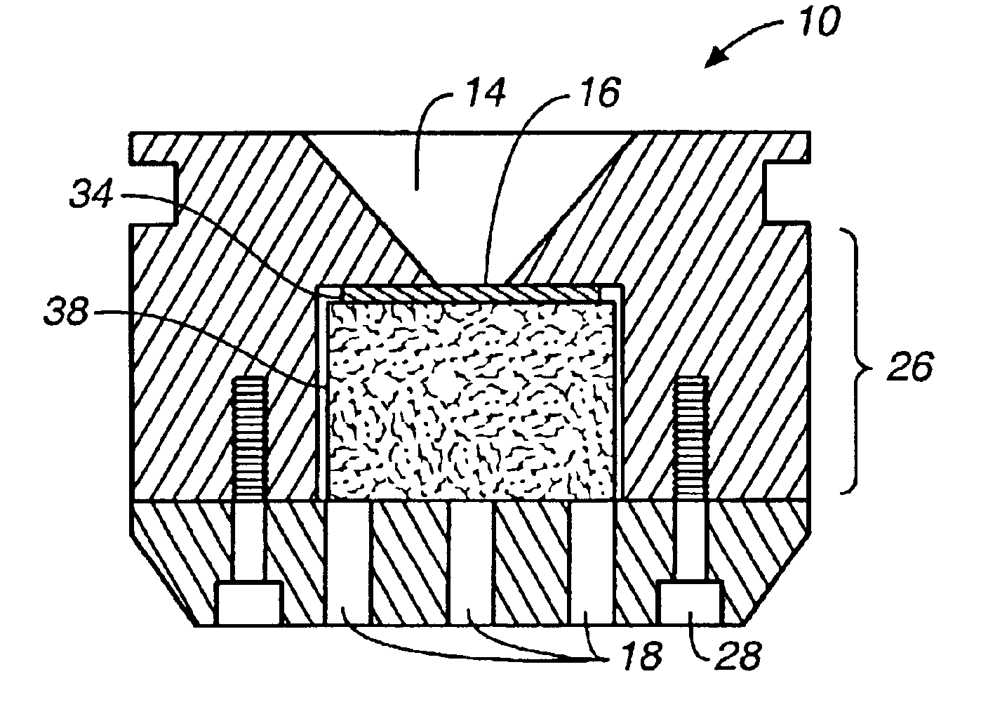
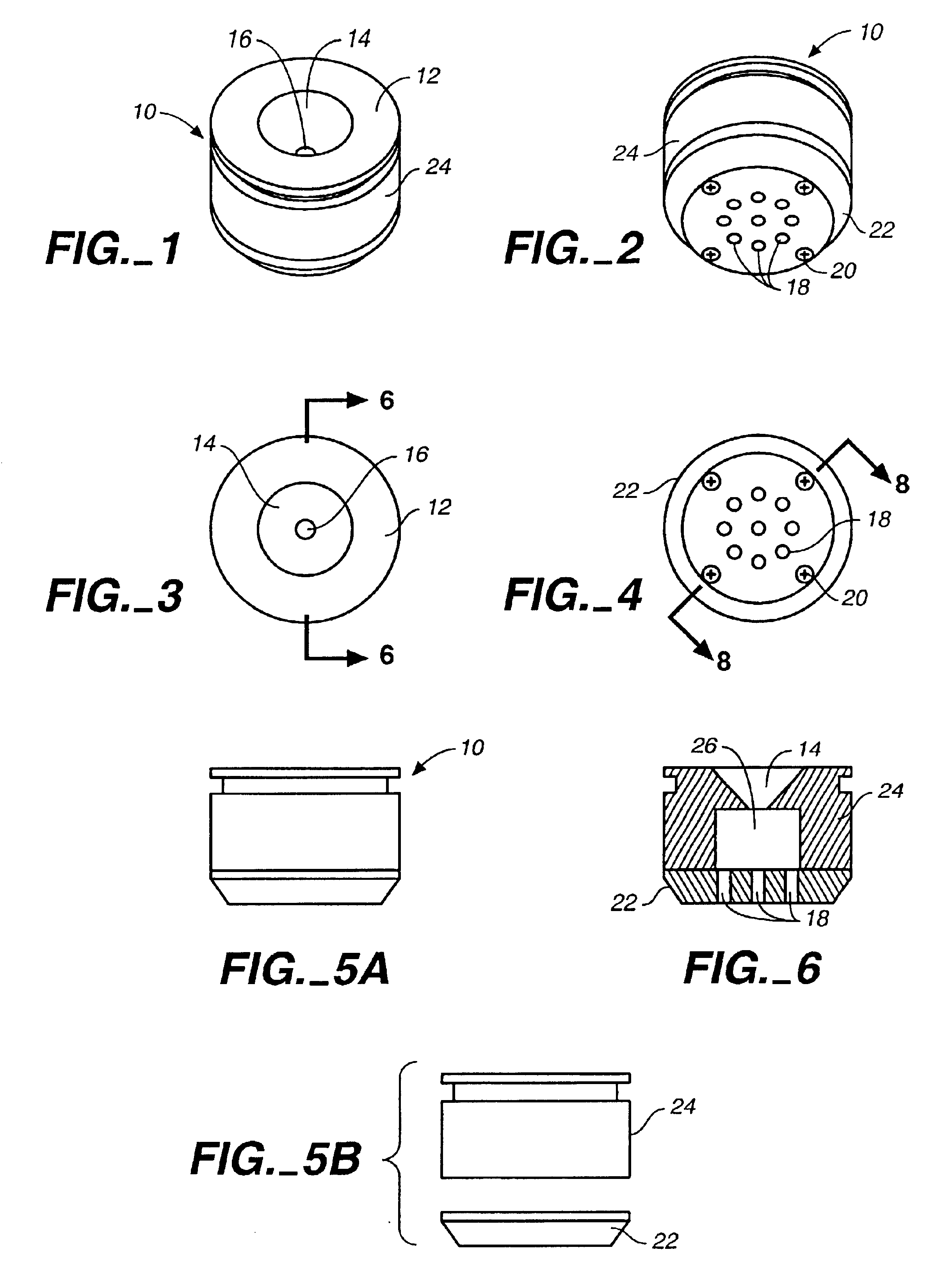
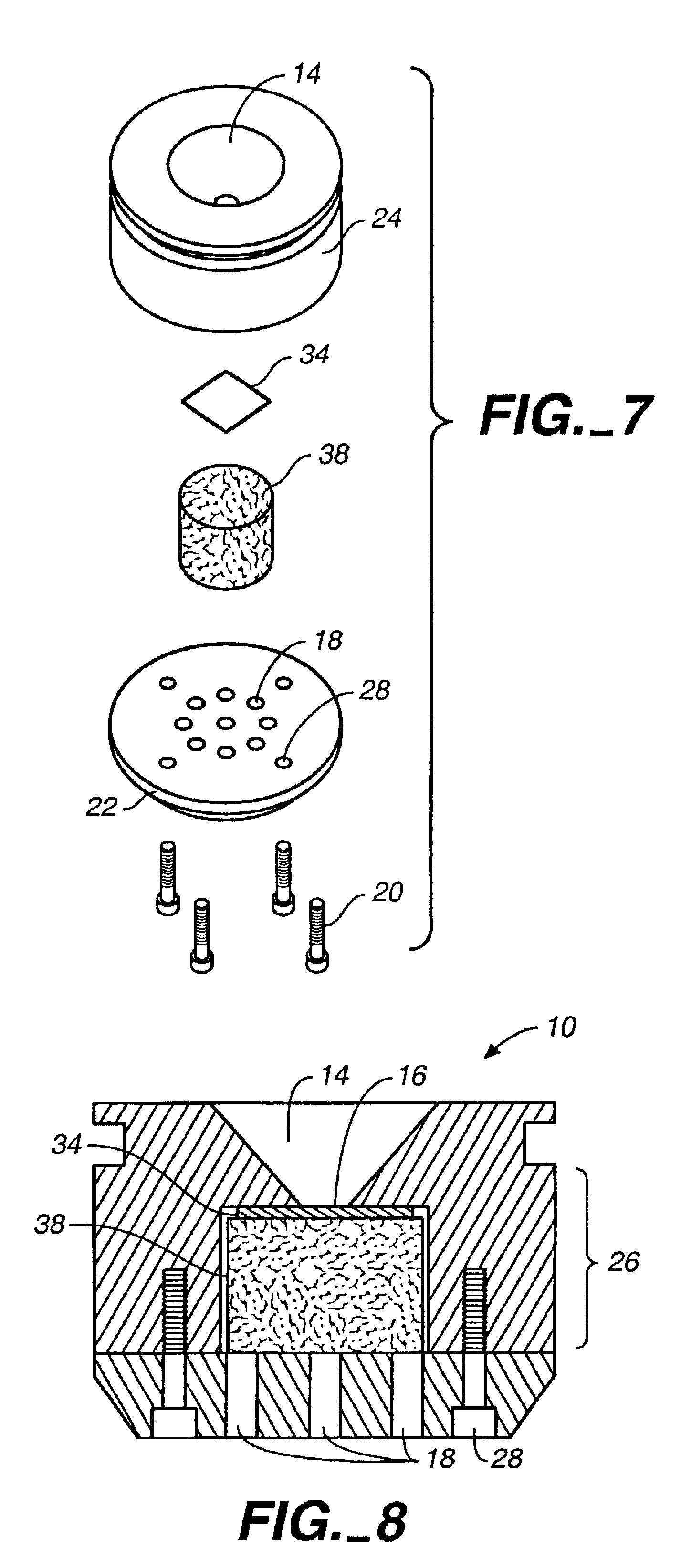
The concentration device (“device”) 10 is shown in FIG. 1. Specimens are placed in cone 14 in the top surface 12 of the device 10. The specimen flows through the opening 16 at the bottom of cone 14; cells of interest are trapped on a membrane at the opening 16, which matches the field of view of the imaging system being used to count the cells trapped on the membrane. The fluid in the specimen, which is not trapped on the membrane, flows into the second chamber of the device 10 (see FIG. 6, below). Both cone 14 and the second chamber are contained in the main body 24 of the device 10.

As shown in FIG. 2, the bottom portion 22 of the device 10 is attached to the main body 24 by screws 20. The bottom portion 22 of the device 10 also features a vacuum port 18 where a vacuum system may be attached.

An overhead view of the device is provided in FIG. 3. A cone 14 which receives specimens is cut into the top surface 12. The walls of cone 14 are smooth and treated to prevent cell adhesion and...


PUM

PropertyMeasurementUnit
Lengthaaaaaaaaaa
Lengthaaaaaaaaaa
Pressureaaaaaaaaaa

Abstract

An apparatus and method for concentrating and measuring low levels of cells in biological samples. The apparatus, or concentration device, consists of two chambers with an optically level collection membrane intermediating between the chambers. The collection membrane filters the biological sample, trapping cellular elements of interest. A vacuum may be attached to the device to assist in filtration. The surface area of the collection membrane matches the view field of a standard imaging system and the device can be mounted on a standard microscope stage. All the cells in the sample volume are collected onto the membrane. The view field provides a fixed volumetric area for cell counting. Since the volume of sample tested is known, the total number of cells in the original sample may be calculated. The sample reservoir of the concentration device may also be used for sample preparation. The concentration device is fully-contained; therefore, the investigator does not have to handle the sample once it is placed in the sample reservoir.

Description

FIELD OF THE INVENTIONThis invention relates to the collection and subsequent measurement of low numbers of cells in biological samples.BACKGROUND OF THE INVENTIONAccurate and rapid counting of cellular elements in biological fluids is a necessity in the biomedical, pharmaceutical, and biological research fields. While there are many automated and semi-automated instruments for counting and examining cells, these instruments cannot measure low levels of cells (i.e., 100 cells / μL or less) or require pre-concentration or other time consuming steps to reach this detection level. Various biological fluids have a concentration of cells below 100 cells / μL and numerous procedures and protocols require that the low levels of cells in these fluids be measured. For instance, the current legal standards for transfusion products allow less than 20 white cells / μL in the United States and less than 4 white cells / μL in Europe before the products can be administered to humans. The concentration of ...

Claims


Application Information

Patent Timeline
IPC IPC(8): G01N1/40
CPCG01N1/40Y10T436/255
Inventor CHAN, ANTHONYROCCO, RICHARD M.
Owner INOVX
Why Eureka
Try Eureka