
Manufacturing therapeutic enclosures

a technology of enclosures and therapeutics, applied in the field of tissue healing and regeneration, can solve the problems of impaired wound healing, inability to properly function, and large physical, emotional, social, and psychological distress of components, and achieve the effect of enhancing the healing of chronic wounds, safe, effective, and interactiv

Inactive Publication Date: 2005-05-10
RGT UNIV OF MICHIGAN +1
View PDF21 Cites 3 Cited by
  • Summary
  • Abstract
  • Description
  • Claims
  • Application Information

AI Technical Summary

Benefits of technology

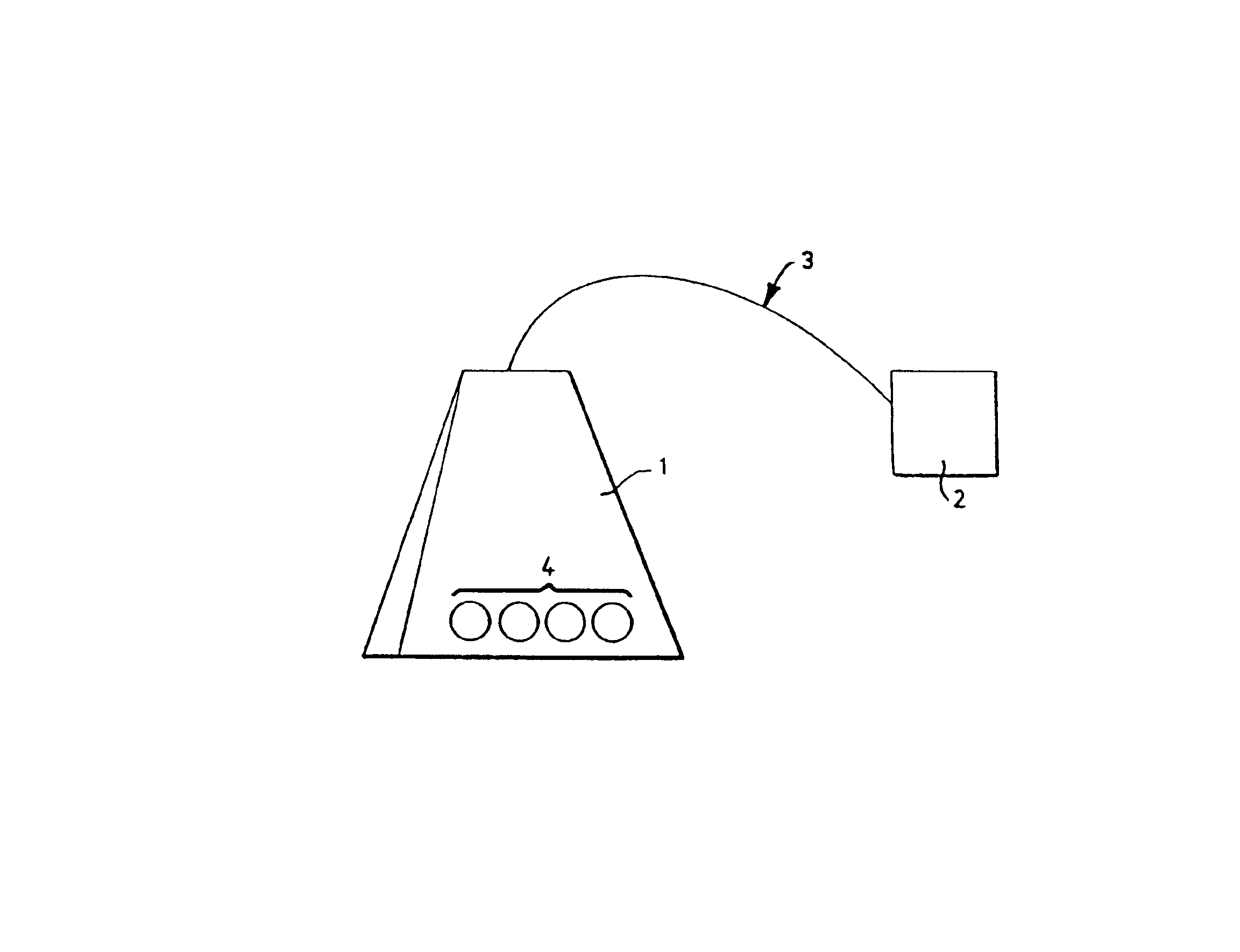
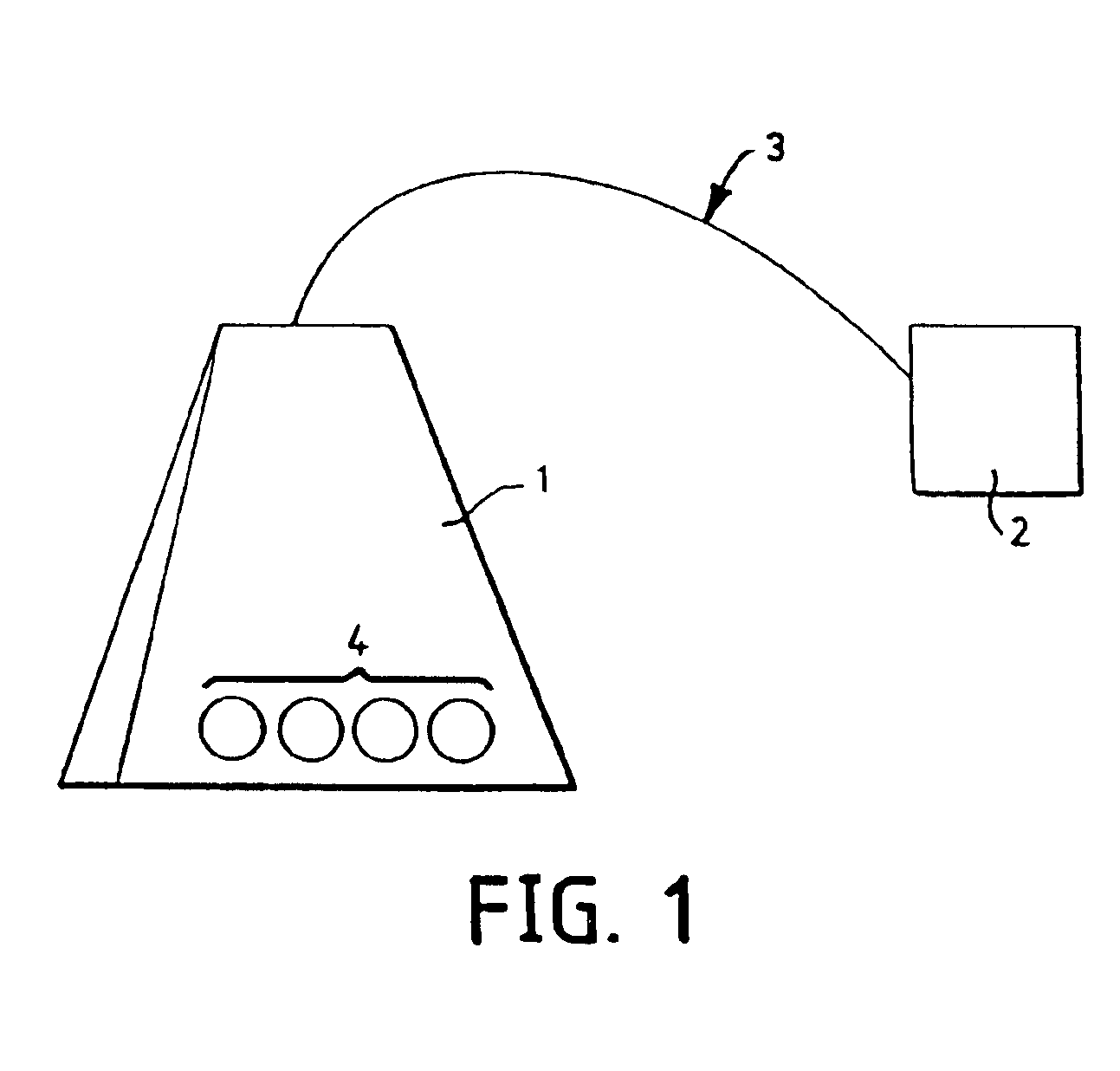
The present invention is about a system and method for enhancing the healing of wounds, especially chronic wounds, using cultured keratinocytes. The invention involves the use of a solid support, such as collagen-coated beads, placed in an enclosure that is placed at the wound site. The enclosure can be a mesh material or a biocompatible membrane. The system can also include a porous extracapular matrix or a handle or string attached to the enclosure. The invention promotes wound healing by inducing the formation of granulation tissue and epithelialization. The invention can be used in the treatment of wounds, including partial thickness wounds and deep wounds.

Problems solved by technology

Wound healing is impaired when these components, either individually or as a whole, do not function properly.
Wounds which do not readily heal can cause the subject considerable physical, emotional, and social distress as well as great financial expense [see, e.g., Richey et al., Annals of Plastic Surgery 23(2):159-165 (1989)].
Indeed, wounds that fail to heal properly and become infected may require excision of the affected tissue.
Unfortunately, certain types of wounds (e.g., diabetic ulcers, pressure sores) and the wounds of certain subjects (e.g., recipients of exogenous corticosteroids) do not heal in a timely manner (or at all) with the use of such dressings.
Thus, except in very limited circumstances, the promotion of wound healing with these agents has met with little success.

Method used


Image

  • Manufacturing therapeutic enclosures
  • Manufacturing therapeutic enclosures

Examples


example 1

[0059]The experiments of this example demonstrate that human culture keratinocytes grown on macroporous microcarriers and contained in a porous enclosure improve healing in surgically created wounds in mice.

A. Experimental Methodology

Preparation of Human Keratinocytes

Isolation and Growing of Human keratinocytes: Human keratinocytes (AATB certified; University of Michigan cultured keratinocyte program) were isolated at The University of Michigan Burn / Trauma Unit from split thickness skin.

[0060]Trypsinization of the split thickness skin was effected as follows. The skin was placed dermis-side down in 150 mm Petri dishes. The pieces were cut into smaller pieces (about 2 cm×about 0.3 cm) and were soaked in a sterile solution of 30 mM HEPES, 10 mM glucose, 3 mM KCl, 130 mM NaCl, 1 mM Na2HPO4 buffer, pH 7.4 containing 50 units of Penicillin and 50 μg Streptomycin (Sigma, P-0906). After soaking for 1-2 hr at 4° C. the buffer was aspirated off, and 0.09% trypsin (Sigma, Type 1X) in a Penici...

example 2

[0086]The experiments of this example demonstrate that human culture keratinocytes grown on macroporous microcarriers and contained in a porous enclosure that is then covered with a wound dressing material improve healing in surgically created wounds in mice.

A. Experimental Methodology

[0087]The experiments of this example were performed as described in Example 1, with the following exceptions. The group of mice that received the keratinocyte-coated CYTOLINE 1™ macroporous microcarrier beads (Pharmacia Biotech) (i.e., the beads / bags group) comprised five animals, while the group that received only the bags (i.e., the bags only group) comprised four animals. (They are labelled 2 to 5 because Mouse 1 expired during anesthesia.) In this example the bags from both the beads / bags group and the bags only group were covered with a polyurethane film dressing (TEGADERM™, 3M Health Care, St. Paul, Minn.) with a cellophane product.

[0088]More specifically, the wounds were dressed either with hum...


PUM

PropertyMeasurementUnit
sizeaaaaaaaaaa
pore sizesaaaaaaaaaa
pHaaaaaaaaaa

Abstract

Devices and methods for enhancing the healing of wounds, especially chronic wounds (e.g., diabetic wounds), involving the use of keratinocytes are described. Keratinocytes are grown on a transplantable solid support (e.g., collagen-coated beads), and the keratinocyte-coated solid support is placed in an enclosure. The enclosure, in turn, is placed in the wound for use as an interactive wound healing promoter.

Description

[0001]The present Application is a Continuation of U.S. patent application Ser. No. 09 / 919,516, filed Jul. 31, 2001, now U.S. Pat. No. 6,440,452, which is a Continuation of U.S. patent application Ser. No. 09 / 747,742, filed Dec. 22, 2000, now U.S. Pat. No. 6,299,898, which is a continuation of U.S. patent application Ser. No. 09 / 338,413, filed Jun. 22, 1999, now U.S. Pat. No. 6,197,330, which is a Continuation of U.S. patent application Ser. No. 08 / 840,804, filed Apr. 16, 1997, now U.S. Pat. No. 5,972,332.FIELD OF THE INVENTION[0002]The present invention relates generally to tissue healing and regeneration and, more particularly, to methods and systems for wound healing.BACKGROUND OF THE INVENTION[0003]The primary goal in the treatment of wounds is to achieve wound closure. Open cutaneous wounds represent one major category of wounds and include burn wounds, neuropathic ulcers, pressure sores, venous stasis ulcers, and diabetic ulcers. Open cutaneous wounds routinely heal by a proce...

Claims


Application Information

Patent Timeline
Patent Type & Authority Patents(United States)
IPC IPC(8): A61L15/40A61L15/16A61L27/60A61L27/38A61L27/00C12N5/06A61K35/12A61K38/00A61L27/56C12N5/071
CPCA61L15/40A61L27/3813A61L27/56C12N5/0629A61L27/60A61K35/12A61K38/00C12N2533/54C12N2510/00C12N2510/02C12N2531/00Y10T442/2525
Inventor REES, RILEY S.MARCELO, CYNTHIAADAMSON, BELINDARHODES, LENOREMARCHANT, BEVERLYLINDBLAD, WILLIAM J.GILMONT, ROBERTGARNER, WARRENZUCCARO, CYNTHIATADDONIO, THOMAS E.
Owner RGT UNIV OF MICHIGAN
Learn More