Gamma camera workflow automation

a gamma camera and workflow technology, applied in the field of diagnostic imaging arts, can solve the problems of high labor intensity, time-consuming and labor-intensive daily quality control procedures, and up to 15 minutes of setup procedures, so as to reduce the likelihood of operator error, reduce the number of personnel needed, and reduce the effect of setup tim

Inactive Publication Date: 2005-06-14
KONINKLIJKE PHILIPS ELECTRONICS NV
View PDF7 Cites 12 Cited by
  • Summary
  • Abstract
  • Description
  • Claims
  • Application Information

AI Technical Summary

Benefits of technology

[0018]One advantage of the present invention resides in substantial reduction in setup time for a nuclear imaging session.
[0019]Another advantage of the present invention resides in a reduced likelihood of operator error producing non-optimal imaging or other difficulties.
[0020]Another advantage resides in a reduction of personnel needed to conduct a patient imaging session.
[0021]Yet another advantage of the present invention resides in simplification of the quality control procedures, imaging session setup process, and other operations related to a nuclear imaging facility.
[0022]Numerous additional advantages and benefits of the present invention will become apparent to those of ordinary skill in the art upon reading the following detailed description of the preferred embodiment.

Problems solved by technology

These set-up procedures can take up to 15 minutes or more, and involve substantial intervention of the operator.
Daily quality control procedures which verify and maintain camera alignment and the like are similarly time-consuming and labor-intensive.

Method used


Image

  • Gamma camera workflow automation
  • Gamma camera workflow automation
  • Gamma camera workflow automation

Examples


Embodiment Construction

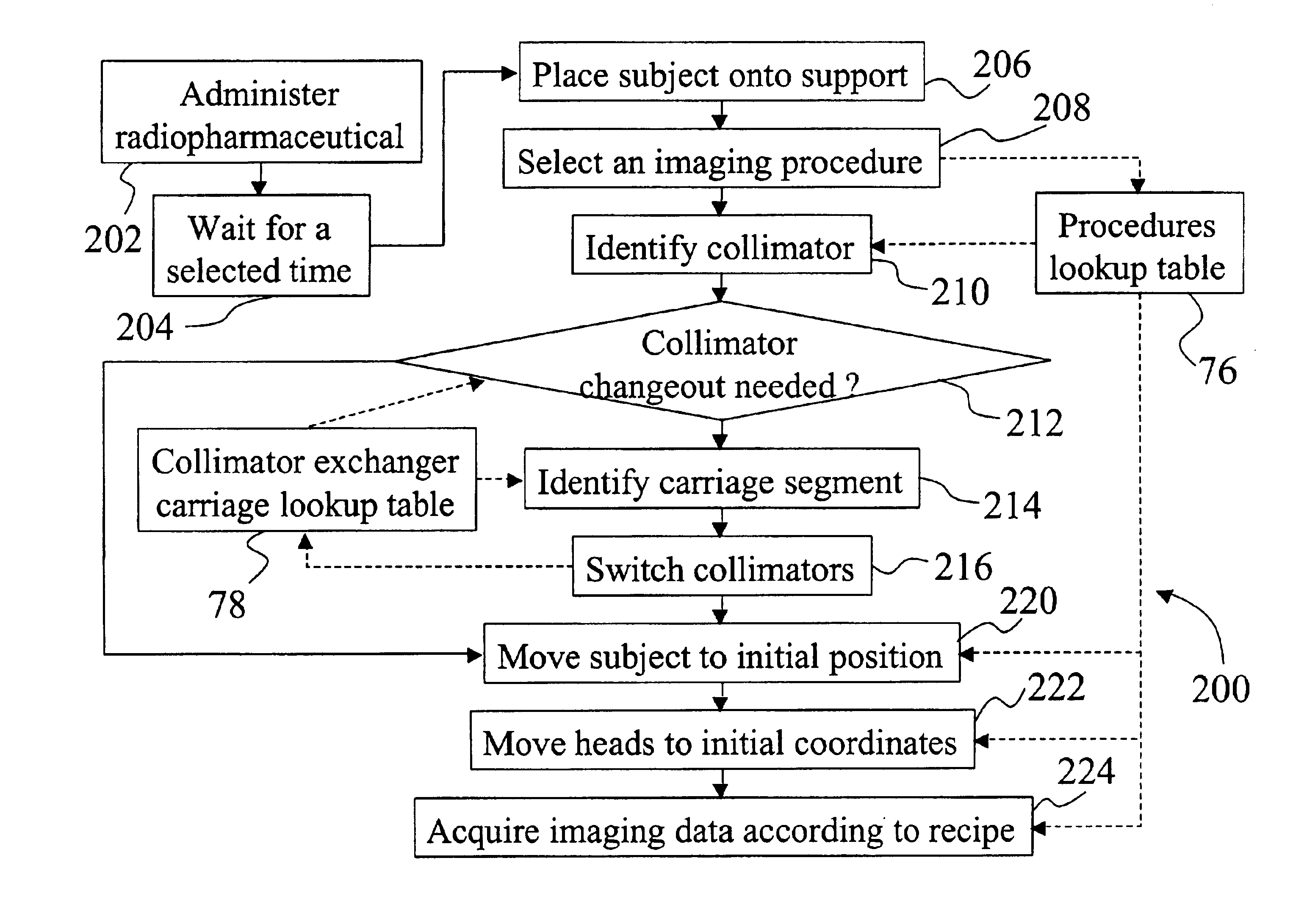
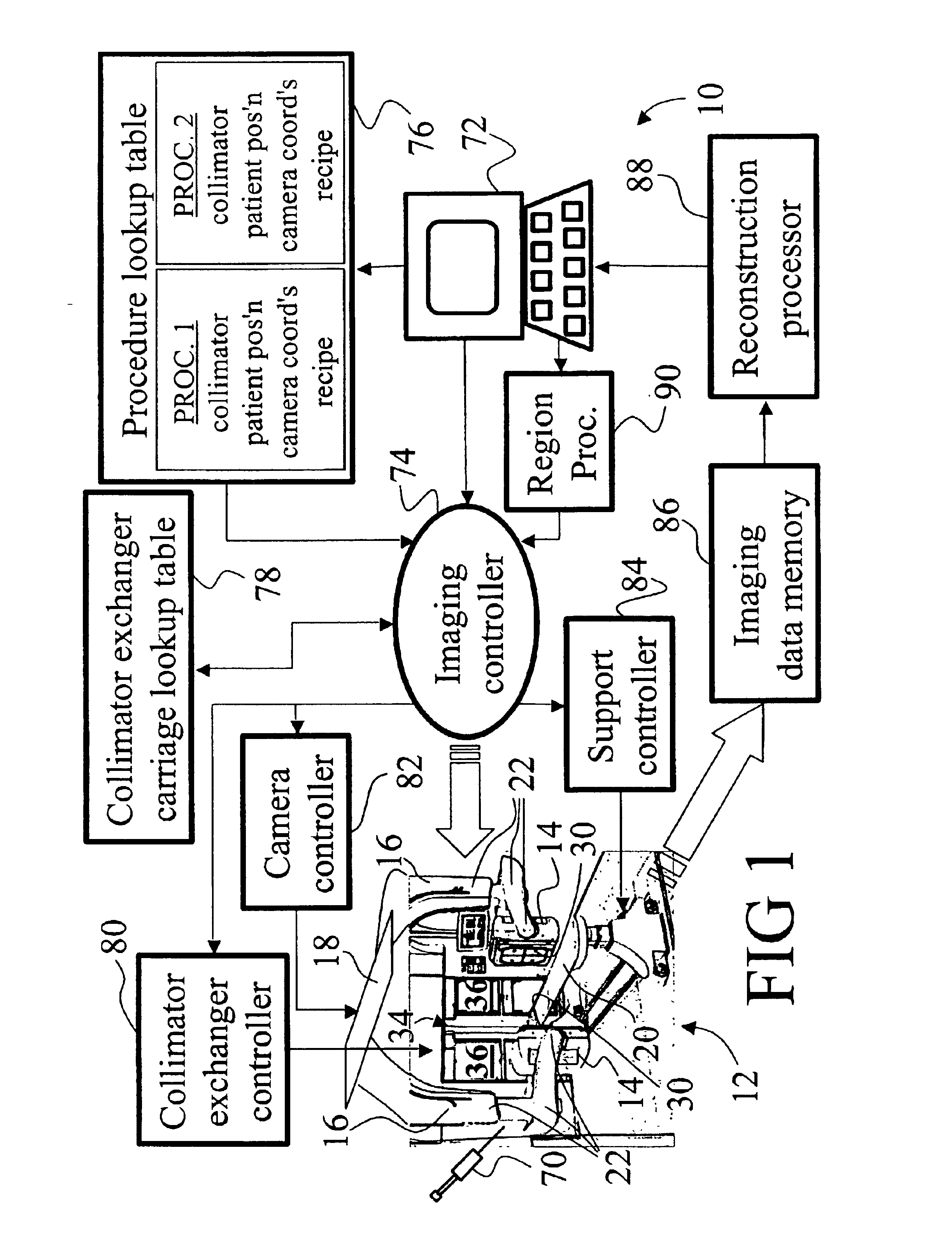
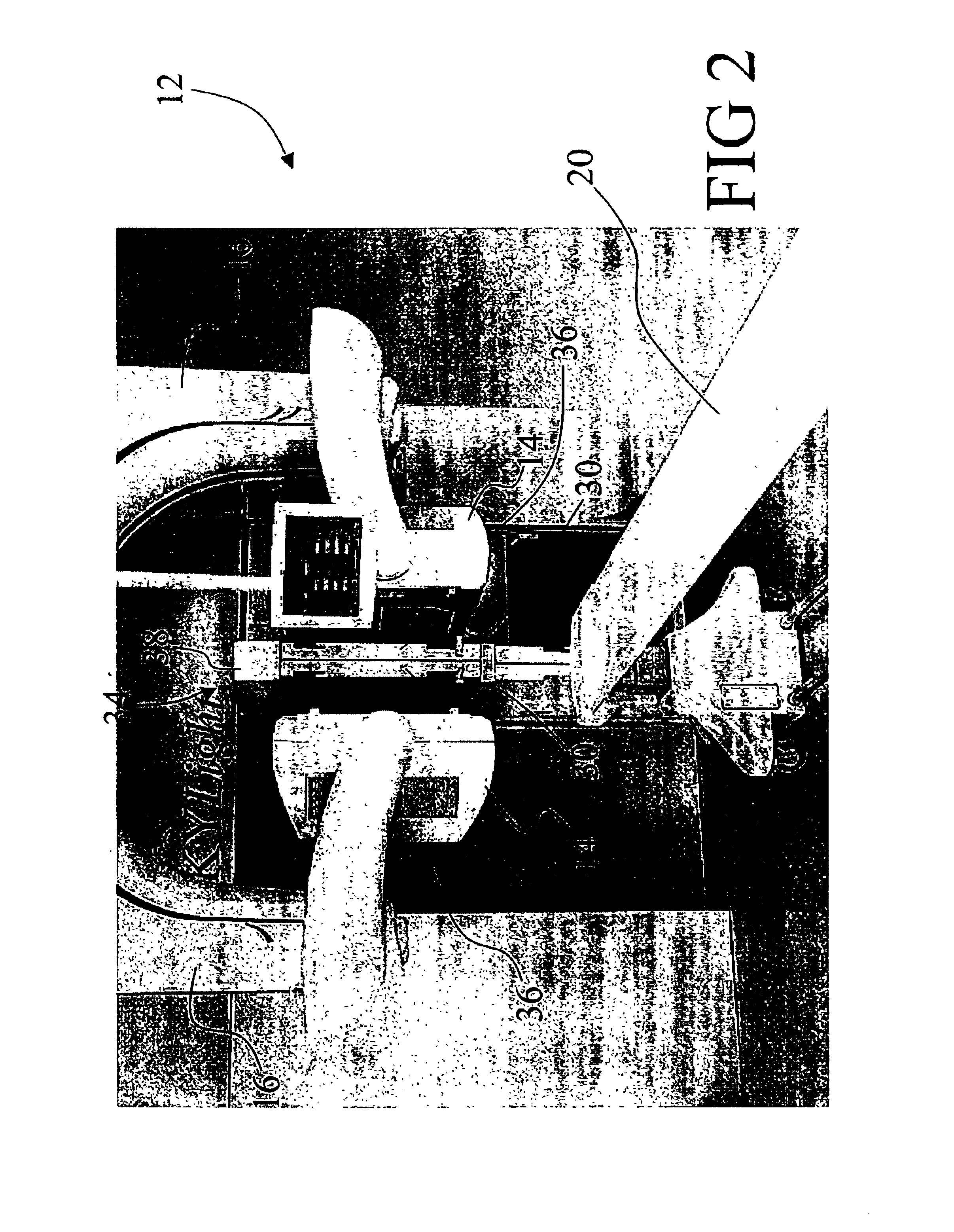
[0030]With reference to FIG. 1, a nuclear imaging system 10 includes a gamma camera 12. One or more detector heads 14, e.g. two heads in FIG. 1, are mounted on robotic arms 16 that in turn mount to an overhead mechanical track 18, e.g. a ceiling track, that moves the robotic arms 16 and attached heads 14 linearly relative to a subject support 20. Each robotic arm 16 includes a plurality of joints 22 that provide a plurality of degrees of movement freedom for the attached head 14, such as rotation about an axis through the head, vertical motion, tomographic rotation about the subject support 20, and the like.

[0031]In a preferred embodiment, each detector head 14 includes a scintillator and an array of photomultiplier tubes arranged to view the scintillator and detect optical flashes or scintillations resulting from impingement of radiation particles on the scintillator. Instead of photomultiplier tubes, photodiodes or other optical detectors can be used. Based upon the relative inten...


PUM


Abstract

A method (200) for setting up and performing a medical imaging session of a subject using a gamma camera (12, 52) includes selecting (208) an imaging procedure on an input terminal (72). A procedure table (76) is electronically accessed to retrieve at least one of: (1) an identification (210) of a collimator (30) to be used with a detector head (14, 54) in the selected imaging procedure; and (2) an initial imaging position of the detector head (14, 54) for the selected imaging procedure. A currently mounted collimator that is currently attached to the detector head (14, 54) is electronically identified. At least one of: (1) loading (216) the identified collimator onto the detector head (14, 54); and (2) positioning (220) the detector head in the identified initial imaging position, is automatically controlled.

Description

BACKGROUND OF THE INVENTION[0001]The present invention relates to the diagnostic imaging arts. It particularly relates to nuclear medical imaging using gamma cameras each including at least a two-dimensional scintillation detector and a collimator, and will be described with particular reference thereto. However, the invention will also find application in conjunction with other imaging modalities.[0002]In nuclear medical imaging, a subject is administered a radiopharmaceutical by ingestion, injection, or another delivery pathway. The radiopharmaceutical is preferably absorbed by one or more organs of interest, such as bone tissue, the liver, the heart, the vascular system, tumors, or other organs or tissues. Radiation generated by the radiopharmaceutical is emitted from the body and detected by one or more detector heads of a gamma camera. Each detector head typically include a scintillation crystal or crystal array facing the subject and an array of photomultiplier tubes, photodio...

Claims


Application Information

Patent Timeline
Patent Type & Authority Patents(United States)
IPC IPC(8): G01T1/164G01T1/00G01T1/161
CPCG01T1/1648
Inventor GARRARD, JODY L.HALLETT, JEFF A.STEPHENS, GARY L.
Owner KONINKLIJKE PHILIPS ELECTRONICS NV
Why Eureka
Try Eureka