Quantitative pulmonary imaging

a pulmonary imaging and quantitative technology, applied in the field of noninvasive methods, can solve the problems of limited spatial resolution, clinically significant limitations, and well-documented existing techniques, and achieve the effects of reducing the number of patients with advanced emphysema, and reducing the number of patients

Inactive Publication Date: 2005-07-05
THE TRUSTEES OF THE UNIV OF PENNSYLVANIA
View PDF7 Cites 21 Cited by
  • Summary
  • Abstract
  • Description
  • Claims
  • Application Information

AI Technical Summary

Benefits of technology

[0013]Using an inhaled noble gas, preferably laser-hyperpolarized helium-3 (3He) gas (HP-3He), three-dimensional MRI images are rapidly obtained. The HP-3He acts as a contrast agent, thereby providing unparalleled, high resolution images defining the air spaces.

Problems solved by technology

However, existing techniques have well-documented, clinically significant limitations.
Moreover they are unfortunately limited in spatial resolution, and they involve the mandatory exposure of both the patient and the practitioner to radioactive agents.
They also require prolonged breathing from an increased resistance closed system through a tight-fitting facemask, an effort that some patients with advanced emphysema are unable to tolerate.
Likewise, without blood flow to an alveolus, wasted ventilation or dead space occurs.
Pulmonary functional imaging methods include qualitative tests that provide valuable clinical information, but each currently available test has a limitation.
The radionuclide V / Q scan has inherently poor spatial relations, lacks the ability to image cross-sectional views, and deposits radiolabeled aerosols in the central airways of the patient, causing errors.
It is the gold-standard physiological test of lung V / Q relationships, but even MIGET has characteristic and problematic limitations, which have prevented its widespread clinical application.
To date, no available technique met all of these requirements.

Method used


Image

  • Quantitative pulmonary imaging
  • Quantitative pulmonary imaging
  • Quantitative pulmonary imaging

Examples


examples

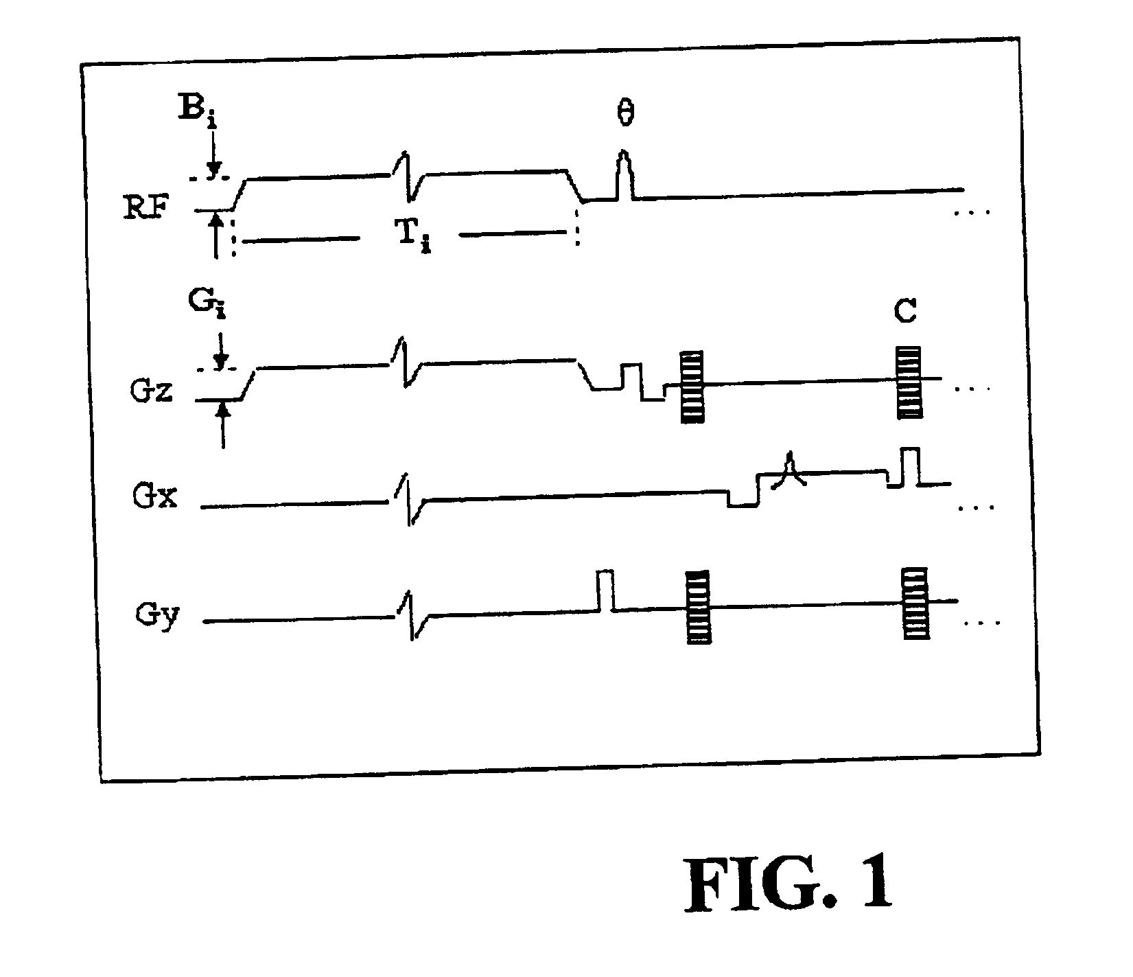
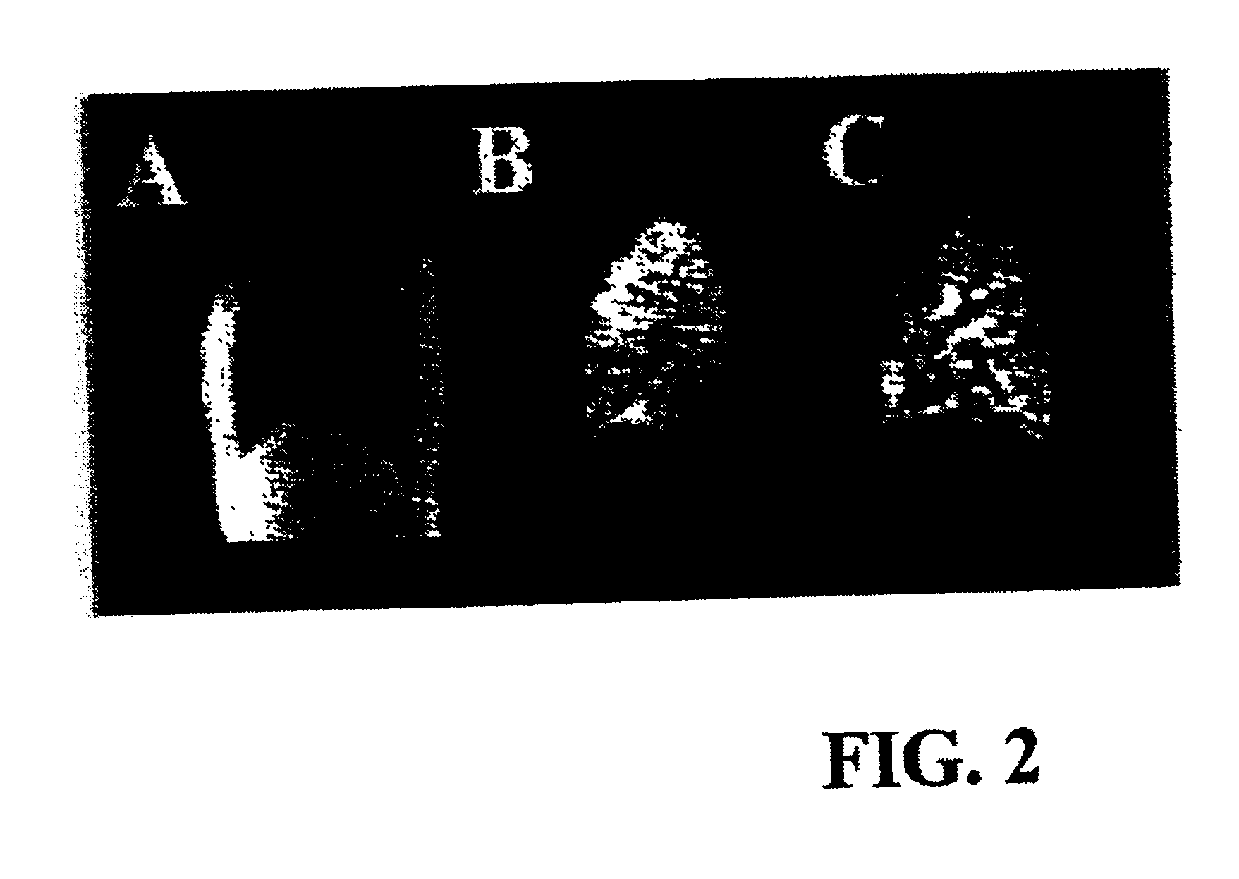
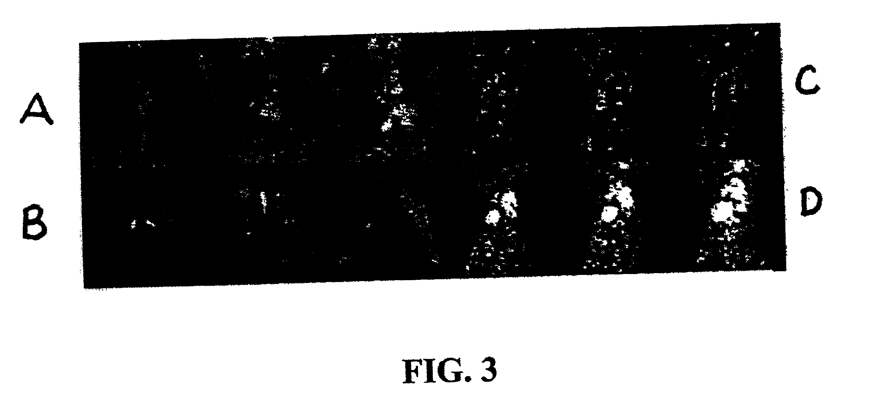
[0075]To confirm the effectiveness of the disclosed method, experiments were performed after informed consent was obtained from the patients using a medically approved protocol. Six patients were imaged. Three normal subjects, two patients with advanced emphysema, and one woman with a pulmonary embolus (PE), documented by history and V / Q scan, were selected to undergo magnetic resonance imaging. The two emphysema patients had each previously undergone single orthotopic lung transplantation (6 months and 5 years prior to imaging). Neither transplant patient had a baseline oxygen requirement, and both were free of respiratory symptoms at the time of imaging. Single lung transplant patients were chosen so that a direct comparison could be made between a severely obstructed and a relatively normal lung in the same patient. In all of the patients, chest radiographs showed no infiltrates or other active disease. The patient with PE was receiving intravenous heparin at the time of imaging....


PUM


Abstract

The present invention comprises imaging and quantitative measurement of lung ventilation, particularly in a human lung. Methods for quantitative imaging of lung ventilation, and the further provided systems and algorithmic tools therefore, comprise three primary components: the combined MRI ventilation/perfusion (V/Q) imaging techniques using hyperpolarized helium-3 (3He) gas (H3He); the three-dimensional quantitative imaging of absolute lung perfusion (Q) and collection of local magnetic resonance image data therefrom to produce an absolute lung perfusion image data; and the algorithmic co-registration of the two image data sets, (HP-3He MRI image of V/Q and MR imaging of quantitative perfusion (Q) in the lung). From the data acquired in the combined data sets and their spatial co-registration, absolute ventilation (V) is computed.

Description

REFERENCE TO RELATED APPLICATIONS[0001]This application claims priority to No. 60 / 267,282 filed Feb. 8, 2001, herein incorporated by reference in its entirety.GOVERNMENT INTERESTS[0002]This invention was supported in part by Grant Nos. K23 HL04486, RR02305, and R01-HL-64741 from the U.S. National Institutes of Health. The Government may have certain rights in this invention.FIELD OF THE INVENTION[0003]The present invention provides a novel non-invasive method to quantitatively evaluate pulmonary ventilation and perfusion, combining the use of a hyperpolarized noble gas for ventilation MR images and arterial spin-tagging for perfusion MR imaging. Also provided are the algorithmic tools necessary to calculate the quantitative imaging of lung ventilation.BACKGROUND OF THE INVENTION[0004]The accurate measurement of pulmonary ventilation (V), perfusion (Q), and V / Q distribution is of great importance in medicine. V / Q relationships are used to diagnose and treat pulmonary disorders, inclu...

Claims


Application Information

Patent Timeline
Patent Type & Authority Patents(United States)
IPC IPC(8): G01R33/54
CPCG01R33/5601
Inventor BAUMGARDNER, JAMESLIPSON, DAVIDRIZI, RAHIMROBERTS, DAVIDSCHNALL, MITCHELL
Owner THE TRUSTEES OF THE UNIV OF PENNSYLVANIA
Why Eureka
Try Eureka