

Detection and typing of human papillomavirus using PNA probes
a technology of human papillomavirus and pna probes, which is applied in the field of detection and typing of human papillomavirus using pna probes, can solve the problems of large quantities of reagents and sample dna, large amount of reagents, and large amount of probes, and achieve the effect of reducing the amount of probes needed
- Summary
- Abstract
- Description
- Claims
- Application Information
AI Technical Summary
Benefits of technology
Problems solved by technology
Method used
Image

Examples
example 1
Constructing and Testing of PNA Probes.
[0057]Biotin-labeled PNA probes complimentary to several high-risk types of HPV DNA were generated. The PNA probe sequences were based on areas of homology found in the L1 consensus region amplified by the MY09 / MY11 (SEQ ID NOS: 6 and 7) degenerate primer set with information from the GENBANK database. Five PNA sequences, PNA Probes I-V, are identified (SEQ ID NOS: 1-5). Biotin-labeled PNA, 14 to 15 bases in length were synthesized to specification by PerSeptive BioSystems (Framingham, Mass.).
[0058]HPV-infected gynecological samples were stored in methanol-based PreservCyt solution (Cytyc Corporation, Boxborough, Mass.). DNA from these samples was isolated and purified using Epicenter's Buccal swab DNA extraction kit (Madison, Wiss.) with the following steps: a 2 ml aliquot of the PreservCyt cell suspension was pelleted by centrifugation at 8,000×g for 5 minutes. The supernatant was removed and the cell pellet resuspended in Epicenter's DNA ext...
example 2
[0068]Step 1: Slide Preparation:
[0069]Cells fixed on ThinPrep slides are digested with proteolytic enzyme (i.e., 500 ul of 10 ug / ml proteinase K in 2×SSC, or with 500 ul of 0.2 N hydrochloric acid solution containing 0.05%-0.15% pepsin) at 37° C. for 30 minutes. The slides are then washed in 2×SSC for 2 minutes, ethanol dehydrated and air dried. Fifteen to 20 ul of the hybridization mixture containing 1 to 20 nM PNA probes are applied to each slide. Slides are then covered with glass coverslip and sealed.
[0070]Step 2: Hybridization
[0071]The PNA probes and target DNAs are co-denatured by placing the slides inside a thermocycler (MJ Research, Inc. Watertown, Mass.) and quickly bringing and maintaining the temperature of the thermocycler at 80° C. for three minutes. The interior temperature is afterwards rapidly dropped to 37° C., and the incubation is allowed to proceed from 15 minutes to 2 hours. Next, the slides are removed from the thermocycler, the coverslip a...
example 3
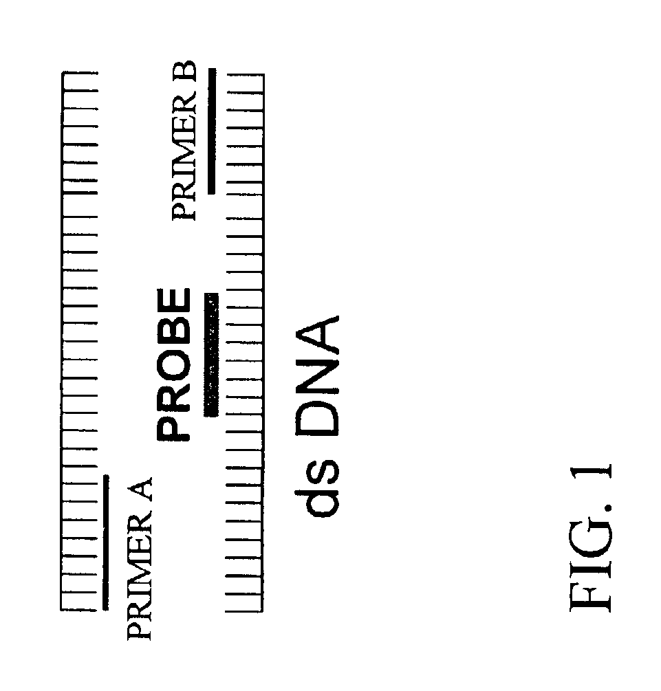
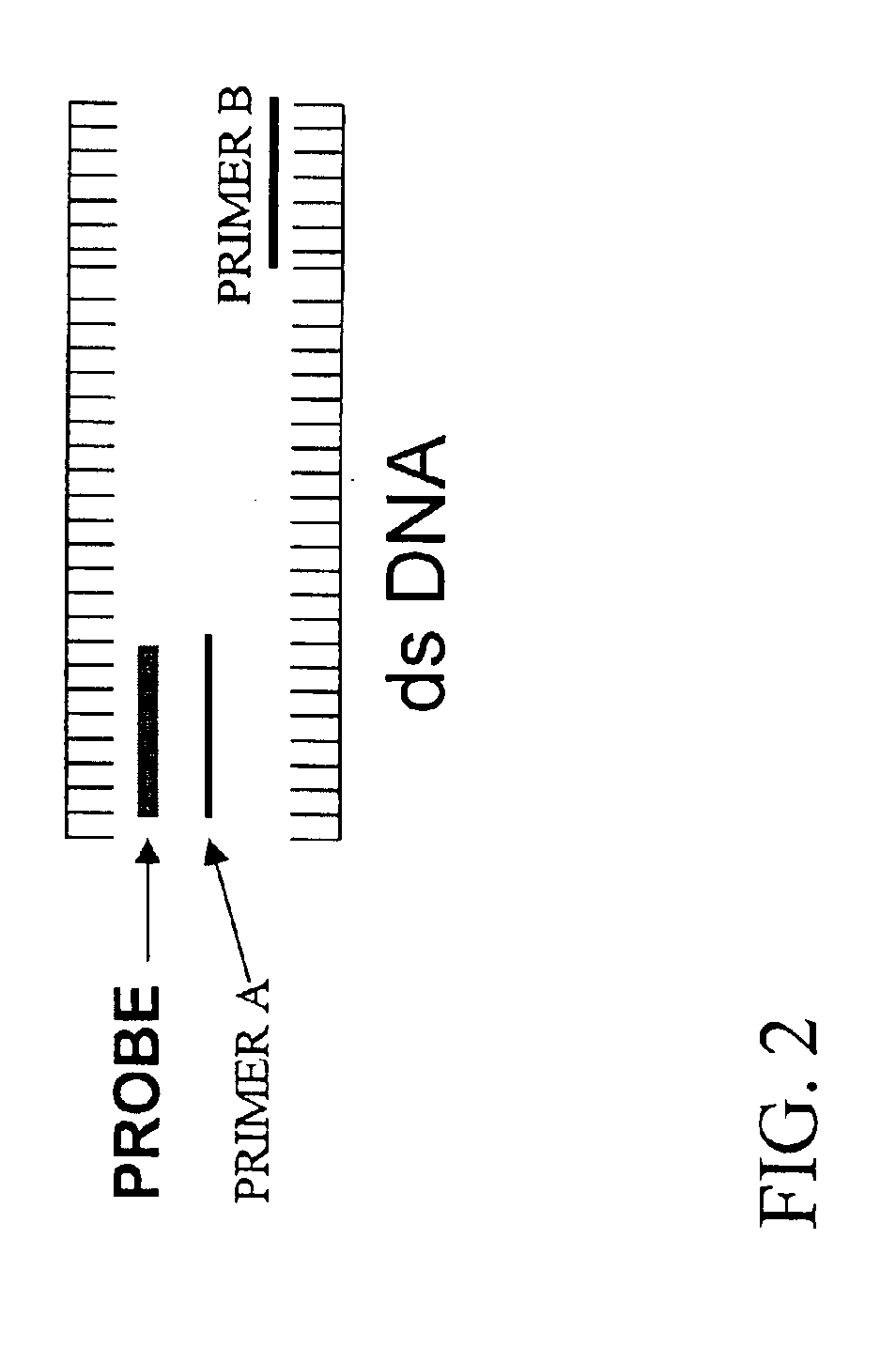
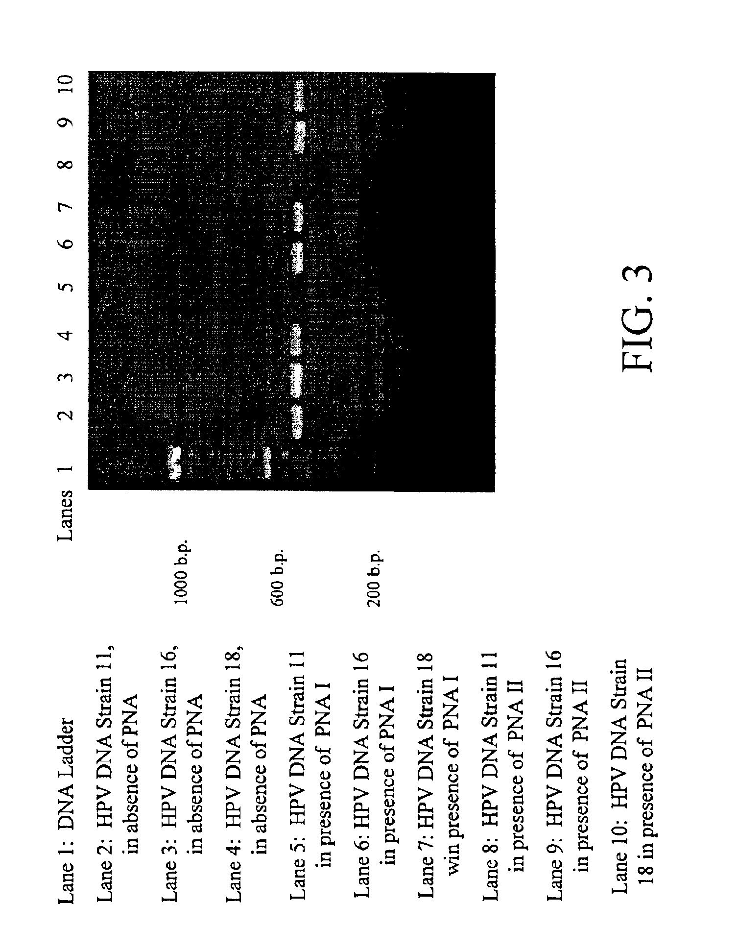
Testing PNA Probes Constructed According to Embodiments Illustrated by FIG. 1
[0084]PNA probes VI and VII (SEQ ID NOS: 23 and 24) were constructed in accordance with embodiments illustrated by FIG. 1. The sequences of both PNA probes VI and VII are substantially complementary to a homologous portion of low-risk HPV strains 6 and 11. Both probes bind to a portion between the binding sites for primer sets MY09 / MY11.
[0085]DNA solutions of a known HPV strain were used to test the selective blocking capabilities of both probes in accordance with the present invention. As shown in FIG. 3, no PCR amplicon was observed in DNA samples of HPV strain 11 when either HPV probe VI and VII was used. PCR amplicon of the expected length was detected through ethidium bromide staining of the DNA of HPV strains 16 or 18 in the presence of either probe VI and VII. These results indicated that probes VI and VII, as they were designed for, were able to block amplification of HPV strain 11 and not 16 or 18....
PUM
| Property | Measurement | Unit |
|---|---|---|
| Length | aaaaa | aaaaa |
| Strain point | aaaaa | aaaaa |
Abstract
Description
Claims
Application Information