Optical tomography of small objects using parallel ray illumination and post-specimen optical magnification

a technology of optical tomography and parallel beam, applied in the direction of material analysis using wave/particle radiation, fluorescence/phosphorescence, instruments, etc., can solve the problems of limited signal-to-noise ratio that can be achieved, high cost, and inability to use parallel beam x-ray sources in the portions of x-ray spectrum used in clinical and scientific imaging applications. achieve the signal-to-noise ratio significantly higher, easy and reproducible fabrication, and easy control

Inactive Publication Date: 2005-09-13
VISIONGATE
View PDF64 Cites 71 Cited by
  • Summary
  • Abstract
  • Description
  • Claims
  • Application Information

AI Technical Summary

Benefits of technology

[0016]Another advantage of the present invention, relative to a similar system employing divergent cone beam illumination geometry, is that it provides a PBOT system wherein a submicron source diameter is not required.
[0017]Another advantage of the present invention, relative to a similar system employing divergent cone beam illumination geometry, is that it provides a PBOT system wherein intensity distribution through a beam cross section can be more easily controlled and made more uniform or more nearly gaussian.
[0018]Another advantage of the present invention, relative to a similar system employing divergent cone beam illumination geometry, is that it provides a PBOT system wherein illumination intensity, herein also called fluence, at the specimen is increased by orders of magnitude.
[0019]Another advantage of the present invention, relative to a similar system employing divergent cone beam illumination geometry, is that it provides a PBOT system wherein signal-to-noise ratios achievable in the projection and reconstructed images is significantly higher.
[0020]Another advantage of the present invention, relative to a similar system employing divergent cone beam illumination geometry, is that it provides a PBOT system wherein required illumination sources can be more easily and reproducibly fabricated.
[0021]Another advantage of the present invention, relative to a similar system employing divergent cone beam illumination geometry, is that it provides a PBOT system wherein geometrical constraints and spatial tolerances required in terms of the location of system components relative to the imaged sample, most importantly the source-to-specimen distance, are considerably relaxed.

Problems solved by technology

Short exposures limit the signal to noise ratio that can be attained when imaging moving objects.
Except for synchrotrons, which are elaborate and expensive devices inaccessible to most research and healthcare professionals, parallel-beam x-ray sources are not available in the portions of the x-ray spectrum usually employed in clinical and scientific imaging applications.
Unfortunately, synchrotron-based microtomography devices are very expensive.
In such systems, divergence of the beam and small cone angle subtended by a specimen placed remotely from the source in order to approximate the parallel geometry result in very low fluence at the specimen and commensurately long scan times of, for example, hours to days.
For example, the achievable spatial resolution is limited by the source size, thus mandating a sub-micron source for microscopic and cellular imaging.
Further, the fluence or number of photons per unit area in the beam available from a sub-micron point source is very low, thereby placing stringent demands on the sensitivity and noise characteristics of the detector if adequate image quality and signal-to-noise ratio are to be obtained in the projection images.
It is challenging to produce the sub-micron source size necessary to provide sub-micron resolution for cone beam imaging.
Reproducibly fabricating such sub-micron light sources that produce relatively uniform or gaussian beam intensity profiles presents a significant challenge.
Differing source-to-specimen distances between the source-detector pairs may result in degradation of the reconstructed image quality.
Because projection magnification varies through the object space in cone beam imaging, the two-dimensional projection images or shadowgrams may be difficult to interpret.
For example, it may be difficult to extract diagnostically-relevant features from the projection images directly.
Cone beam projection geometry also requires 3D image reconstruction algorithms and computer programs that are complex and computationally intensive.

Method used


Image

  • Optical tomography of small objects using parallel ray illumination and post-specimen optical magnification
  • Optical tomography of small objects using parallel ray illumination and post-specimen optical magnification
  • Optical tomography of small objects using parallel ray illumination and post-specimen optical magnification

Examples


Embodiment Construction

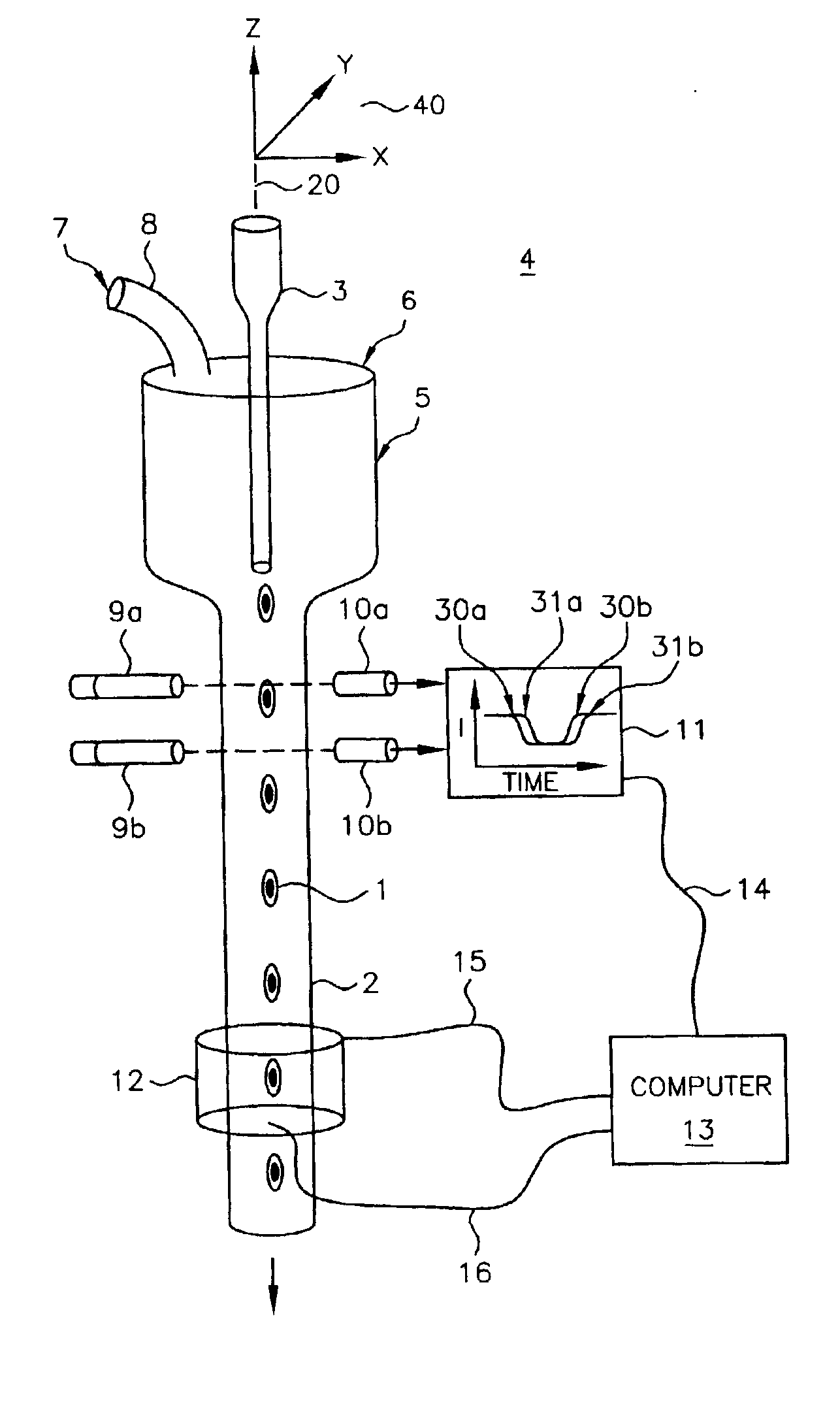
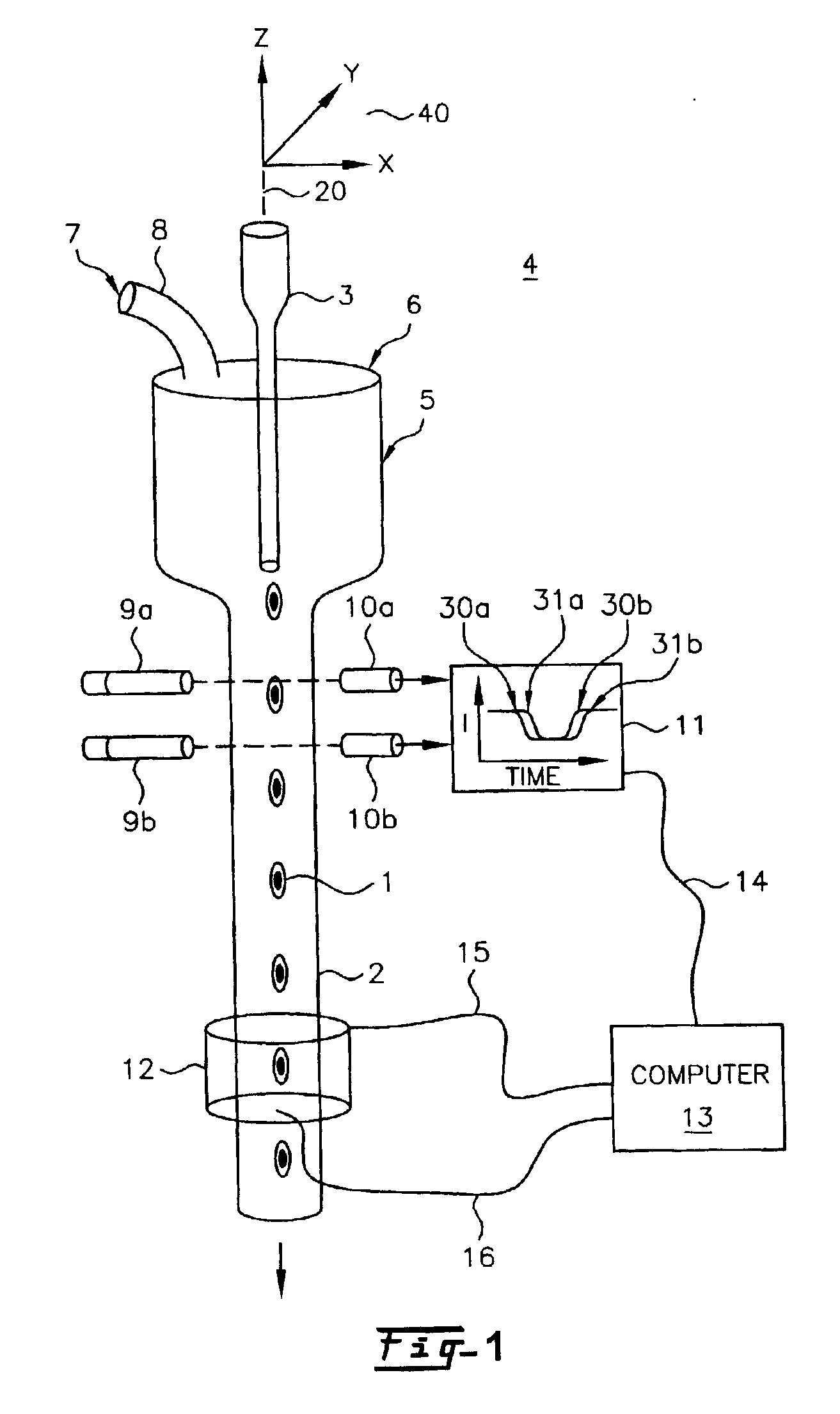
[0039]The invention is described herein with respect to specific examples relating to biological cells. It will be understood, however, that these examples are for the purpose of illustrating the principals of the invention, and that the invention is not so limited. In one example, constructing a three dimensional distribution of optical densities within a microscopic volume enables the quantification and the determination of the location of structures, molecules or molecular probes of interest. By using tagged molecular probes, the quantity of probes that attach to specific structures in the microscopic object may be measured. For illustrative purposes, an object such as a biological cell may be labeled with at least one stain or tagged molecular probe, and the measured amount and location of this probe may yield important information about the disease state of the cell, including, but not limited to, various cancers such as lung, breast, prostate, cervical and ovarian cancers.

[004...


PUM

PropertyMeasurementUnit
pixel sizeaaaaaaaaaa
diameteraaaaaaaaaa

Abstract

A parallel-beam optical tomography system for imaging an object of interest includes a parallel ray beam radiation source that illuminates the object of interest with a plurality of parallel radiation beams. After passing through the object of interest the pattern of transmitted or emitted radiation intensities is magnified by a post specimen optical element or elements. An object containing tube is located within an outer tube, wherein the object of interest is held within or flows through the object containing tube. A motor may be coupled to rotate and/or translate the object containing tube to present differing views of the object of interest. One or more detector arrays are located to receive the emerging radiation from the post specimen magnifying optic. Two- or three-dimensional images may be reconstructed from the magnified parallel projection data.

Description

RELATED APPLICATION[0001]This application is a continuation-in-part of co-pending U.S. application Ser. No. 09 / 927,151 of Alan C. Nelson, filed Aug. 10, 2001, issued on Feb. 18, 2003 as U.S. Pat. No. 6,522,775, that is in turn related to the provisional application of Alan C. Nelson, Ser. No. 60 / 279,244, filed Mar. 28, 2001, both entitled “APPARATUS AND METHOD FOR IMAGING SMALL OBJECTS IN A FLOW STREAM USING OPTICAL TOMOGRAPHY.”[0002]This application is also related to U.S. Pat. No. 6,591,003, issued Jul. 8, 2003 to Chu, entitled “OPTICAL TOMOGRAPHY OF SMALL MOVING OBJECTS USING TIME DELAY AND INTEGRATION IMAGING.”FIELD OF THE INVENTION[0003]The present invention relates to optical tomographic (OT) imaging systems in general, and, more particularly, to parallel-beam optical tomography (PBOT) where a small object, such as a biological cell, for example, is illuminated by an intense, parallel beam in the visible or ultraviolet portion of the electromagnetic spectrum and magnified tran...

Claims


Application Information

Patent Timeline
Patent Type & Authority Patents(United States)
IPC IPC(8): G01N15/14G01N23/02G01N23/04G06T11/00G01N21/05G01N21/03G01N21/17G01N21/59G01N21/64G01N21/78G01N33/543G01N33/554G01N33/60G06T1/00G06T3/00
CPCG01N15/14G01N15/147G01N21/0303G01N21/4795G01N21/6428G01N21/6456G01N23/046G06T11/006G01N21/05G01N2021/0307G01N2021/0392G01N2021/052G01N2021/1772G01N2021/1787G01N2021/5919G01N2021/5923G01N2021/5957G01N2201/0696G01N2223/419
Inventor JOHNSON, ROGER H.NELSON, ALAN C.
Owner VISIONGATE
Why Eureka
Try Eureka