Class characterization of circulating cancer cells isolated from body fluids and methods of use

Inactive Publication Date: 2005-11-01
GPB SCI
View PDF6 Cites 163 Cited by
  • Summary
  • Abstract
  • Description
  • Claims
  • Application Information

AI Technical Summary

Benefits of technology

[0032]In another embodiment, the presence of more terminal cancer cells in the second isolati

Problems solved by technology

The definitive classification of a malignant cell found in a body fluid sample remains challenging.
Due to a previous lack of specific identification criteria for circulating cancer cells, these cells could not be easily distinguished from blood cell precursors, a typical forms of cells normally found in the blood, or cells foreign to the blood.
Still, with current techniques and knowledge, the pathway of secondary tumor fo

Method used


Image

  • Class characterization of circulating cancer cells isolated from body fluids and methods of use
  • Class characterization of circulating cancer cells isolated from body fluids and methods of use
  • Class characterization of circulating cancer cells isolated from body fluids and methods of use

Examples


example 1

[0126]Cancer Cell Isolation Methodology and Idendfication

[0127]This example illustrates the cancer cell isolation methodology and identification of micrometastasis and other circulating cancer cell populations in prostate blood samples of patients diagnosed with prostate cancer.

Blood Collection

[0128]With informed consent, 10-20 ml of blood was drawn from the antecubital veins of control subjects and patients with prostate and breast cancer into Vacutainer tubes (Becton Dickinson; Franklin Lakes, N.J.) containing acid citrate dextrose (ACD) solution A as an anticoagulant. The samples were processed at room temperature via the isolation procedure described below within 24-hour hours, including the transport time. All protocols and consent forms were IRB (Institutional Review Board) by collaborating institutions.

Double Density Gradient Centrifugation (Prostate)

[0129]About 10 to 20 ml of blood were diluted to 30 ml with PBS at room temperature in a 50 ml polypropylene tube. Tubes were c...

example 2

Morphological Analyses of Circulating Cancer Cells

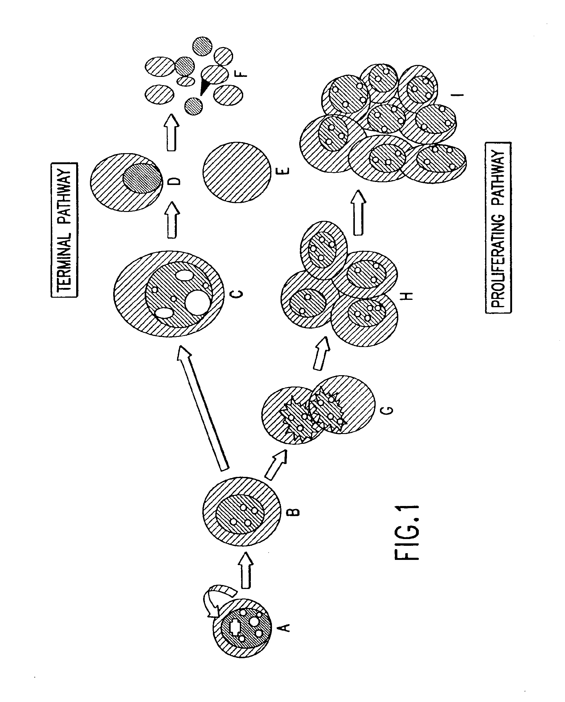
[0140]This example shows the data for the neoplastic developmental pathway. The identification of cell types of circulating cancer cells for characterization is based upon cytological analysis to assess whether the cells are terminal or proliferative. The role of these isolated cells in the formation of circulating microtumors and resultant metastases is also of interest.

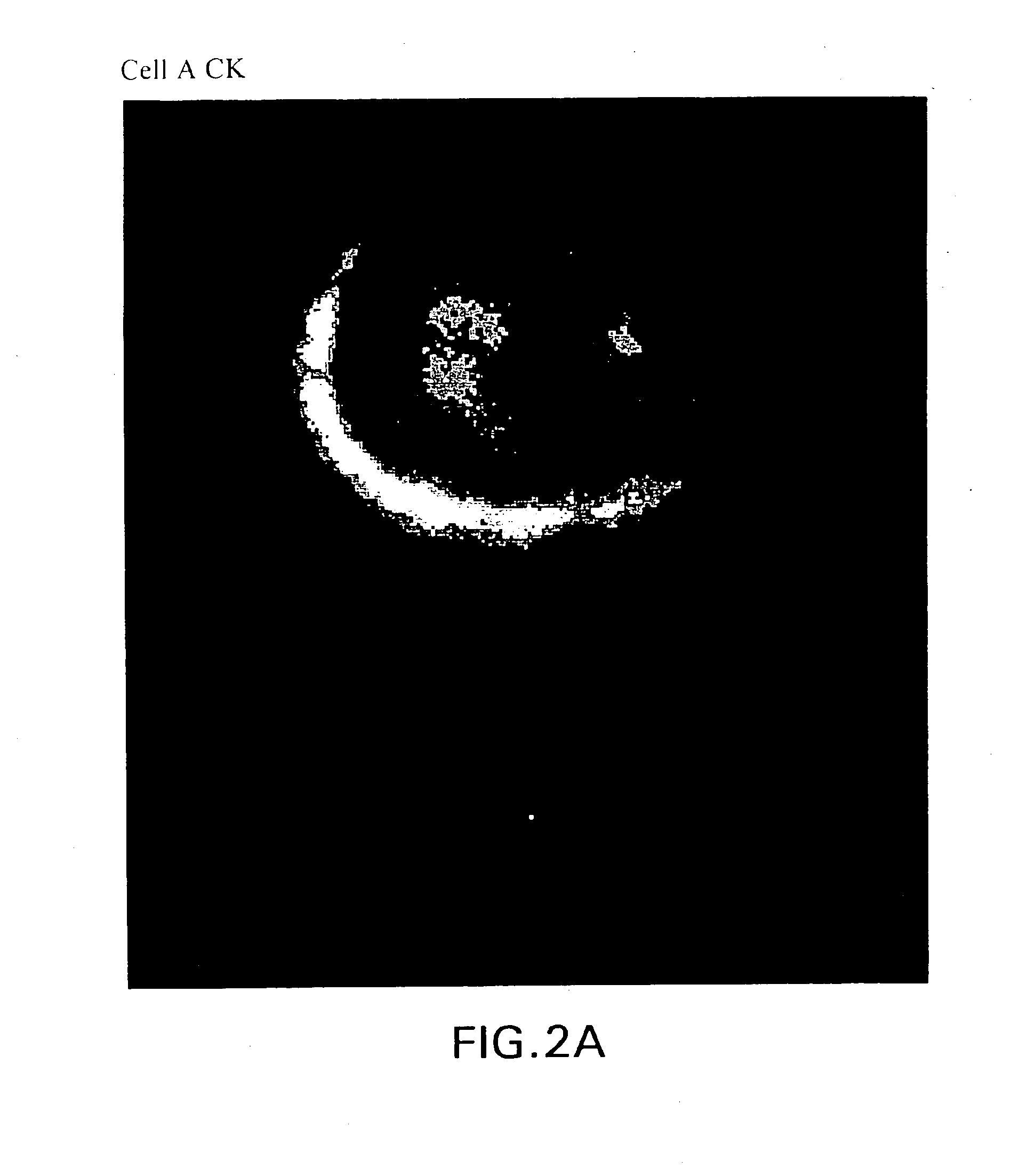
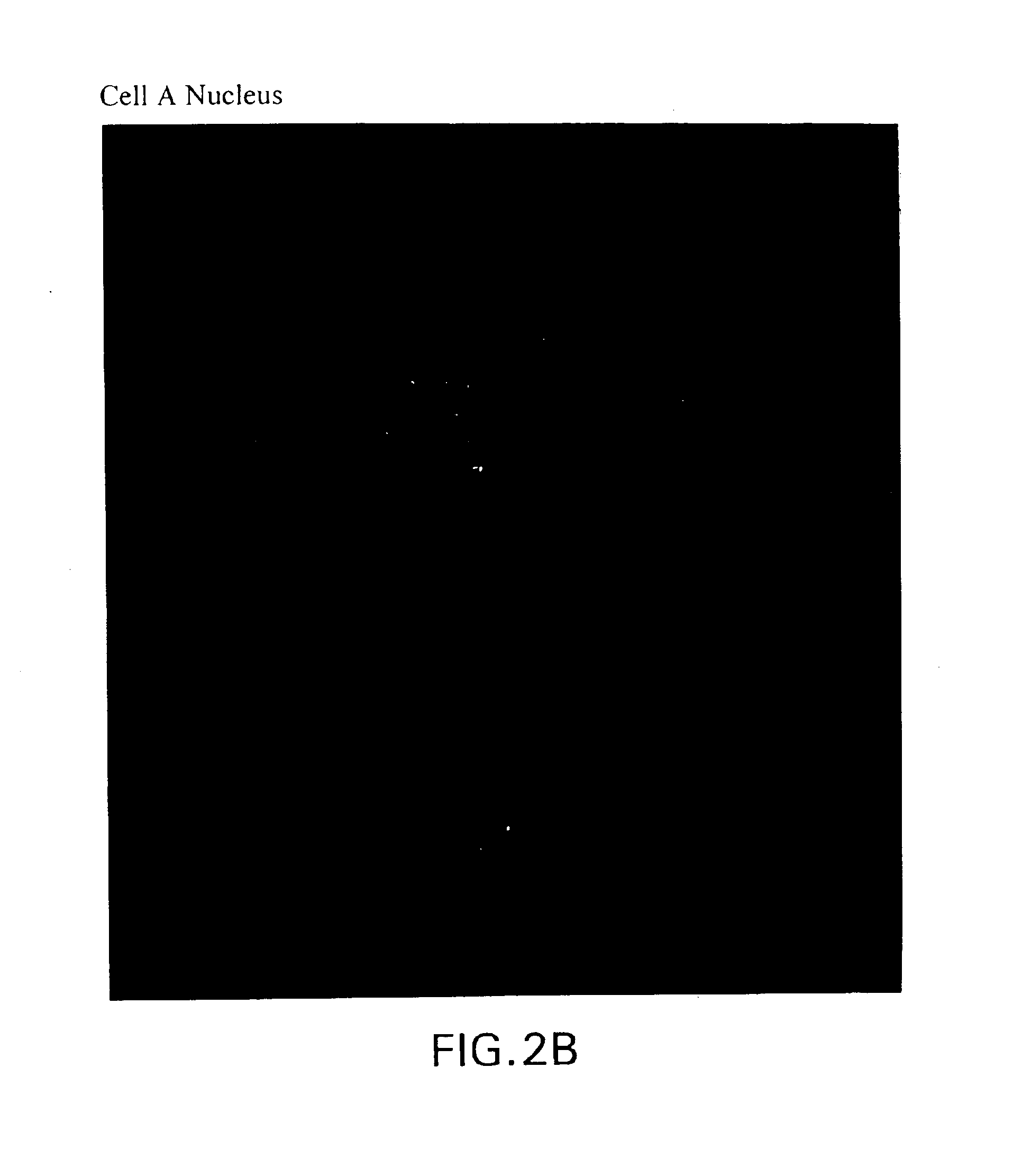
[0141]Cancer cells from 18 breast cancer patients were isolated according to the methodology set forth in Example 1. Based on the dynamic neoplastic developmental pathway, the cells were imaged and viewed using a computerized fluorescent microscope to determine the cell types are set forth in FIGS. 1A-1IFIGS. 2A-2S are monochrome images of the cell types depicted in FIGS. 1A-1I. FIGS. 2 through 1I depict monochrome images of cellular cytokeratin staining (“CK”) (CAM 5.2 labeled with FITC), thymnidylate synthetase staining (TS monoclonal antibody conjugated to FITC), ...

example 3

[0146]Determination of Aneuploidy via DNA Quantification Method

[0147]This example illustrates a method to measure the nuclear DNA content in single cancer cells in comparison to white blood cells found within the same sample as a measure of aneuploidy. The fluorochrome, 4′,6-diamidino-2-phenylindole (DAPI), binds to DNA with high specificity and the complex exhibits intense fluorescence (IFI—Integrated Fluorescent Intensity or # of pixels (area) multiplied by average fluorescence / pixel (density 1 μm)). This has permitted the measurement of DNA in nuclei and viral particles (Rao, JY et al, Cancer Epidemiology, Biomarkers &Prevention, 7: 1027-1033 (1998)), and in breast cancer cells (Coleman, AW, et al, J. Histochem.&Cytochem. 29: 959-968 (1981)).

[0148]The basis for the quantitative fluorescence image assay is a comparison of the DNA content of a reference cell, white blood cells (WBC) in this case, with the circulating epithelial cells (CEC) in question. Circulating WBCs are usually ...


PUM


Abstract

The present invention relates to the identification and characterization of classes and subclasses of circulating cancer cells, including microtumors from body fluid samples using molecular, cytological, and morphological analyses, and methods for staging patients and measuring the efficacy of medical treatments.

Description

CROSS-REFERENCE TO RELATED APPLICATIONS[0001]This application claims the benefit of U.S. Provisional Application No. 60 / 119,460, filed Feb. 10, 1999, and U.S. Provisional Application No. 60 / 159,558, filed Oct. 15, 1999, the contents of each which are fully incorporated by reference herein.STATEMENT AS TO THE RIGHTS TO INVENTIONS MADE UNDERFEDERALLY-SPONSORED RESEARCH AND DEVELOPMENT[0002]Part of the work performed during development of this invention utilized U.S. Government funds. The U.S. Government has certain rights in this invention pursuant to Grant No. DOD #980996.BACKGROUND OF THE INVENTION[0003]1. Field of the Invention[0004]The present invention is directed to the identification and characterization of classes and subclasses of circulating cancer cells, including microtumors, from body fluid samples.[0005]2. Related Art[0006]Metastases are established potentially by cancer cells, derived from a primary or a secondary tumor site, that are released into the circulation via t...

Claims


Application Information

Patent Timeline
IPC IPC(8): G01N33/50G01N33/574
CPCG01N33/5091G01N33/57496
Inventor WANG, ZHENG-PINTS'O, PAUL O. P.
Owner GPB SCI
Why Eureka
Try Eureka