

Methods, devices, arrays and kits for detecting and analyzing biomolecules
a biomolecule and array technology, applied in the field of biomolecule detection and analysis methods, can solve the problems of low biomolecule specificity and high affinity of certain membranes used in such methods, and achieve the effect of increasing specificity
- Summary
- Abstract
- Description
- Claims
- Application Information
AI Technical Summary
Benefits of technology
Problems solved by technology
Method used
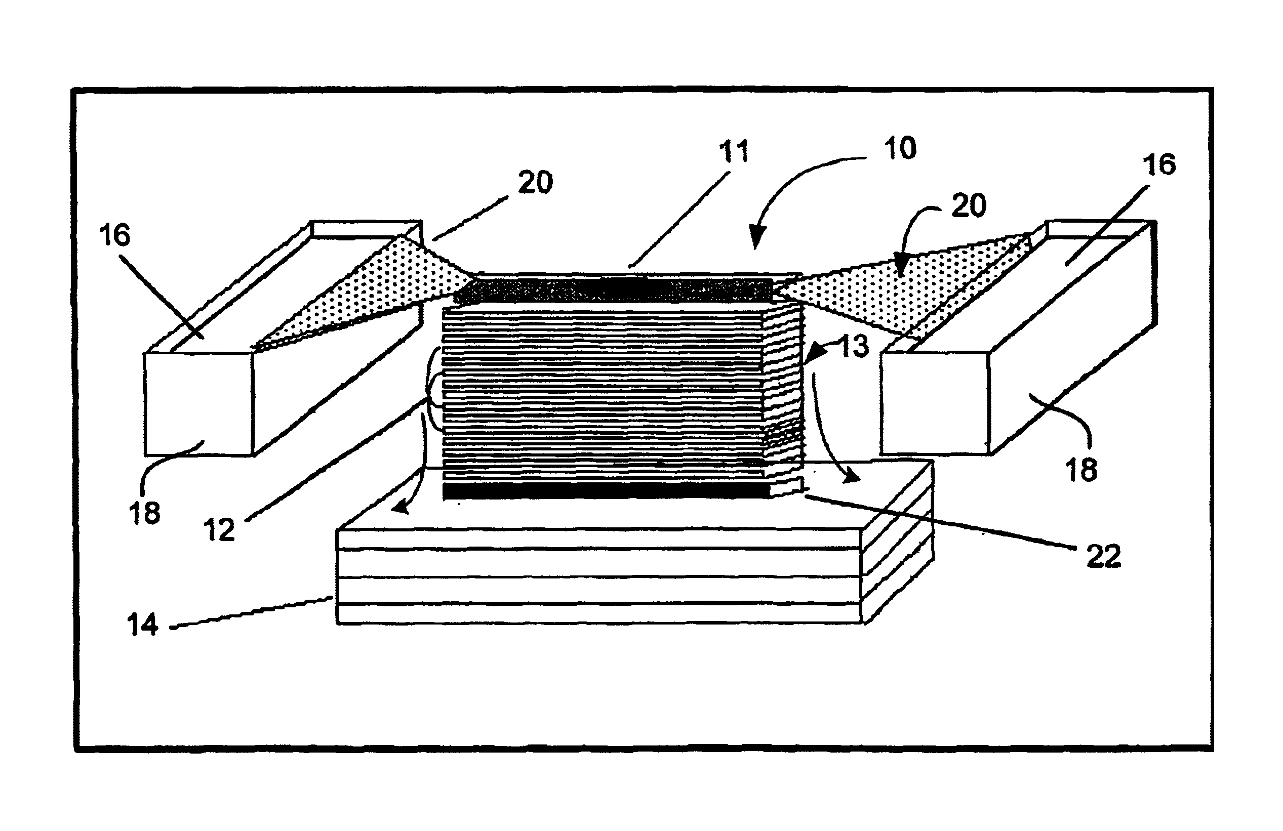
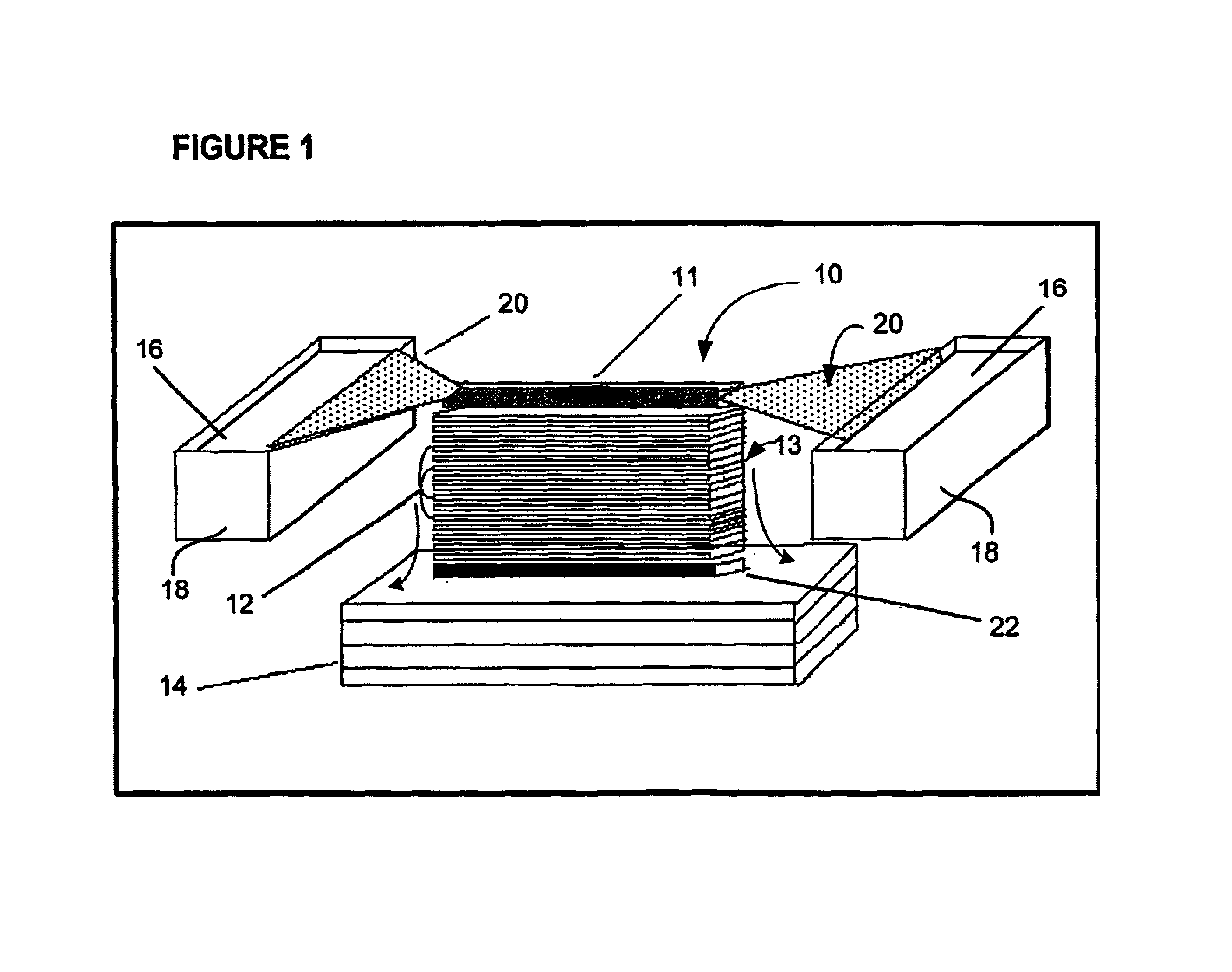
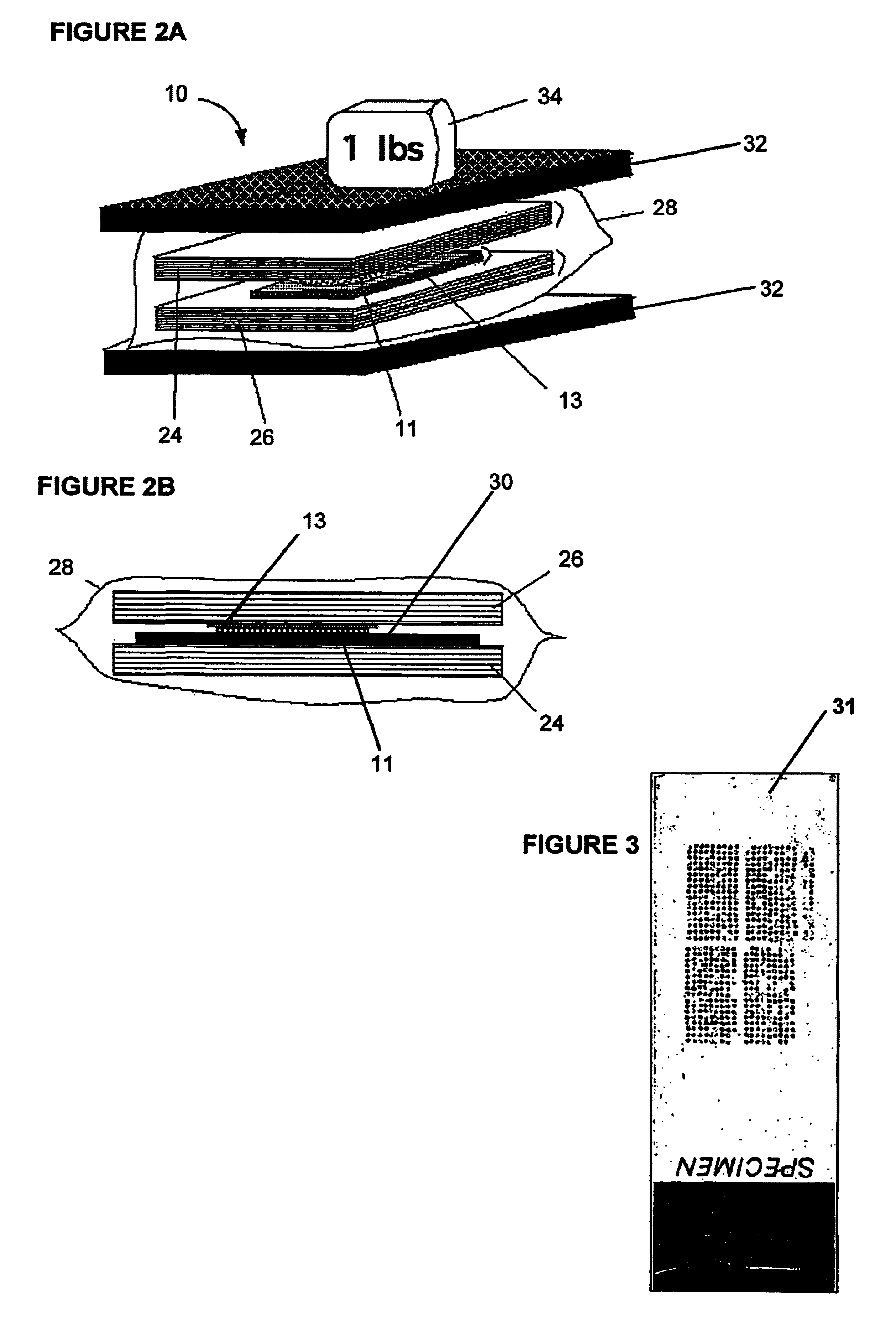
Image

Examples
example detection
Chemistries with Detector Cocktails
[0241]In certain embodiments, after proteins have been transferred through the membrane stack, individual membranes layers are separated and each is incubated in a separate antibody (or other detector molecule) cocktail. A key advantage of creating multiple replicate blots is that many more detector molecules (e.g., antibodies) can be usefully employed than if all of the detectors had to be crowded onto a single blot.
[0242]An exemplary process for designing the ligand cocktails—and for determining which proteins will be identified on each membrane layer—is provided below. First the panel of proteins of interest is selected. These can be randomly selected proteins and / or proteins that are not directly related to one another or may be groups of known proteins previously implicated to play a role in one or more particular cellular phenomena (e.g. apoptosis, cell cycle progression) or a particular disease (e.g. prostate cancer specific antigen, PSA). T...
example 1
Construction of Polycarbonate Membranes for Protein Binding
[0287]Native, non-coated polycarbonate membrane (Millipore, Mass.) has low affinity and low binding capacity for proteins. To improve its protein binding characteristics, polycarbonate membranes were coated with either poly-L lysine (referred to as PC+Lysin in FIG. 26) or nitrocellulose (referred to as PC+NC in FIG. 26). Membranes (177 square centimeters) were immersed for one minute in 5 ml of either aqueous solution of 0.1% poly-L-lysine or 0.1-1.0% nitrocellulose solution in 100% methanol. After coating, membranes were suspended in vertical position and air-dried at room temperature for 5-10 minutes. Poly-L-lysine treated membranes were before use additionally baked for two hours at 50° C. Small squares (0.25 square centimeters) of both treated and non-treated membranes were incubated in TBST solution (50 mM TRIS pH 8.0, 150 mM NaCl and 0.05% Tween-20) with 1.0-100.0 ng / μl of goat immunoglobulin labeled with Cy3 fluoresce...
example 2
Testing the Porosity of Prepared Polycarbonate Membrane Layers
[0289]To demonstrate porosity of manufactured layers, native, poly-L-lysine or nitrocellulose coated membranes were blocked in 5% bovine serum albumen solution in 50 mM TRIS pH 8.0 to prevent any protein binding. Fifty-one square centimeter pieces were cut out and stacked together to make a pile. A non-blocked pure nitrocellulose layer was used at the bottom to capture proteins (NC-trap). Three adjacent 20 micrometer thick frozen sections of normal breast tissue were collected on a polycarbonate membrane with 5.0 um pore size and embedded in a 2% agarose gel and transferred side by side through each stack. Between 50 and 100 milliliters of TBST buffer was used per square centimeter of the membrane stack with average length of the transfer being 1 hour. After transfer, proteins remaining in the tissue sections and total proteins on the NC-trap were visualized by Ponceau S staining (SIGMA, Mo.).
[0290]As shown in FIG. 27, th...
PUM
| Property | Measurement | Unit |
|---|---|---|
| thickness | aaaaa | aaaaa |
| thickness | aaaaa | aaaaa |
| thick | aaaaa | aaaaa |
Abstract
Description
Claims
Application Information