Methods for delivering biologically active molecules into cells

a technology of biologically active molecules and cells, applied in the field of methods, can solve the problems of low transfection efficiency in primary cells, difficult production of clinical grade virions, and inability to achieve clinical grade virions,

Inactive Publication Date: 2005-12-13
US DEPT OF HEALTH & HUMAN SERVICES
View PDF5 Cites 17 Cited by
  • Summary
  • Abstract
  • Description
  • Claims
  • Application Information

AI Technical Summary

Problems solved by technology

However, the production of clinical grade virions remains arduous.
While these lipid and bioconjugate vectors are relatively simple to prepare, their stability in vivo and transfection efficiency in primary cells remains low.
Delivery of DNA to cancer cell lines via these folate receptors has been demonstrated, but transfection is limited to cells expressing the receptor at high levels

Method used


Image

  • Methods for delivering biologically active molecules into cells

Examples


example 1

Methods

A. Biotinylation of Cell Surfaces

1. Direct Biotinylation of Cell Surfaces

[0031]Human chronic myelogenous leukemia cells, line K562, are biotinylated as follows: 106 cells are incubated in a final concentration of 0.5 ng sulfo-NHS-Biotin per cell (Pierce Chemicals, Rockford, Ill.) in phosphate buffered saline (PBS) for 30 minutes at 4° C. and washed twice with PBS. For human erythroleukemia (HEL) cells, 5×10⁵ cells are incubated in a final concentration of 1.0 ng sulfo-NHS-Biotin per cell, and for buffy coat cells, 5×10⁶ cells are incubated in a final concentration of 0.1 ng sulfo-NHS-Biotin per cell. After biotinylation, cells are washed with PBS and transferred into 24-well plates in 1.5 ml of adequate cell media. Non-biotinylated cells are used as a control.

[0032]Human cells in situ are biotinylated by injecting sulfo-NHS-Biotin into the target tissue or organ at an amount of at least approximately 1 ng per cell. Human blood cells treated ex vivo using apheretic techniques ...

example 2

Avidin-FITC Complexes undergo Endocytosis after Binding to Biotinylated Cells

A. Directly Biotinylated Cells

[0043]Untreated K562 cells show very little autofluorescence or nonspecific binding of Av-FITC as assessed by flow cytometry and fluorescence microscopy. When Av-FITC is added to biotinylated K562 cells, Av-FITC is evenly distributed on cell surfaces. Thirty minutes after addition, fluorescein is detected inside endosomal compartments in all cells. The fluorescence gradually shifts from the cell surface to the cell interior during the next 24 hours, which is demonstrated by comparing the fluorescence and phase images of the same microscopic field. The fluorescein remains inside the cells without being recycled to the surface. No internalization is observed when cells are incubated at 4° C., and no exocytosis of fluorescent complexes is detected. A similar pattern and time-course can be observed using K562 and Jurkat cell lines.

[0044]Flow cytometry is used to determine whether a...

example 3

PEI-Avidin Conjugates Condense DNA and Efficiently Transfect Cultured Cells

A. Directly Biotinylated Cells

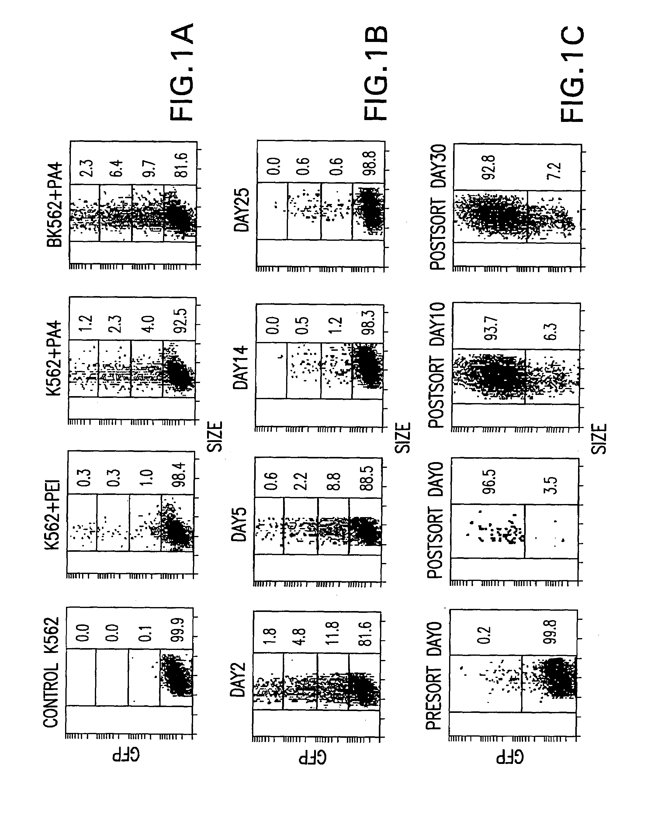
[0046]Three PEI-avidin conjugates were prepared with increasing content of avidin as described in the Materials and Methods. PA4, PA8 and PA16 define conjugates with avidin to PEI molar ratios of 4, 8 and 16 respectively. As assessed by gel retardation, cationic binding by PEI and all the PA conjugates completely neutralized the anionic charge of the plasmid DNA to prevent its electrophoresis at the N:P ratio of 3.2. Due to the additional cationic charge of avidin (see Savage, M D et al. 1994, Avidin-biotin Chemistry: A Handbook, 2nd Ed., Pierce Chemical Co., Rockford, Ill.) and possible differences in the protonation profile of the conjugated PEI, the PA conjugates caused gel retardation at a proportionally lower PEI level. As expected, the PA16 conjugates are most cationic with complete DNA gel retardation at an N:P ratio of 0.8.

[0047]Gene transfer efficiencies in K562 and biot...


PUM

PropertyMeasurementUnit
Surfaceaaaaaaaaaa

Abstract

Methods for delivering a biologically active molecule into a cell by linking a molecule to the cell surface, wherein the molecule can act as a surface receptor, then complexing the biologically active molecule with a ligand for the surface receptor, and finally contacting the biologically active molecule-ligand complex with the cell surface are disclosed. Delivery of any biologically active molecule, e.g. proteins, enzymes, nucleic acids, hormones, nucleic acids, and oligonucleotides, is contemplated. The use of biotin or biotinylated antibodies as the surface receptor is disclosed. The use of PEI and PEI-avidin conjugates complexed with oligonucleotides for delivery into a directly or indirectly biotinylated cell surface, along with the PEI-avidin-nucleic acid compositions, are disclosed. Primary and cultured cells with a covalently linked surface receptor molecule, such as biotin, on their surfaces are also disclosed.

Description

[0001]This application claims benefit to Provisional Application 60 / 116,240, filed Jan. 15, 1999.FIELD OF THE INVENTION[0002]This invention is in the field of methods and compositions for delivering biologically active molecules into cells.BACKGROUND OF THE INVENTION[0003]With the rapid increase in knowledge of the cellular mechanisms and processes that lead to disease or that are disrupted by genetic mutations, scientists can envisage potential solutions that involve the delivery of molecules into a cell that can then affect a change in a mechanism or process for therapeutic or prophylactic value. For example, several technologies to deliver nucleic acids, particularly DNA, into a cell have been described. Researchers have identified at least three requirements for accomplishing DNA delivery: 1) DNA has to be in a form that will associate with the cell membrane and be taken up by the cell; 2) the DNA has to be coupled with a molecule that is targeted to the correct types of cells, ...

Claims


Application Information

Patent Timeline
IPC IPC(8): A61K48/00C12N5/02C12N5/09C12N15/87
CPCA61K48/0008A61K48/0041C12N5/0694C12N15/87
Inventor MILLER, JEFFERY L.WOJDA, URSZULAGOLDSMITH, PAUL K.
Owner US DEPT OF HEALTH & HUMAN SERVICES
Why Eureka
Try Eureka