Grooved brachytherapy sources

a radioactive source and groove technology, applied in the field of radiotherapy, can solve the problems of difficult to achieve accurate placement of sources, adversely affect the accuracy of source placement, unsatisfactory side effects in patients, etc., and achieve the effect of enhancing the ultrasound visibility of radioactive sources, facilitating the detection of devices, and enhancing ultrasound reflection

Inactive Publication Date: 2006-08-01
GE HEALTHCARE LTD
View PDF18 Cites 67 Cited by
  • Summary
  • Abstract
  • Description
  • Claims
  • Application Information

AI Technical Summary

Benefits of technology

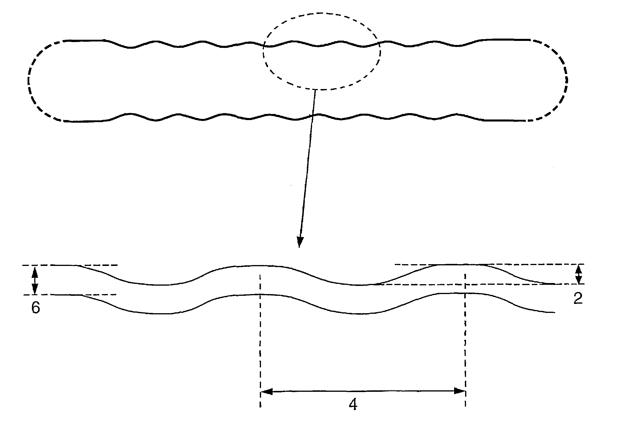
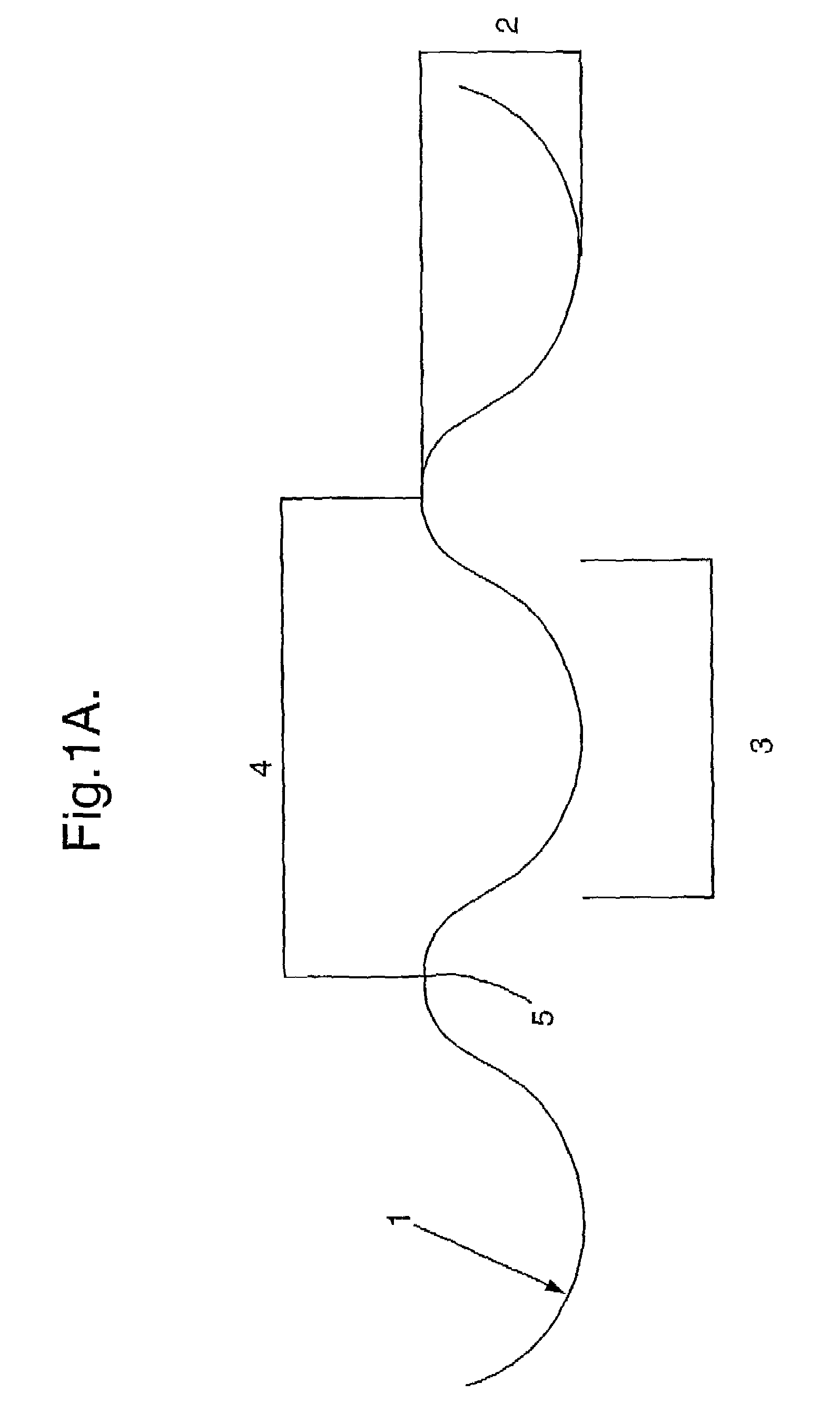
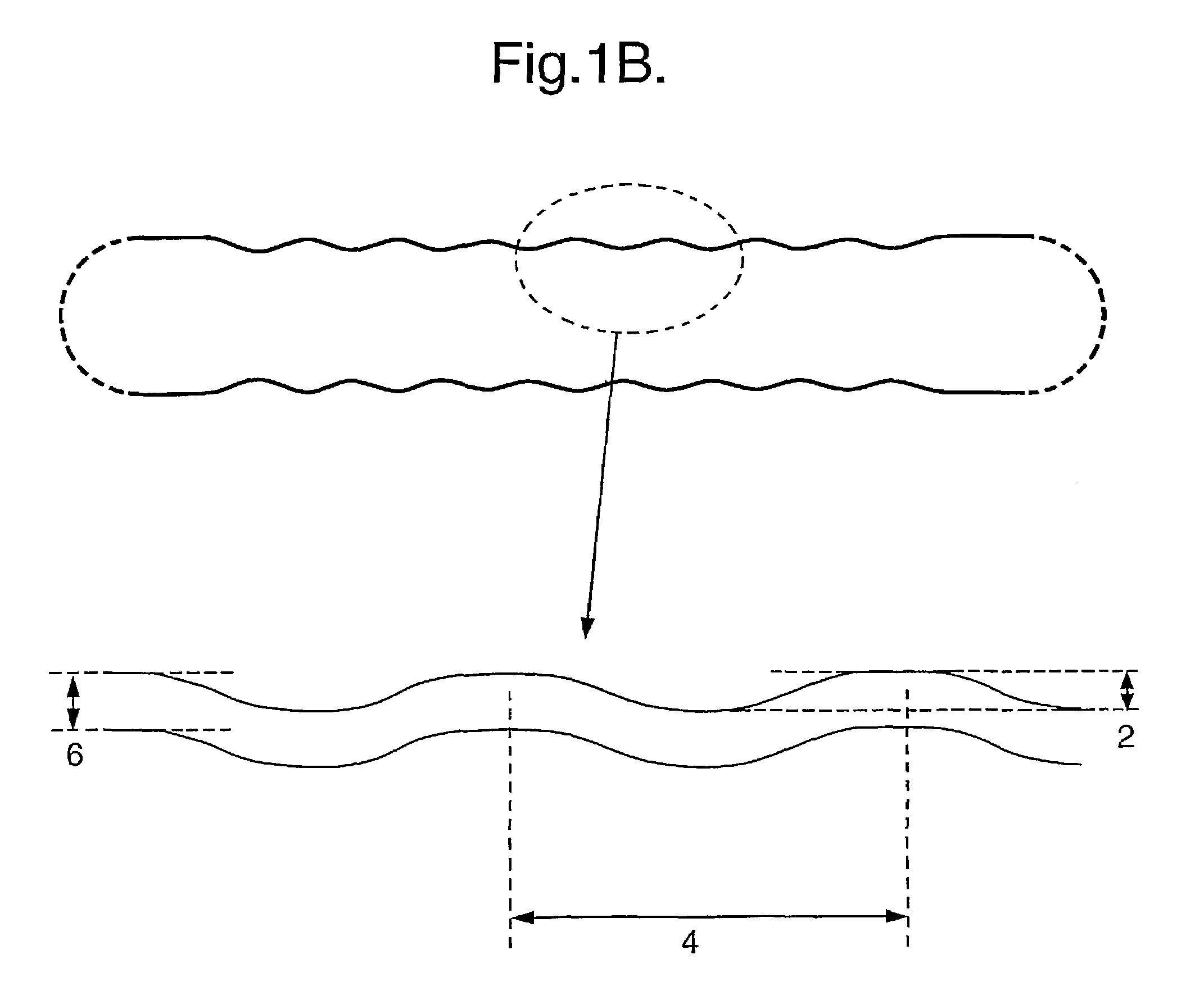
[0029]According to another aspect of the invention, there is provided a method for increasing the ultrasound visibility of a radioactive source for use in brachytherapy with a radioisotope and a sealed biocompatible container, comprising providing the outer surface or part of the outer surface the container with grooves of specific dimensions and arrangement, effective to enhance reflection of ultrasound to thus facilitate detection of the device in vivo.

Problems solved by technology

Such seeds are only suitable for use with radioisotopes which emit radiation which can penetrate the chamber / container walls.
However, the procedure is relatively invasive and often leads to undesirable side effects in the patient.
However, difficulties may arise during the radioactive source placement procedure which may adversely affect the accuracy of the placement of the source if only pre-placement images are used to guide the source placement.
Thus, it is difficult to achieve accurate placement of sources to achieve a desired dosage profile in brachytherapy using only knowledge of tissue anatomy and position that was obtained prior to the placement procedure.
Theory and practical experiments show that even at an angle of 8 degrees between the long axis of the seed and the ultrasound transducer (a deviation of 8° from orthogonal), the signal intensity drops by a factor of 100 (20 dB), and the seed becomes difficult to detect.
At an orientation of 10 degrees the seed is not possible to detect against a tissue background.
Thus, the relatively small size of current brachytherapy radioactive sources and the specular reflection properties of their surfaces makes them very difficult to detect by ultrasound imaging techniques.
While five of the seven catheter materials had good to excellent sonographic detection, nylon and polyethylene catheters were poorly visualised.
This is highly undesirable from a clinical perspective, as it may lead to underdosing of a tumour or other diseased tissue and / or unnecessary exposure of healthy tissue to radiation.

Method used


Image

  • Grooved brachytherapy sources
  • Grooved brachytherapy sources
  • Grooved brachytherapy sources

Examples


example 1

[0093]A wide band imaging ultrasound transducer ATL L10-5 was mounted in the wall of a water tank. The transducer was connected to an ATL HDI 5000 ultrasound scanner and imaging was performed at 6.5 MHz, a typical imaging frequency used in clinical transrectal ultrasound.

[0094]A brachytherapy seed was mounted on a holder located 50 mm from the transducer surface, which could be rotated to defined angles in relation to the direction of the ultrasound beam. The seed was glued on to the tip of a needle protruding from the specimen holder with cyanoacrylate glue so that the seed's centre of gravity coincided with the rotational axis of the holder. The angular rotation could be set with half a degree accuracy, which is of great importance given the high angular dependency of the ultrasound backscatter. The holder could also be adjusted by translation to position the seed in the focal point of the transducer and fixed throughout the experiments.

[0095]A series of measurements mapping the u...

example 2

[0096]A smooth steel rod and a rod with a square surface pattern was imaged in vitro as described in Example 1. Images are acquired at 0, 20 and 40 degrees rotation, and are shown in FIG. 2. The upper series of images is a smooth 0.8 mm diameter, 6.5 mm length steel rod while the lower series is a similar steel rod with a cut surface with 0.1 mm wide helical square grooves, having a spacing of 0.54 mm and with a depth of 0.05 mm.

example 3

[0097]An excised dog prostate was imaged in a water tank with an ATL HDI 5000 scanner using an imaging frequency of 6.5 MHz. Two steel rods as described in Example 2 were implanted using an 18G needle. The prostate with the rods implanted was then rotated and imaged at different angles—see FIG. 3.


PUM


Abstract

Radioactive sources, preferably radioactive seeds, for use in brachytherapy comprising a radioisotope within a sealed biocompatible container, wherein at least one part of the outer surface of the container is grooved, preferably with a curved groove. The grooved outer surface is preferably substantially free from angularities. Such grooves enhance the echogenicity of the source using medical ultrasound at a greater range of angles to the ultrasound probe, thus enhancing the ultrasound visibility of the source. Preferred radioisotopes are palladium-103 and iodine-125.

Description

FIELD OF INVENTION[0001]This invention relates to radiotherapy. More particularly, it relates to radioactive sources for use in brachytherapy, and in particular to grooved radioactive sources, with improved ultrasound imaging visibility.BACKGROUND OF INVENTION[0002]Brachytherapy is a general term, covering medical treatment which involves placement of a radioactive source near a diseased tissue, and may involve the temporary or permanent implantation or insertion of a radioactive source into the body of a patient. The radioactive source is thereby located in proximity to the area of the body to be treated. This has the advantage that a high dose of radiation may be delivered to the treatment site with relatively low dosages of radiation to surrounding or intervening healthy tissue.[0003]Brachytherapy has been proposed for use in the treatment of a variety of conditions, including arthritis and cancer, for example breast, brain, liver and ovarian cancer, and especially prostate cance...

Claims


Application Information

Patent Timeline
Patent Type & Authority Patents(United States)
IPC IPC(8): A61N5/00G21G4/08A61B19/00A61M36/04A61M36/12A61N5/10
CPCA61N5/1027A61B2019/5425A61N2005/1024A61B2090/3925
Inventor TORNES, AUDUNERIKSEN, MORTEN
Owner GE HEALTHCARE LTD
Why Eureka
Try Eureka