Optical methods and systems for rapid screening of the cervix

a technology of optical methods and systems, applied in the direction of fluorescence/phosphorescence, spectroscopy, instruments, etc., can solve the problems of increasing the overall cost of unnecessarily overloading the health care system, and difficulty in, if not impossible, to see the surface changes caused by the presence of lesions

Inactive Publication Date: 2006-10-24
LUMA IMAGING CORP
View PDF359 Cites 36 Cited by
  • Summary
  • Abstract
  • Description
  • Claims
  • Application Information

AI Technical Summary

Benefits of technology

[0014]Embodiments of the invention may include one or more of the following features. The computed distance may be a Mahalanobis distance, which may be computed by including Quadratic Discriminant Analysis (QDA) and / or Regularized Discriminant Analysis (RDA) to eliminate problems caused by singularities. The source of the optical radiation, which may be the cervical probe, and / or the detector may be placed so as not to contact the cervical tissue. The at least two pathological classes may include CIN (cervical intraepithelial neoplasia) and NED (squamus normal or no evidence of disease) tissue. The source of optical radiation may be an ultraviolet (UV) source, such as a UV laser, for example, a frequency-tripled Nd:YAG laser, a nitrogen laser or a He-Cd laser, emitting at a wavelength between 300 and 400 nm. Alternatively or in addition, the source of optical radiation is a broadband light source emitting in the visible / infrared spectral range, such as a xenon lamp, the detector may includes at least two detection systems, with a first system detecting the fluorescence response and a second system detecting the backscatter response. The response signal may be a normalized signal obtained, for example, by scaling the fluorescence response and / or the backscatter response with respect to the other of the backscatter response and / or fluorescence response. The backscatter response may be calibrated against a target having a known spectral reflectivity, for example, a reflectivity of approximately 10%. The reference signals may be stored in a memory.

Problems solved by technology

Unfortunately, this method suffers from excessive errors and results in too many false positive as well as false negative determinations, which increases the overall cost to the health care system.
In the case of false negative determinations, additional colposcopic examinations and biopsies of the cervix are carried out on patients with normal tissue, overloading the health care system unnecessarily.
Since the location of abnormal tissue is often difficult to determine by eye, this approach tends to require highly trained practitioners to determine normal tissue.
In the cervix, however, surface changes caused by the presence of lesions are often difficult, if not impossible, to see.
Other spectroscopic methods employ multiple fluorescence excitation wavelengths and use discrimination functions, which frequently cannot easily classify the health of the tissue, even when comparing spectra from the same cervix.
Accordingly, these spectroscopic methods have little predictive power when used with other cervixes since each “standard” (normal tissue) invariably may have to be determined for each cervix screened.

Method used


Image

  • Optical methods and systems for rapid screening of the cervix
  • Optical methods and systems for rapid screening of the cervix
  • Optical methods and systems for rapid screening of the cervix

Examples


Embodiment Construction

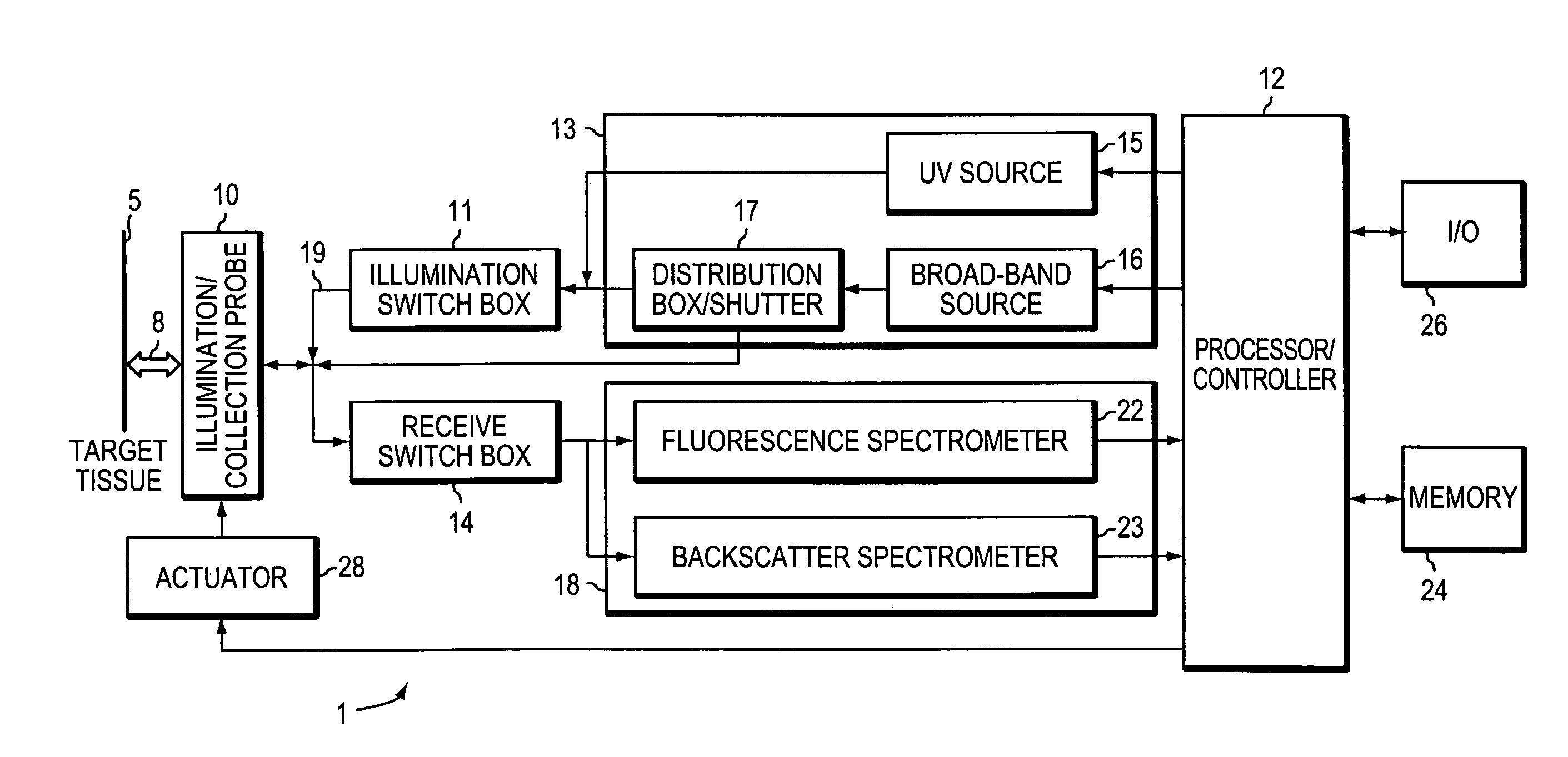
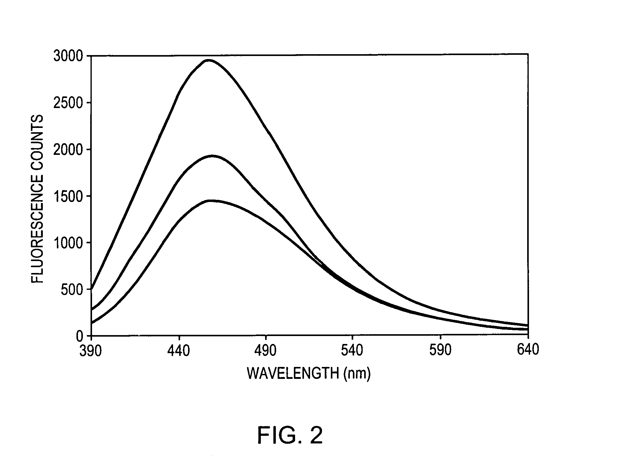
[0029]The present invention comprises systems and methods of using algorithms on optical responses of cervical tissue obtained from target tissues in subject population to be screened. The data are taken from a plurality of different sites within the target tissue, typically at least 100 such sites covering the region of interest in the tissue are probed. The optical response consists of fluorescence from the tissue induced by illumination with ultraviolet (UV) light and / or backscattering of white light from substantially the same tissue sites.

[0030]By way of background information, spectra are taken from the screened tissue, each from a different point in a given cervix. An algorithm, which classifies the detected spectra as either NED (No Evidence of Disease) or CIN (Cervical Intraepithelial Neoplasia), is applied to each of these spectra. Pathologists classify CIN into the three subgroups: CIN I, CIN II, and CIN III, depending of the stage or grade of the CIN. In addition, interm...


PUM

PropertyMeasurementUnit
wavelengthaaaaaaaaaa
spectral reflectivityaaaaaaaaaa

Abstract

A method and a system is provided for discriminating between healthy cervical tissue and pathologic cervical tissue based on the fluorescence response of the tissue to laser excitation (LIF) and the backscatter response to illumination by white light (in the spectral range of 360 to 750 nm). Combining LIF and white light responses, as well as evaluating a spatial correlation between proximate cervical tissue sites in conjunction with a statistically significant “distance” algorithm, such as the Mahalanobis distance between data sets, can improve the discrimination between normal and abnormal tissue. The results may be displayed in the form of a map of the cervix representing the suspected pathology.

Description

REFERENCE TO RELATED APPLICATIONS[0001]This application is a continuation of application Ser. No. 09 / 471,700, filed on Dec. 23, 1999, now U.S Pat. No. 6,427,082, issued Jul. 30, 2002, which claims priority to and the benefit of provisional patent application Ser. No. 60 / 113,624 filed on Dec. 23, 1998, the entire disclosures of which are incorporated by reference herein.FIELD OF THE INVENTION[0002]The present invention relates to optical devices and methods for screening of cervical tissue abnormalities.BACKGROUND OF THE INVENTION[0003]Early detection of a variety of pathologies has been shown to dramatically improve the therapeutic outcome for such pathologies. This is particularly true with cancer, and specifically with cancer of the cervix. In the case of cancer of the cervix and other cervix anomalies, the pap smear test has been used for many years as a screening method for the detection of pre-cancerous conditions as well as for the detection of cancer. Unfortunately, this meth...

Claims


Application Information

Patent Timeline
Patent Type & Authority Patents(United States)
IPC IPC(8): A61B6/00G01N21/64A61B5/00A61B10/00G01N21/27G01N21/35
CPCA61B5/0071A61B5/0075A61B5/0084A61B5/4331A61B5/0086
Inventor NORDSTROM, ROBERTMODELL, MARK
Owner LUMA IMAGING CORP
Why Eureka
Try Eureka