Method of analyzing and displaying blood volume using myocardial blood volume map

a technology of myocardial blood volume and analysis method, which is applied in the direction of ultrasonic/sonic/infrasonic image/data processing, instruments, applications, etc., can solve the problems of reducing the amount of blood supplied to the heart, affecting the heart's action, and requiring a long period of examination

Inactive Publication Date: 2007-10-23
YD +2
View PDF13 Cites 6 Cited by
  • Summary
  • Abstract
  • Description
  • Claims
  • Application Information

AI Technical Summary

Benefits of technology

[0015]In view of the foregoing, an object of the present invention is to provide a method for analyzing blood perfusion within the myocardium on the basis of a contrast echo image obtained by use of ultrasound diagnostic equipment, to thereby provide a diagnostician with data for visually and quantitatively determining the perfusion.
[0030]In an overlap sliding window method, a contrast echo image is finely divided into calculation regions in a manner that is basically the same as that used in the equal-interval sliding window method. However, the overlap sliding window method differs from the equal-interval sliding window method in the following points. In order to further reduce the influence of variation in acoustic field intensity, every time the intensity difference between the myocardium calculation region and the cardiac chamber calculation region is calculated for coloring the myocardium calculation region, the acoustic field lines are shifted by a small distance d3 (e.g., d3=0.5 mm) along a vertical axis perpendicular to the acoustic field lines. In this method, since the shifting distance is small, the calculation regions partially overlap. The overlap portion of the preceding calculation region is colored by a new color corresponding to the calculation value (intensity difference) of the succeeding calculation region. The reason while the vertical dimension of each calculation region is set to d1 despite the shifting distance being set to d3, which is smaller than d1, is to eliminate influence of noise (the influence of noise increases as the area of each calculation region decreases).

Problems solved by technology

X-ray diagnostic equipment, X-ray CT equipment, MRI equipment, or nuclear medical diagnostic equipment enables diagnosis of interior tissues of a human body without surgical operation, but such equipment involves the following problems: a subject is exposed to X-rays or nuclear radiation, and examination requires a long period of time.
When the coronary arteries are occluded or stenosed due to formation of thrombi or occurrence of coronary artery disorder, the amount of the blood supplied to the heart is reduced, causing adverse effects on action of the heart.
This symptom is called “myocardial infarction.” When the amount of the blood supplied to the myocardium is reduced as a result of narrowing of the coronary arteries, although occlusion of the arteries does not occur, the heart is adversely affected.
However, echo signals obtained by use of an ultrasound contrast agent predominantly containing microbubbles include non-fundamental wave components attributable to non-linear behaviors.
Therefore, difficulty is encountered in correctly determining the state of perfusion of blood on the basis of contrast echo images of the myocardium and the cardiac chambers.
However, in the conventional image processing technique, intensity measurement is performed without correction of variation in intensity, which variation occurs due to the difference in acoustic field or focusing of ultrasound waves.
Therefore, in the conventional technique, difficulty is encountered in determining whether the difference in intensity between different sites in the cardiac chambers or the myocardium is caused by the difference in acoustic field or by the difference in perfusion.
Meanwhile, diagnosing the state of perfusion of the cardiac chambers or the myocardium on the basis of a contrast echo image of the heart depends on the skill of a diagnostician, and an unskilled diagnostician encounters difficulty in correctly diagnosing such perfusion in a simple manner.
This is because image processing has not conventionally been performed objectively, and quantitative analysis of the contrast echo image has not been carried out.

Method used


Image

  • Method of analyzing and displaying blood volume using myocardial blood volume map
  • Method of analyzing and displaying blood volume using myocardial blood volume map
  • Method of analyzing and displaying blood volume using myocardial blood volume map

Examples


Embodiment Construction

[0044]An embodiment of the present invention will be described with reference to the appended drawings. In the present invention, the state of blood perfusion of the myocardium is analyzed on the basis of contrast echo images obtained by use of ultrasound diagnostic equipment. A computer unit is employed as hardware for analysis. Contrast echo images obtained by use of ultrasound diagnostic equipment are stored in a high-capacity external storage medium such as a CD or MO, and image analysis is performed by use of the thus-stored images.

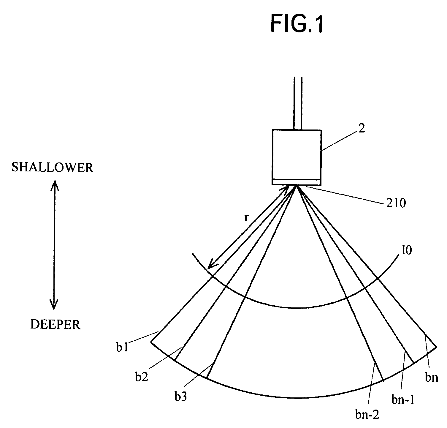
[0045]FIG. 1 shows scanning of the body by use of an ultrasound probe. Ultrasound beams b1, b2, . . . bn which are transmitted (actually, continuously transmitted) from a tip portion 210 of an ultrasound probe 2 applied to the body surface are reflected by an object, and the thus-reflected waves are received by the probe 2. The thus-received waves are converted to a digital contrast echo image, which is then stored in a memory or a storage device. In...


PUM


Abstract

A contrast echo image of the myocardium and a cardiac chamber obtained by use of ultrasound diagnostic equipment is analyzed so as to determine the volume of blood within the myocardium. The contrast echo image is divided into a plurality of calculation regions, each being considered to have a uniform acoustic field intensity and covering a portion of a myocardium image region and a corresponding portion of a cardiac chamber image region. For each calculation region, a difference in intensity between the myocardium image region and the cardiac chamber image region is obtained, and the myocardium image region is colored in accordance with the difference.

Description

BACKGROUND OF THE INVENTION[0001]1. Field of the Invention[0002]The present invention relates to a contrast echo image analysis method employed for detection or treatment of ischemic heart diseases or similar diseases, in which a contrast echo image of the myocardium and cardiac chambers—which image is obtained by use of ultrasound diagnostic equipment after injection of a microbubble ultrasound contrast agent into blood vessel—is analyzed so as to visually and quantitatively determine the volume of blood flowing through the myocardium on the basis of intensity values of the myocardium region and the cardiac chamber regions of the image.[0003]2. Background Art[0004]X-ray diagnostic equipment, X-ray CT equipment, MRI equipment, or nuclear medical diagnostic equipment enables diagnosis of interior tissues of a human body without surgical operation, but such equipment involves the following problems: a subject is exposed to X-rays or nuclear radiation, and examination requires a long p...

Claims


Application Information

Patent Timeline
Patent Type & Authority Patents(United States)
IPC IPC(8): A61B8/14A61B8/06A61B8/00A61B8/08G06T1/00
CPCA61B8/481
Inventor ITO, HIROSHINAKAJIMA, YASUHIRO
Owner YD
Why Eureka
Try Eureka