
Active tissue augmentation materials and method

a soft tissue and active technology, applied in the field of soft tissue augmentation, can solve the problems of limited long-term use, long-term prosthetic devices, limitations and drawbacks of each existing treatment, etc., to prevent absorption by macrophages, increase viscosity and apparent density, and prevent circulatory insufficiency

Inactive Publication Date: 2008-02-05
TORAX MEDICAL
View PDF49 Cites 24 Cited by
  • Summary
  • Abstract
  • Description
  • Claims
  • Application Information

AI Technical Summary

Benefits of technology

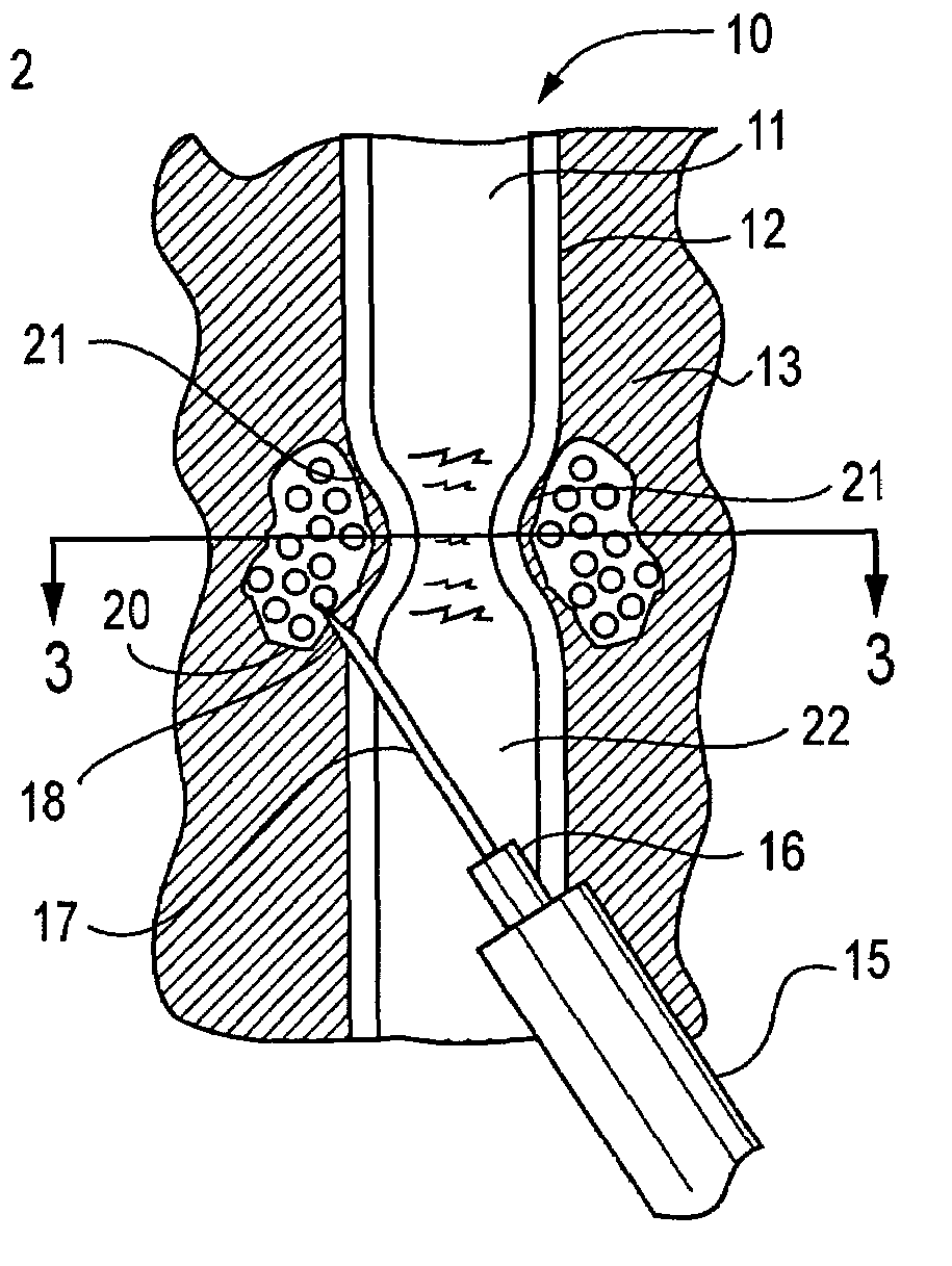
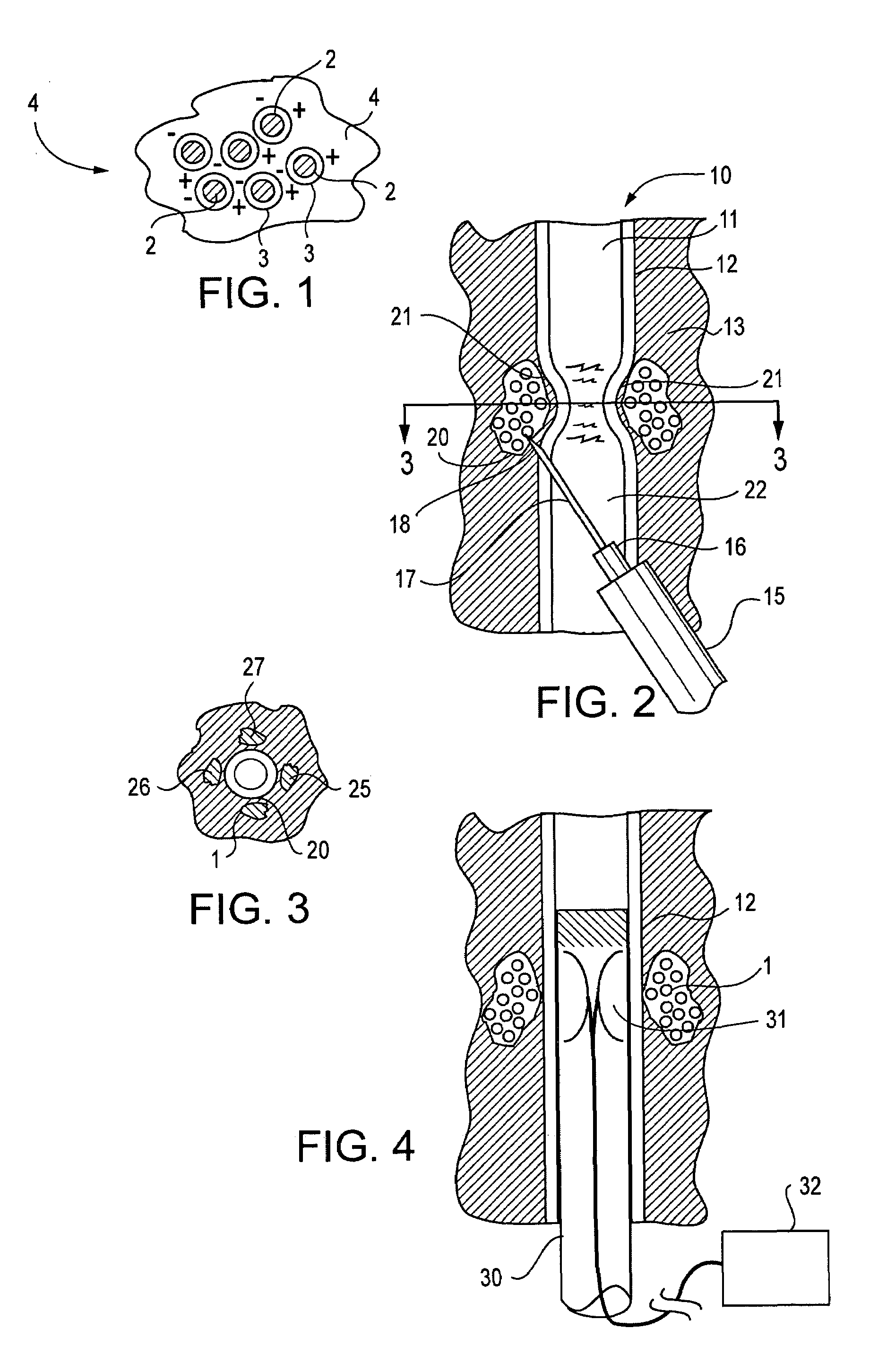
[0024]A suspension of magnetically active particles according to the invention provides an active device that utilizes magnetic particles in such a way as to prevent absorption by macrophages while providing a compliant surface to the urethra in order to prevent circulatory insufficiency. When deposited in tissue surrounding a lumen, for example the urethral lumen, the small, permanent magnetic fields created by the magnetically active particles can cause an artificial increase in the viscosity and apparent density of the suspension.
[0025]Thus, in one embodiment, the invention provides an injectible, magnetically active control media and method providing a unique solution to the growing number of people suffering from urinary incontinence due to sphincter insufficiency. The injectible magnetically active media addresses the shortcomings of many existing products and procedures available to physicians and supplies a unique solution to restoring individual control over bladder function.

Problems solved by technology

However, each of the existing treatments have limitations and drawbacks.
Injectible implants of collagen have proven to be successful in mild cases, but are of limited long term use as the collagen is absorbed by the body over a relatively short period of time and must constantly be replaced.
The use of a long term prosthetic device has, historically, been the least desirable as these devices can cause irritation to the of the urethra and ultimately tissue necrosis if the prosthetic device causes circulatory insufficiency.
In addition, the application of the obturating collars of some older devices required that the urethra be severed and reattached, further complicating the surgical procedure and causing additional trauma to the tissue.
However, undesirable subsequent particle migration and granulomatous reactions have commonly resulted.
While these materials create immediate augmentation, the result is generally short-lived as the material also has a tendency to migrate and / or be reabsorbed from the injection site by the host tissue.
In creating the localized pressure necessary to overcome the material back pressures, the cuff type devices also tend to obstruct the normal flow of blood in the conduit walls, and thus long term usage of cuff-type artificial sphincters is not viable.
The continuous ring of pressure without adequate blood flow can result in scarring and localized tissue damage.
While the device is made to apply forces only on two opposite sides of the conduit that it is surrounding, rather than in a noose-like complete ring, in order to prevent necrosis, the unit is still difficult to operate, and varying the closing pressure is difficult because the magnet used is for actuation of an over center actuating device.
It can be seen that such devices have limitations in the force that can be generated, and require the use of external controls for operation.
Also, the coils can cause closure which is too tight and thus, can cause damage to the tissue as well.
While some of these approaches have enjoyed modest success, relief has been for the most part, only temporary in those patients where success is noted.

Method used


Image

  • Active tissue augmentation materials and method
  • Active tissue augmentation materials and method

Examples


Embodiment Construction

[0031]The present invention is directed to active tissue augmenting agents, compositions and procedures for active tissue augmentation when the agents are deposited at selected locations within or near a tissue to be augmented. In some embodiments, passive tissue augmentation can also occur due to the bulk of the compositions deposited within the tissue. The invention is particularly advantageous for amelioration of sphincter disorders in humans and animals.

[0032]As used herein, to “augment” a tissue means to cause the tissue to have an increase in size or a change in configuration relative to the size or configuration of the tissue prior to augmentation. “Active augmentation” means that the tissue configuration is altered as a result of an attracting or repelling force exerted by an agent deposited in or near the tissue. In contrast, “passive augmentation” means that the tissue configuration is altered as a result of volumetric displacement of the tissue contours due to the volume ...


PUM

PropertyMeasurementUnit
sizeaaaaaaaaaa
sizeaaaaaaaaaa
diameteraaaaaaaaaa

Abstract

Active tissue augmenting agents, compositions and methods for use are disclosed. In a typical embodiment, the active augmenting agents of the invention can be used to form an artificial sphincter around a lumen of a human or animal body. In one embodiment, the active augmenting agent comprises magnitizable particles which can provide occlusion of a lumen, such as the urethral lumen, by circumferential attraction of the injected material toward the center of the lumen by the inherent magnetic flux field created from the magnetic dipoles of the magnetic particles.

Description

Cross-Reference to Related Applications[0001]This application is a continuation of U.S. patent application Ser. No. 10 / 371,012, filed Feb. 20, 2003 now abandoned, which is a continuation of U.S. patent application Ser. No. 09 / 634,414, filed Aug. 8, 2000 now abandoned, both of which applications are incorporated herein by reference in their entireties.FIELD OF THE INVENTION[0002]The present invention relates to soft tissue augmentation. In particular the invention provides materials and methods for active augmentation of soft tissue structures. The invention can be particularly advantageous for the treatment of sphincter disorders including urological disorders such as incontinence, vesicoureteral reflux; and gastro-intestinal disorders including gastric fluid reflux, anal sphincter incontinence, etcBACKGROUND OF THE INVENTION[0003]Urinary incontinence is a dysfunction of the bladder's ability to securely retain urine until an individual intends to void. Causes for urinary incontinen...

Claims


Application Information

Patent Timeline
Patent Type & Authority Patents(United States)
IPC IPC(8): A61F2/00A61B17/34A61L27/04A61L27/44A61L27/50A61L31/12A61L31/14A61L31/18
CPCA61F2/0036A61L27/04A61L27/446A61L27/50A61L31/128A61L31/14A61L31/18A61B17/3478A61F5/0069
Inventor MILLER, PAUL LEONARD
Owner TORAX MEDICAL
Learn More