Device and method to limit filling of the heart

a technology of limiting devices and heart valves, applied in the field of devices and methods to limit heart valve filling, can solve the problems of progressive dilatation of the heart, ineffective mechanism, and deleterious effect of mechanical devices, so as to prevent further enlargement reduce the size of the diseased heart, and limit the volume of blood

Active Publication Date: 2008-03-11
STARKEY THOMAS DAVID
View PDF53 Cites 42 Cited by
  • Summary
  • Abstract
  • Description
  • Claims
  • Application Information

AI Technical Summary

Benefits of technology

[0026]The invention described in this patent application is a device used to treat heart disease by decreasing the size of a diseased heart, or to prevent further enlargement of a diseased heart. The device works by limiting the volume of blood entering the heart during each cardiac cycle. The device is called a “diastolic volume limiting apparatus”, or “divola” for short. The device partitions blood within the heart, and protects the heart from excessive volume and pressure of blood. This new invention is placed within the interior of the heart, particularly within the left ventricular cavity. The device is a hollow sac, with two openings, which simulates the shape and size of the interior lining of a normal heart. It allows the heart to fill through one opening juxtaposed to the annulus of the mitral valve to a predetermined, normal volume, and limits filling of the heart beyond that volume. It then allows blood to be easily ejected through the second opening through the aortic valve. By limiting the amount of blood entering the heart, the left ventricle is not subjected to the harmful effect of excessive volume and pressure of blood during diastole, the period of the cardiac cycle when the heart is at rest. This allows the heart to decrease in size, or to reverse remodel, and to recover lost function. In some applications, a second device, referred to as a “volume compensating device”, or “VCD” for short, may be simultaneously placed inside the heart to take up excessive space between the heart and the primary device. This invention may be used to treat a variety of heart disease, most importantly diseases affecting the left ventricle. This invention is a significant improvement over mechanical heart assist devices because of its simplicity, durability, and significant cost benefit.

Problems solved by technology

However, this mechanism becomes ineffective, and actually deleterious, beyond a mild degree of dilatation.
Excessive volume of blood and increased filling pressures may actually cause progressive dilatation of the heart.
A vicious cycle of increased blood volume resulting in increased heart failure resulting in increased blood volume, etc., is started.
The heart is particularly susceptible to dilatation from excessive pressure and volume during diastole, the period of the cardiac cycle when the heart is at rest.
Remodeling, a term used to describe abnormal enlargement of the heart, causes the heart to function less efficiently.
None of the modes of conventional cardiac surgery directly prevent progressive or persistent dilatation of the heart.
This report also demonstrates that the heart may shrink in size when excessive volume of blood in the heart is treated.
Unfortunately, not all of the patients in this group had significant of long-term benefit, and some required transplantation.
The left ventricle can rupture in the free wall causing a cardiac tamponade, or it may rupture into the right ventricle, causing an ischemic ventricular septal defect.
Currently there is no medical device that can be used to treat this fatal disease.
This technique surgically decreases the size of the left ventricle, but unfortunately does not prevent progressive and recurrent dilatation of the heart.
There are unfortunately a limited number of donors, and only a very small fraction of patients who would benefit from heart transplantation are able to receive a donor heart.
Heart transplantation requires immunosuppression, which has many serious and sometimes fatal side effects.
Heart transplantation is extremely expensive, with the initial cost of approximately US$150,000 to 200,000, and recurrent yearly costs of approximately US$25,000 to 40,000.
Currently, the use of mechanical cardiac assist devices is limited by frequent and common complications of the implantation and use of the devices.
The complications include thrombosis, thrombotic emboli causing stroke, organ, and limb ischemia, infection, particularly from the driveline, as well as catastrophic mechanical failure of the device.
Furthermore, these devices are extremely expensive, with some costing approximately US$85,000.
An intraventricular rotary blood pump is now in clinical use (Jarvik, U.S. Pat. No. 5,824,070), but with only modest improvement of the patients, and an important rate of complications.
These devices may erode into the vital structure of the heart, possibly even perforating the heart.
The rigid parts of these devices have the potential to erode into the vital structure of the heart, possibly even perforating the heart.
These devices have the potential to disrupt the mitral valve and subvalvular apparatus, as well as the potential to erode into other vital structure of the heart, possibly even perforating the heart.
There is a potential for bleeding from the wires puncturing the left ventricular chamber of the heart.
These devices do not prevent further dilatation of the heart, nor do they direct shrinking or reverse remodeling of the heart.
The device has the potential to slip out of position, to constrict the heart, and to cause formation of scar tissue around the heart.
Of these patients who had removal of a device, some have had sustained benefit of the reverse remodeling, although many unfortunately have recurrent progression of their dilated heart disease.

Method used


Image

  • Device and method to limit filling of the heart
  • Device and method to limit filling of the heart
  • Device and method to limit filling of the heart

Examples


Embodiment Construction

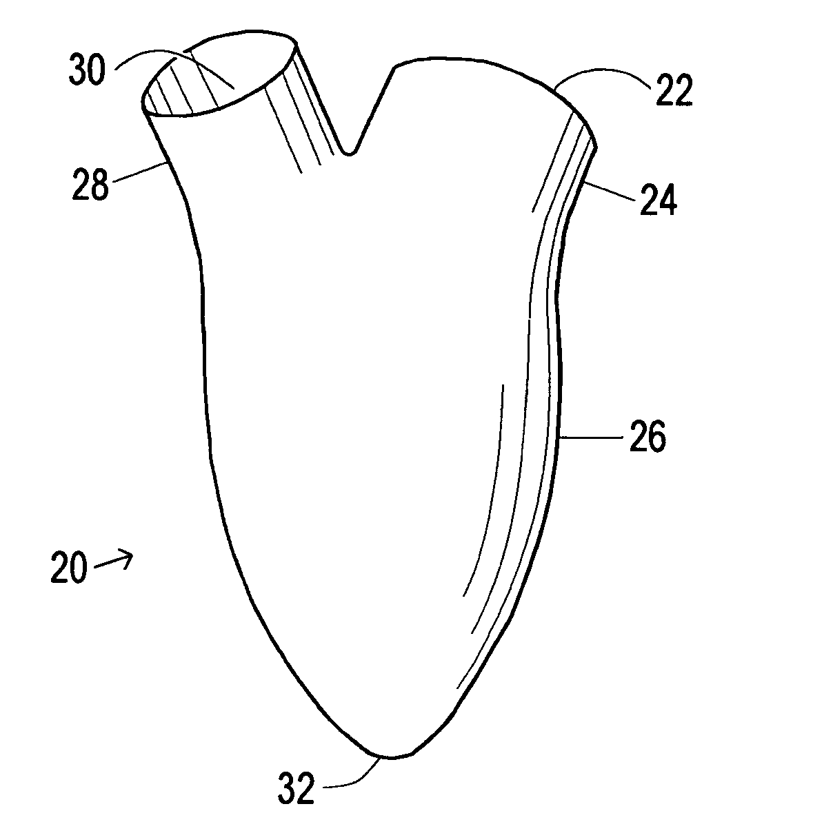
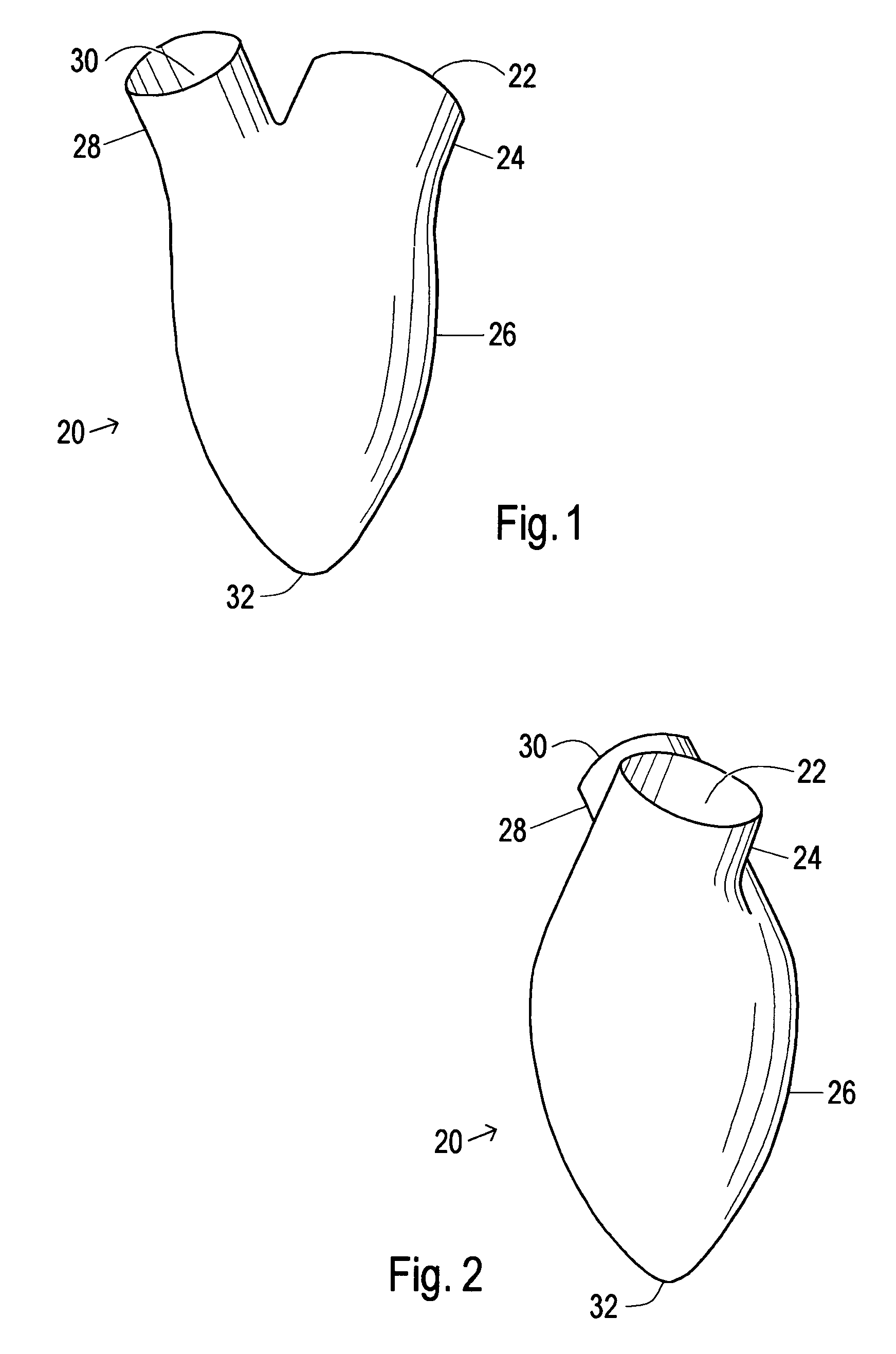
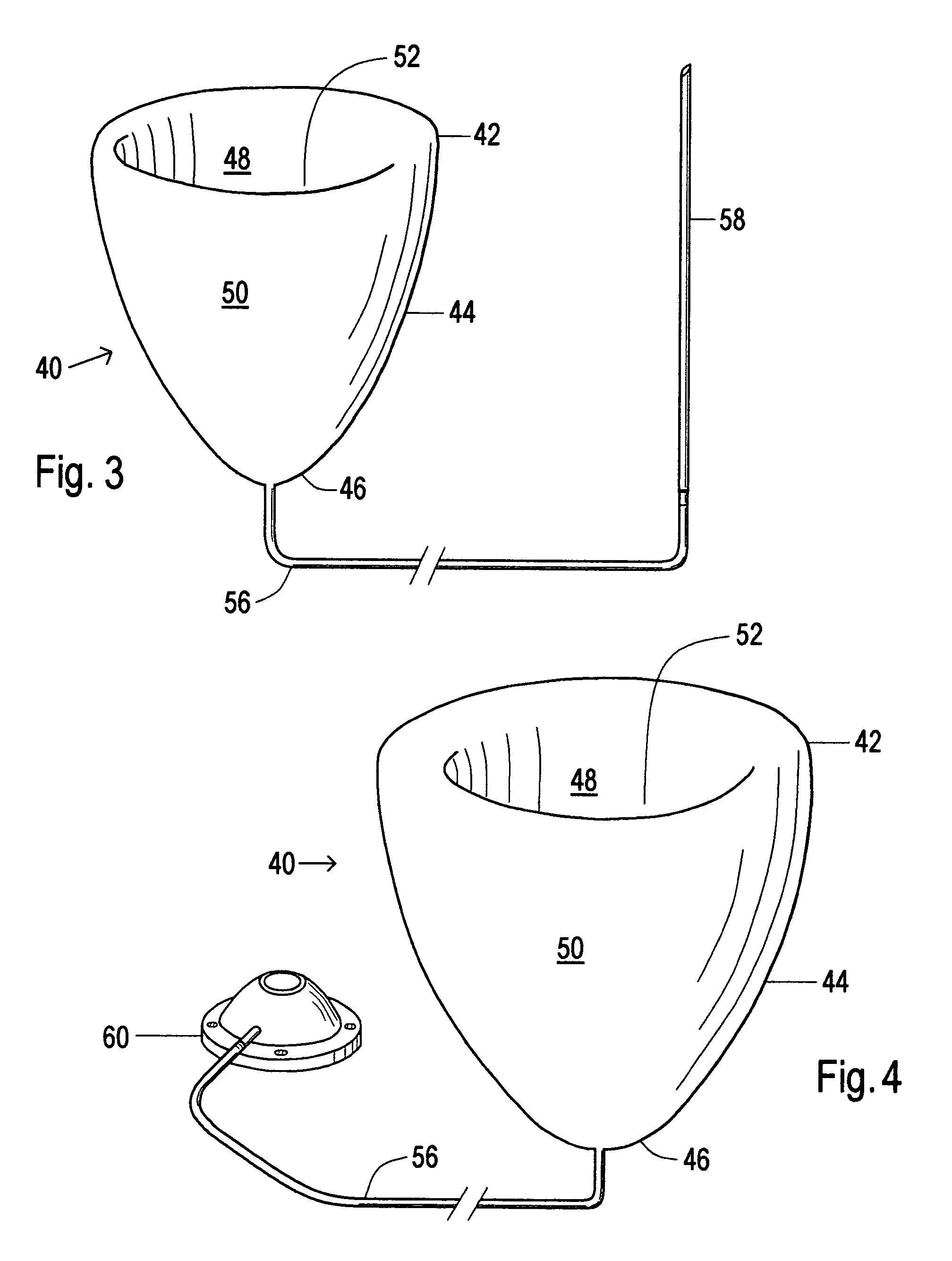
[0044]The purpose of the invention is to allow for recovery of a diseased heart. The invention works by limiting the amount of blood that fills a ventricular cavity during each cardiac cycle. The primary device is called a diastolic volume limiting apparatus, or divola for short. The divola is a hollow plastic sac with two openings. The divola is usually placed within the left ventricular chamber of the heart. The divola is soft and compliant, and fills easily with blood to a certain, predetermined volume. When the divola has reached capacity, no further filling is allowed. By limiting filling of the left ventricle, the heart is not stretched by the excessive volume and pressure of blood. This allows for recovery of a diseased heart, or prevents progression or recurrence of heart disease. In some applications, a secondary device, called a volume compensating device or (VCD) may be simultaneously placed into the ventricle to take up excessive space between the ventricle and the prima...


PUM


Abstract

A device used to treat heart disease by decreasing the size of a diseased heart, or to prevent further enlargement of a diseased heart. The device works by limiting the volume of blood entering the heart during each cardiac cycle. The device partitions blood within the heart, and protects the heart from excessive volume and pressure of blood. The device is placed within the interior of the heart, particularly within a ventricular cavity. The device is a hollow sac, with two openings, which simulates the shape and size of the interior lining of a ventricle of a normal heart. It allows the ventricle to fill through one opening juxtaposed to the annulus of the inflow valve to a predetermined, normal volume, and limits filling of the heart beyond that volume. It then allows blood to be easily ejected through the second opening through the outflow valve. By limiting the amount of blood entering the ventricle, the ventricle is not subjected to the harmful effect of excessive volume and pressure of blood during diastole, the period of the cardiac cycle when the heart is at rest. This allows the heart to decrease in size, or to reverse remodel, and to recover lost function. In some applications, a second device may be simultaneously placed inside the heart to take up excessive space between the heart and the primary device.

Description

[0001]This patent application is filed for the invention by Thomas David Starkey, of Nashville, Tenn. of A Device and Method to Limit Filling of the Heart. The application is based on and claims priority of previously filed Provisional Application Ser. No. 60 / 474,310, filed on May 30, 2003.STATEMENT REGARDING FEDERALLY SPONSORED RESEARCH OR DEVELOPMENT[0002]The research and development leading to the present invention was not federally sponsored.BACKGROUND OF THE INVENTION[0003]The invention described in this application pertains to the class of medical devices used to treat heart disease that function to decrease the size of the heart, or to prevent enlargement of the heart.[0004]Heart disease is common in our society, and the incidence is rapidly increasing. One class of heart disease, heart failure, affects 4 to 5 million patients in the United States, with up to two million hospitalizations per year. This accounts for 2 to 3 percent of the national healthcare budget. The America...

Claims


Application Information

Patent Timeline
Patent Type & Authority Patents(United States)
IPC IPC(8): A61N1/362A61M1/10A61F2/00
CPCA61B17/12122A61B17/12136A61F2/2481
Inventor STARKEY, THOMAS DAVID
Owner STARKEY THOMAS DAVID
Why Eureka
Try Eureka