Methods for treating muscle diseases and disorders
a technology for applied in the field of methods for treating muscle diseases and disorders, can solve the problems of no useful therapy for the promotion of muscle differentiation and survival, and achieve the effects of increasing reducing the number of nuclei in myotubes, and reducing the number of labelled nuclei
- Summary
- Abstract
- Description
- Claims
- Application Information
AI Technical Summary
Benefits of technology
Problems solved by technology
Method used
Image
Examples
example 1
Mitogenic Activity of rhGGF on Myoblasts
[0159]Clone GGF2HBS5 was expressed in recombinant Baculovirus infected insect cells as described in Example 13, infra, and the resultant recombinant human GGF2 was added to myoblasts in culture (conditioned medium added at 40 μl / ml). Myoblasts (057A cells) were grown to preconfluence in a 24 well dish. Medium was removed and replaced with DMEM containing 0.5% fetal calf serum with or without GGF2 conditioned medium at a concentration of 40 μl / ml. Medium was changed after 2 days and cells were fixed and stained after 5 days. Total nuclei were counted as were the number of nuclei in myoblasts (Table 1).
[0160]
TABLE 1Total NumberNuclei inFusionTreatmentof Nuclei / mm2MyotubesIndexControl395 ± 28.3204 ± 9.190.515 ± 0.01GGF 40 μl / ml636 ± 8.5 381 ± 82.70.591 ± 0.15
GGF treated myoblasts showed an increased number of total nuclei (636 nuclei) over untreated controls (395 nuclei) indicating mitogenic activity. rhGGF2 treated myotubes had a greater number ...
example 2
Effect of rhGGF2 on Muscle Cell Mitogenesis
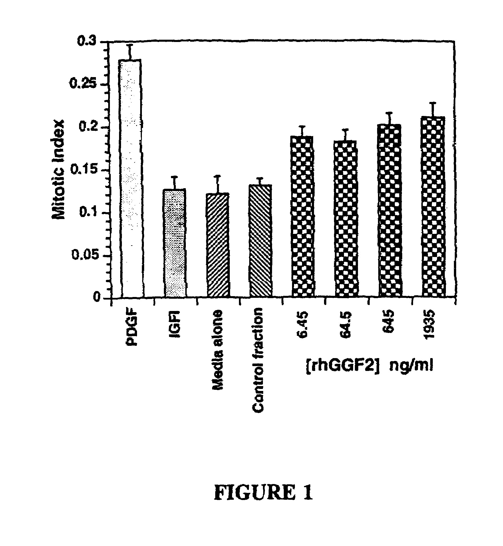
[0162]Quiescent primary clonal human myoblasts were prepared as previously described (Sklar, R., Hudson, A., Brown, R., In vitro Cellular and Developmental Biology 1991; 27A:433-434). The quiescent cells were treated with the indicated agents (rhGGF2 conditioned media, PDGF with and without methylprednisolone, and control media) in the presence of 10 μM BrdU, 0.5% FCS in DMEM. After two days the cells were fixed in 4% paraformaldehyde in PBS for 30 minutes, and washed with 70% ethanol. The cells were then incubated with an anti-BrdU antibody, washed, and antibody binding was visualized with a peroxidase reaction. The number of staining nuclei were then quantified per area. The results show that GGF2 induces an increase in the number of labelled nuclei per area over controls (see Table 2).
[0163]
TABLE 2Mitogenic Effects of GGF on Human MyoblastsLabelledT-TestTreatmentNuclei / cm2p valueControl120 ± 22.4Infected Control103 ± 11.9GGF 5 μl / ml223 ±...
example 3
Effect of rhGGF2 on Muscle Cell Differentiation
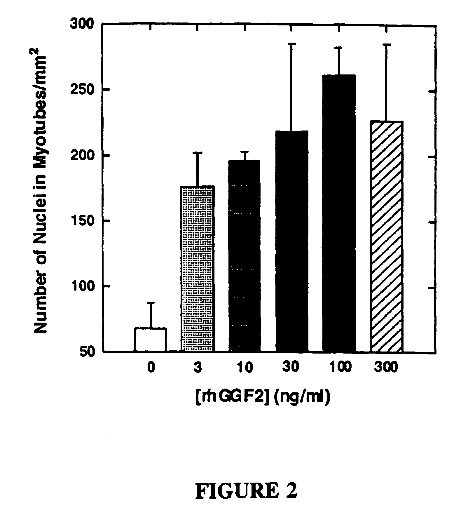
[0166]The effects of purified rhGGF2 (95% pure) on muscle culture differentiation were examined (FIG. 2). Confluent myoblast cultures were induced to differentiate by lowering the serum content of the culture medium from 20% to 0.5%. The test cultures were treated with the indicated concentration of rhGGF2 for six days, refreshing the culture medium every 2 days. The cultures were then fixed, stained, and the number of nuclei counted per millimeter. The data in FIG. 2 demonstrate a large increase in the number of nuclei in myotubes when rhGGF2 is present, relative to controls.
PUM
| Property | Measurement | Unit |
|---|---|---|
| size | aaaaa | aaaaa |
| concentration | aaaaa | aaaaa |
| pH | aaaaa | aaaaa |
Abstract
Description
Claims
Application Information
 - R&D
- Intellectual Property
- Life Sciences
- Materials
- Tech Scout
- Unparalleled Data Quality
- Higher Quality Content
- 60% Fewer Hallucinations
Browse by: Latest US Patents, China's latest patents, Technical Efficacy Thesaurus, Application Domain, Technology Topic, Popular Technical Reports.
© 2025 PatSnap. All rights reserved.Legal|Privacy policy|Modern Slavery Act Transparency Statement|Sitemap|About US| Contact US: help@patsnap.com


