Method for staining corneocytes, method for preparing corneocytes specimen and skin analysis system
a corneocyte and skin analysis technology, applied in the field of corneocyte staining corneocytes, can solve the problems of reducing the staining step, affecting the quality of corneocytes, so as to reduce the time and eliminate the staining step
- Summary
- Abstract
- Description
- Claims
- Application Information
AI Technical Summary
Benefits of technology
Problems solved by technology
Method used
Image
Examples
example 1
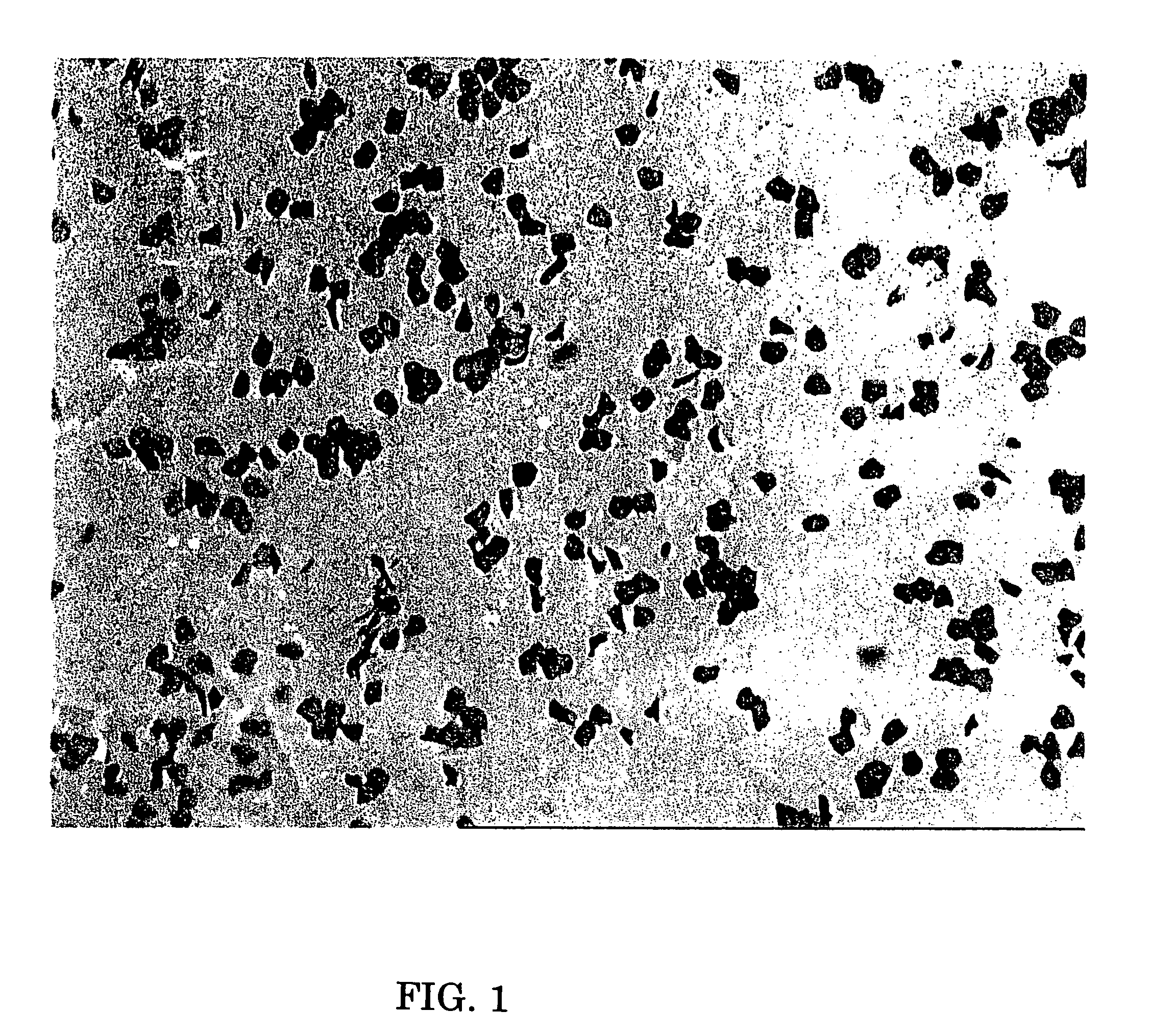
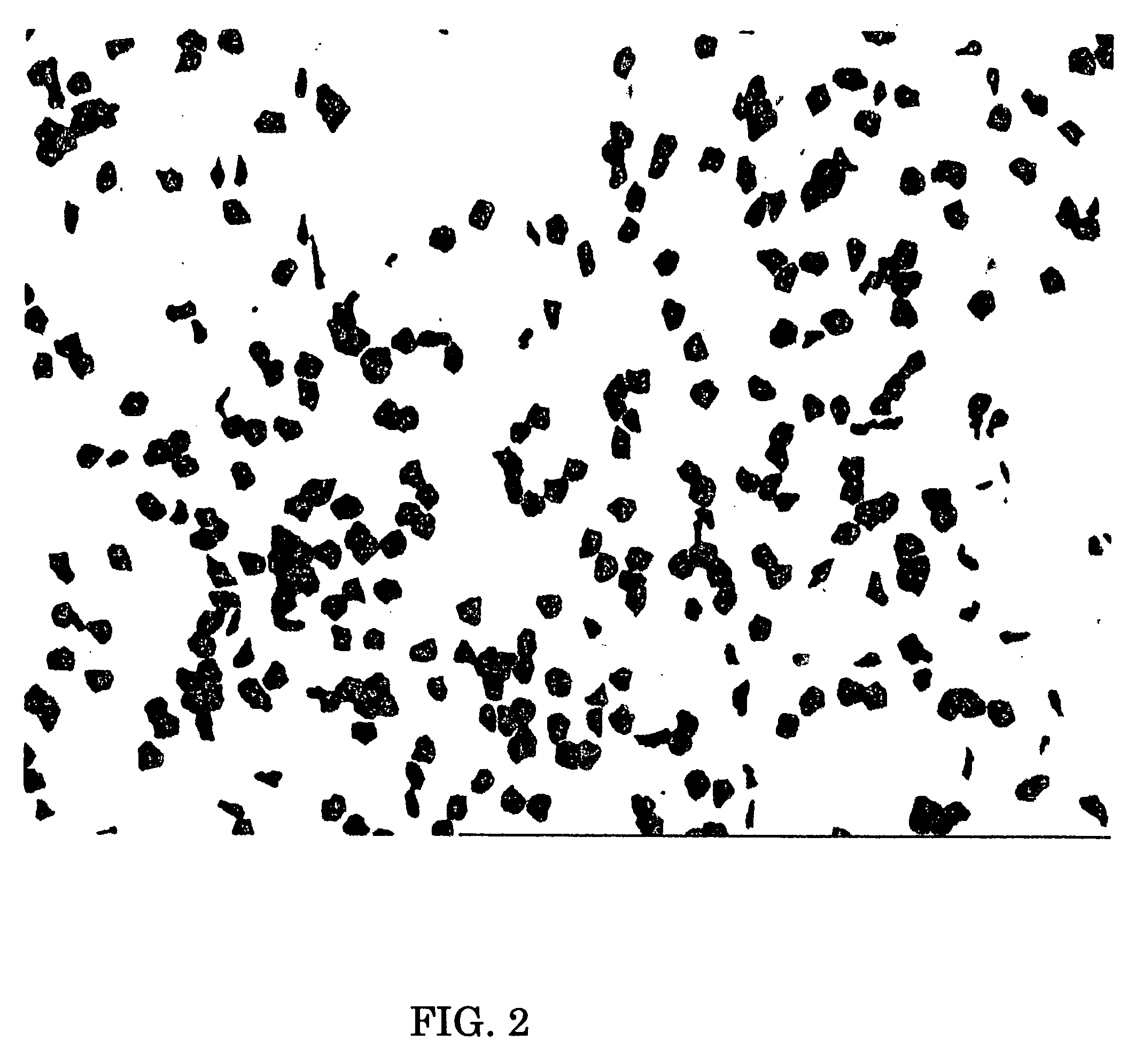
[0076]Three horny cell samples were prepared from one panelist by stripping corneocites off from the surface of his / her skin using an adhesive tape, and two of them were stained according to the inventive staining method. Particularly, the two samples were immersed in a dyeing solution of 3 wt % gentian violet and 1 wt % brilliant green in an aqueous solution of 20% ethanol for 2 minutes and then washed in water sufficiently. One of them was mounted in an ultraviolet-curing resin and then cured by exposure to UV light (Comparative Example 1) while the other was mounted in dimethicone (silicone) according to the present invention (Example 1). The remaining sample was immersed in a dyeing solution of 1 wt % gentian violet and 0.5 wt % brilliant green in water for 10 minutes, washed in water sufficiently, and then mounted in dimethicone (Comparative Example 2). Microscopic photographs of these samples are shown in FIGS. 1-3. As shown in FIGS. 1-3, a low contrast was obtained due to col...
example 2
[0077]Average size was calculated for each of the horny cell specimens obtained by Example 1, Comparative Examples 1 and 2. The image of the horny cell specimen of Example 1 was manually binarized and used to calculate an average size (μm2). Twenty cells were averaged for each example. The results are shown in Table 1 below. Those results show that automatic calculation of size gave the same result as the result obtained by manual calculation according to the present invention. On the other hand, automatic calculation of size gave higher values than the actual results for the specimens with less clear staining or color running.
[0078]
TABLE 1SAMPLEAVERAGE SIZE ± VARIANCEAUTOMATIC DETERMINATION568 ± 121(EXAMPLE 1)AUTOMATIC DETERMINATION721 ± 237(COMPARATIVE EXAMPLE 1)AUTOMATIC DETERMINATION736 ± 175(COMPARATIVE EXAMPLE 2)MANUAL DETERMINATION559 ± 134(EXAMPLE 1)
example 3
[0079]Similar process was repeated as in Example 1 except for using variable amounts of gentian violet. Again, 1% by weight of brilliant green was used. Dimethicone was used for mountion. The clearness of staining were indicated as: ◯=clear; Δ=less clear; X not clear. The results are shown in Table 2 below. Those results show that gentian violet may preferably be present at an amount of 1.5-5% by weight and more preferably 2-4% by weight.
[0080]
TABLE 2CONC. OF GENTIAN VIOLETSTAINING COLOR DEFINITION1.5% BY WEIGHT◯~Δ2.5% BY WEIGHT◯4.5% BY WEIGHT◯
PUM
| Property | Measurement | Unit |
|---|---|---|
| weight | aaaaa | aaaaa |
| size | aaaaa | aaaaa |
| roughness | aaaaa | aaaaa |
Abstract
Description
Claims
Application Information
 - R&D
- Intellectual Property
- Life Sciences
- Materials
- Tech Scout
- Unparalleled Data Quality
- Higher Quality Content
- 60% Fewer Hallucinations
Browse by: Latest US Patents, China's latest patents, Technical Efficacy Thesaurus, Application Domain, Technology Topic, Popular Technical Reports.
© 2025 PatSnap. All rights reserved.Legal|Privacy policy|Modern Slavery Act Transparency Statement|Sitemap|About US| Contact US: help@patsnap.com


